Effect of Heat-Killed Enterococcus faecalis, EF-2001 on C2C12 Myoblast Damage Induced by Oxidative Stress and Muscle Volume Decreased by Sciatic Denervation in C57BL/6 Mice
Sang-Jin Chang1, Myung-Hun Lee1, Wan-Joong Kim1, Yuri Chae1, Masahiro Iwasa1,2, Kwon-Il Han1,2, Wan-Jae Kim2 and Tack-Joong Kim1,3*
1Division of Biological Science and Technology, Yonsei University, Wonju 26493, Korea
2Research & Development Center, Korea BeRM Co. Ltd., Wonju 26361, Korea
3Research & Development Center, Doctor TJ Co. Ltd., Wonju 26493, Korea
Received December 6, 2018 /Revised December 28, 2018 /Accepted December 28, 2018
Muscle dysfunction may arise from skeletal muscle atrophy caused by aging, injury, oxidative stress, and hereditary disease. Powdered heat-killed Enterococcus faecalis (EF-2001) has anti-allergy, anti-in- flammatory, and anti-tumor effects. However, its antioxidant and anti-atrophy effects are poorly characterized. In this study, we examined the effects of EF-2001 on muscle atrophy. To determine the protective effect of EF-2001 on oxidative stress, C2C12 myoblasts were treated with H2O2 to induce oxidative stress. This induced cell damage, which was reduced by treatment with EF-2001. The mecha- nism of EF-2001’s effect was examined in response to oxidative stress. Treatment with EF-2001 re- versed the expression of HSP70 and SOD1 proteins. Also, mRNA levels of Atrogin-1/MAFbx and MuRF1 increased under oxidative stress conditions but decreased following EF-2001 treatment. To evaluate muscle volume, two and three dimensional models of the muscles were analyzed using mi- cro-CT. As expected, muscle volume decreased after sciatic denervation and recovered after oral ad- ministration of EF-2001. Therefore, EF-2001 is a candidate for the treatment of muscular atrophy, and future discovery of the additional effects of EF-2001 may yield further applications as a functional food with useful activities in various fields.
Key words : EF-2001, Heat-killed Enterococcus faecalis, muscle atrophy, oxidative stress, sciatic denervation
*Corresponding author
*Tel : +82-33-760-2242, Fax : +82-33-760-2183
*E-mail : [email protected]
This is an Open-Access article distributed under the terms of the Creative Commons Attribution Non-Commercial License (http://creativecommons.org/licenses/by-nc/3.0) which permits unrestricted non-commercial use, distribution, and reproduction in any medium, provided the original work is properly cited.
Journal of Life Science 2019 Vol. 29. No. 2. 215~222 DOI : https://doi.org/10.5352/JLS.2019.29.2.215
Introduction
Muscle atrophy is defined as the loss of muscle tissue that occurs as a result of damage to muscle-innervating nerves or the muscle itself. The most important cause of atrophy is disuse atrophy, which is caused by the non-use of muscle.
When activity decreases, muscle tone decreases, which grad- ually proceeds to atrophy. The physical behaviors of all the animals, including humans, are related to muscle strength, and dysfunctions of physical behavior have negative effects because they affect the ability to exercise, social life, and daily life. Muscle atrophy usually results from the reduced muscle use that occurs in an immobile state, such as during long- term care, after nerve removal, and during space travel [18].
Under oxidative stress, cells activate various proteins to protect against damage and cell death. For example, super- oxide dismutase 1 (SOD1) is a protein that is capable of scav- enging radicals, such as intracellular reactive oxygen species ROS [9, 19, 20]. When ROS penetrates cells and cause dam- age, the expression of SOD1 is increased to promote homeo- stasis and stability. Heat shock protein 70 (HSP70) protects cells from various apoptotic stimuli, including heat shock, tumor necrosis factor, oxidative stress, ceramides, and radia- tion by mitigating the various effects of stress, thus allowing cells to tolerate such external stimuli, and inhibiting cell death [7, 12, 15, 16, 18, 21]. Muscle ring finger protein 1 (Murf1) is a protein that is expressed in myoblasts and con- tains a weak ring domain [13]. When MuRF1 expression is increased in the myoblasts, vital proteins are ubiquitinated to induce cell death [5].
The heat-killed Enterococcus faecalis (EF-2001) has pre- viously been shown to have several beneficial effects on hu- man health including anti-allergy, anti-inflammatory and anti-tumor activities [3, 4, 6], but so far anti-muscle atrophy effects of this EF-2001 have been unclear.
In the present study, we examined the effects of EF-2001 on muscle atrophy both under oxidative stress in myoblasts and following sciatic denervation in mice, and confirmed that several cellular signal expression levels, including HSP70, and SOD1 are affected by EF-2001 in situations of oxidative stress induced by disuse muscle atrophy.
Materials and Methods
Materials
The EZ-Cytox cell viability kit was purchased from Daeil Lab (Seoul, Korea). Penicillin-streptomycin and fetal bovine serum (FBS) were obtained from Capricorn (Ebsdorfergrund, Germany). N-acetyl-L-cysteine (NAC), TRI-reagent, and trypsin-EDTA solution were purchased from Sigma-Aldrich (St. Louis, MO, USA). Phosphate buffered saline (PBS) was purchased from Gibco Life Technologies, Inc. (Rockville, MD, USA). An antibody against HSP70 was purchased from Enzo Life Sciences (AG, Switzerland). Antibodies against SOD1 and β-actin as well as anti-rabbit IgG and anti-mouse IgG were purchased from Cell Signaling Technology (Dan- vers, MA, USA).
Heat-killed Enterococcus faecalis (EF-2001) EF-2001 is a commercially available probiotic that was originally isolated from healthy human feces. It was sup- plied by Nihon BeRM Co. Ltd., (Tokyo, Japan) as a heat-kil- led, dried powder. One gram of dried E. faecalis is equivalent to 7.5×10¹² colony-forming units (CFU) prior to heat-killing.
Cell culture
C2C12 myoblasts were cultured in Dulbecco’s Modified Eagle’s Medium (DMEM; Sigma-Aldrich) containing 10%
(v/v) FBS and penicillin-streptomycin (100 μg/ml). C2C12 myoblasts were cultured in 100 mm cell culture dishes at 37℃ in a humidified, 5% CO2 incubator.
Cell viability
When C2C12 myoblasts reached 70% confluence, the me- dium was suctioned and the medium was replaced with se- rum-free DMEM containing EF-2001 at various concen- trations (0, 25, 50, 100, 250, and 500 μg/ml) and cultured for 24 hr. Then, the C2C12 myoblasts were treated with 1 mM H2O2 in serum-free DMEM for 2 hr. EZ-Cytox kit re- agent (10 μl) was added to each well of the plate, and then incubated for 1 h. The cell viability was measured at 450 nm with a FLx800 Microplate reader (BioTek Instruments,
Inc., Winooski, VT, USA).
Evaluation of apoptotic cells
C2C12 myoblasts were seeded on a glass cover slip in a 6-well culture plate at a density of 1×10⁵ cells/ml and in- cubated for 24 hr. The medium was replaced with DMEM with or without EF-2001 (500 μg/ml) and incubated for 24 hr. Then, the cells were treated with 1 mM H2O2 in se- rum-free DMEM for 2 hr. After incubation, the C2C12 myo- blasts were fixed with 4% para-formaldehyde in PBS at room temperature for 30 min. The washed C2C12 myoblasts were mounted with mounting medium containing DAPI. The cov- er glass containing the fixed cells was reversed and placed on a glass slide. Sample images were acquired with a Zeiss LSM710 confocal microscope (Carl Zeiss, Oberkochen, Ger- many). During observation, all images were taken at the same setting. Images were acquired at wavelengths of 358 and 461 nm. Apoptotic cells were counted with Image J software.
Western blotting
C2C12 myoblasts were plated in 6-well culture plates at a density of 1×10⁵ cells/ml and cultured in DMEM contain- ing 10%(v/v) FBS at 37℃ for 24 hr, and the medium was replaced with serum-free DMEM with or without EF-2001 (500 µg/ml) for 24 hr. Then treated with 1 mM H2O2 in se- rum-free DMEM for 2 hr. The C2C12 myoblasts were lysed, and protein was extracted using the PRO-PREP protein ex- traction kit (iNtRON Biotechnology, Inc., Korea). After soni- cation, protein was quantified by Bradford assay (Bio-Rad, Hercules, CA, USA). Then, whole cell lysates were separated by using sodium dodecyl sulfate-polyacrylamide gel electro- phoresis (SDS-PAGE) on 10~15% polyacrylamide gels. The separated proteins were transferred to polyvinylidene fluo- ride membrane (PVDF; Bio-Rad) using a Trans-Blot SD Semi- Dry Transfer Cell (Bio-Rad). The membrane was blocked overnight at 4℃ with 5% skimmed milk in Tris-buffered sal- ine containing 0.1% Tween-20 (TBS-T) and then incubated with each primary antibody (1:2,000 dilution). The primary antibodies were anti-SOD1, anti-HSP70, and anti-β-actin.
After incubation with the primary antibody, the membrane was washed three times with TBS-T and then incubated for 2 hr with the secondary antibody at room temperature (1:5,000 dilution). The secondary antibody was detected by enhanced chemiluminescence (ECL) reagent, and each image was analyzed by an ImageQuant LAS 4000 system (GE Healthcare, Buckinghamshire, UK).
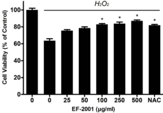
Fig. 1. Effects of Effect of EF-2001 on the viability of C2C12 myo- blasts induced by oxidative stress. C2C12 myoblasts were seeded in 96-well culture plates at a density of 1×10⁵ cells/ml and incubated for 24 hr. Then the cells were pretreated with EF-2001 for 24 hr. Next, the cells were pretreated with H2O2 in serum-free DMEM for 2 hr and washed. NAC (N-acetyl-L-cysteine; an antioxidant com- pound) was included as a control. Bars represent mean±
SD (n=4). *p<0.05 indicates values that are significantly different from the EF-2001 untreated group in H2O2. Real-time polymerase chain reaction
C2C12 myoblasts were seeded in a 6-well cell culture plate in DMEM containing 10%(v/v) FBS and incubated for 24 hr. After incubation, the medium was replaced with DMEM with or without EF-2001 (500 μg/ml) for 24 hr. The cells were treated with 1 mM H2O2 in serum-free DMEM for 2 hr. C2C12 myoblasts were lysed by using TRI-reagent (Sigma-Aldrich), and total RNA was extracted according to the manufacturer’s instructions. After the RNA concen- tration was measured, the extracted mRNA was used as a template to synthesize cDNA. Real-time polymerase chain reaction (Real-time PCR) analysis was performed by using SYBR Green 1, a LightCycler® 96 instrument (Roche, Basel, Switzerland), and the following primers: mouse Atrogin-1/
MAFbx sense 5'-CCATCCTCTTTCTTGCCCGT-3' and anti- sense 5'-ATCACTGTCCAACCTGGCTG-3'; mouse MuRF1 sense 5'-TGGGACAGATGAGGAGGAGG-3' and antisense 5'- TTTACCCTCTGTGGTCACGC-3'; and mouse GAPDH sense 5'-AGGTCGGTGTGAACGGATTTG-3' and antisense 5'-TGT AGACCATGTAGTTGAGGTCA-3'. The cycling conditions were as follows: 40 cycles of denaturation at 94℃ for 30 sec, annealing at 60℃ for 60 sec, and extension at 72℃ for 60 sec.
Animal experiments
Male C57BL/6 mice were purchased from Orient Bio (Gangneung, Korea). The mice were housed in wire cages and maintained under constant temperature (20-22℃) and humidity (40-50%) conditions to minimize the animals’
discomfort. This animal experiment protocol was approved by the Institutional Animal Care and Use Committee (IACUC, YWC-160217-1) at Yonsei University (Wonju, Korea).
The sciatic nerve in the right leg of each mouse was surgi- cally removed to induce immobilization, except in mice in the control group. The surgery caused immobilization and muscle atrophy in the mice. Muscle atrophy was induced for 7 days. Then, EF-2001 (at 3 mg/kg and 30 mg/kg) was orally administered 10 times over 2 week period. The total experimental period was 21 days. Then, 21 days after sciatic nerve denervation, the mice were sacrificed.
Measurement of muscle volume using micro-computed tomography
Micro-Computed tomography (micro-CT) image data was obtained 14 days after denervation and before sacrifice (at 21 days after denervation) using a SkyScan 1076 micro-CT (Bruker, Germany) at a resolution of 30 μm, with the follow-
ing parameters: 100 kV, 100 mA, 790 ms, and a rotation step of 1.2°. During rotation and scanning, all mice were under anesthesia. The beam-hardening errors were corrected to im- prove the quality of the images by flat-field correction before scanning and beam-hardening correction during recon- struction. To evaluate muscle volume, two-dimensional models and three-dimensional models of the muscles were generated by CT-Analyzer 1.11 (Bruker).
Statistical analysis
Statistical analysis of data was carried out using the SAS statistical software (SAS Institute, Cary, NC, USA). Multiple group data were analyzed using one-way analysis of var- iance followed by Dunnett’s multiple range tests. All results are expressed as the mean ˘ standard deviation of com- parative fold differences. Data are representative of three in- dependent experiments. Significance was set at p<0.05.
Results
Effect of EF-2001 on the viability of C2C12 myo- blasts induced by oxidative stress
Oxidative stress caused by muscle disuse and damage re- sults in muscle atrophy. So, to determine the protective effect of EF-2001 in oxidative stress, the cell viability was analyzed by the EZ-Cytox kit. In the Fig. 1, the viability of the H2O2-
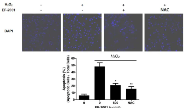
Fig. 2 Effect of EF-2001 on apoptosis in C2C12 myoblasts induced by oxidative stress. C2C12 myoblasts were cultured in 6-well culture plates and replaced with DMEM with or without EF-2001 (500 μg/ml). Next, the medium was pretreated with 1 mM H2O2 in DMEM. Then, the myoblasts were fixed with 4% paraformaldehyde in PBS for 30 min and mounted using mounting medium containing DAPI. Image data were collected with a confocal microscope. DAPI was detected at 358 and 461 nm. Bars represent mean ± SD (n=4). *p<0.05 indicates values that are significantly different from the EF-2001 untreated group in H2O2.
Fig. 3. Effect of EF-2001 on HSP70 and SOD1 protein expression in C2C12 myoblasts induced by oxidative stress. C2C12 myoblasts were cultured in 6-well culture plates and re- placed with DMEM with or without EF-2001 (500 μg/
ml). Next, the medium was pretreated with 1 mM H2O2
in DMEM. Then, HSP70, SOD1, and β-actin protein lev- els were analyzed by Western blotting.
treated C2C12 myoblasts decreased with 63.51±2.37%. The pre-treatment of EF-2001 reduced the viability of the cells in a dose dependent manner. The viability of cells was 75.45
±1.19, 78.46±1.46, 82.87±0.90, 83.72±1.92, and 87.05±1.08, at EF-2001 concentration of 25, 50, 100, 250, and 500 μg/ml.
Effect of EF-2001 on apoptosis in C2C12 myoblasts induced by oxidative stress
Furthermore, to verify the reduced effect of EF-2001 in the H2O2-treated C2C12 myoblast damage, DAPI staining was used. DAPI fluorescence was increased in H2O2-treated cells (48.18±5.38%) compared to that in untreated control cells (6.19±1.95%). The fluorescence in cells pre-treated with 500 μg/ml of EF-2001 was 20.99±2.73%, which is lower than that in the only H2O2-treated group (Fig. 2).
Effect of EF-2001 on the expression of muscle atro- phy-related proteins under oxidative stress in C2C12 myoblasts
HSP70 and SOD1 show muscle-protective activity during muscle injury. To examine the mechanism of EF-2001 effect in response to oxidative stress, the expression of HSP70 and SOD1 proteins was used by Western blotting. The expression
of HSP70 and SOD1 was increased by H2O2 treatment.
However, the expression of these proteins was decreased in the presence of EF-2001 (Fig. 3).
Effect of EF-2001 on the expression of muscle atro- phy-related genes under oxidative stress in C2C12 myoblasts
A B
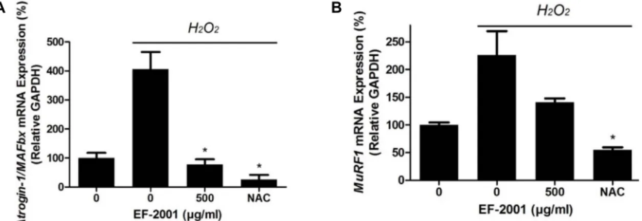
Fig. 4. Effect of EF-2001 on Atrogin-1/MAFbx and MuRF1 mRNA expression in C2C12 myoblasts induced by oxidative stress. C2C12 myoblasts were cultured in 6-well culture plates and replaced with DMEM with or without EF-2001 (500 μg/ml). Next, the medium was pretreated with 1 mM H2O2 in DMEM. Then, the mRNA expressions of Atrogin-1/MAFbx and MuRF1 were analyzed by real-time PCR. Bars represent mean ± SD (n=4). *p<0.05 indicates values that are significantly different from the EF-2001 untreated group in H2O2.
We also examined the level of mRNAs related to sarcope- nia. MuRF1 is a ring finger protein containing a zinc finger domain that is involved in muscle cell death by ubiquitinat- ing proteins [1, 13]. In addition, the transcriptional regu- lation of Atrogin-1/MAFbx mRNA is induced during muscle atrophy, similar to MuRF1 [14]. Therefore, we analyzed the mRNA expression of MuRF1 and Atrogin-1/MAFbx by using Real-time PCR.
C2C12 myoblasts treated with H2O2 showed increased mRNA levels of Atrogin-1/MAFbx (415.16±59.12%, with lev- els in untreated cells set to 100%) and MuRF1 (226.65±
43.13%, with levels in untreated cells set to 100%). However, in cells pre-treated with 500 μg/ml of EF-2001, Atrogin-1/
MAFbx expression was decreased to 77.07±18.87%(Fig. 4A) and MuRF1 expression was decreased to 162.18±7.96%(Fig.
4B).
Effect of EF-2001 on muscle volume caused by sciatic denervation in C57BL/6 mice
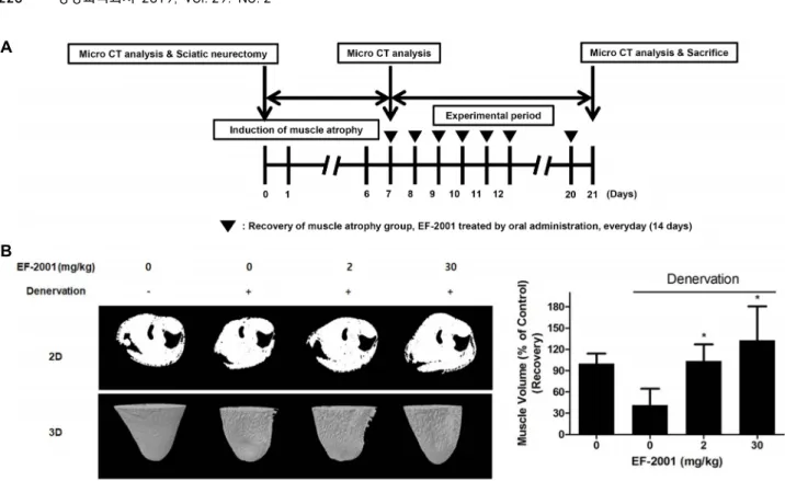
Because muscle atrophy is caused by various factors, it is difficult to test it using the human. A sciatic denervation is done with research animal models which can imitate vari- ety of conditions that induce human skeletal muscle atrophy [2]. To induce sarcopenia in C57BL/6 mice, the sciatic nerve in the right leg of each mouse was surgically removed to induce immobilization. Sarcopenia was induced by denerva- tion for 7 days. Then, EF-2001 was orally administered by everyday (14 days). To evaluate muscle volume, two-dimen- sional (2D) models and three-dimensional (3D) models of the muscles were analyzed by micro-CT (Fig. 5A). The mus-
cle volume of denervated mice was 41.25±23.34% of that in the control mice (100%). In contrast, the mice orally adminis- trated with EF-2001 at 3 mg/kg and 30 mg/kg reduced the decreased muscle mass in a dose-dependent manner (Fig.
5B). This result indicates that EF-2001 can restore sarcopenia caused by oxidative stress in vitro and in vivo. Thus, EF-2001 is a candidate for the treatment of sarcopenia, and future discovery of the additional effects of EF-2001 is expected to open up more possibilities as a drug or functional food with useful activities in various fields.
Discussion
We confirmed that EF-2001 prevented oxidative stress-in- duced cell death in muscle cells. This conclusion can be drawn from the observation that EF-2001, at 0-500 μg/ml, increased the survival of C2C12 myoblasts following in- duction of cell death by H2O2 in a dose-dependent manner (Fig. 1). Muscle atrophy is caused by cell death following muscle damage, muscle non-use, and excessive exercise. If muscle loss occurs due to sustained muscle atrophy, muscle use and movement will be limited [10, 11]. Therefore, our result suggests that EF-2001 has prevent effects in the dam- age of oxidative stress-induced C2C12 myoblasts. The mech- anism of EF-2001 effect was examined in response to oxida- tive stress. SOD1, which inhibits ROS damage by scavenging ROS from cells [17], was highly expressed in H2O2-treated cells and was decreased following EF-2001 treatment. HSP70 is a heat shock protein that prevents cells from dying in the presence of external stress [8]. HSP70 was also expressed
A
B
Fig. 5. Effect of EF-2001 on muscle atrophy induced by sciatic denervation in C57BL/6 mice. (A) Male C57BL/6 mice were used after acclimation to the environment. Three CT scans were performed: before the sciatic neurectomy, 7 days after atrophy induction, and before sacrifice (at 21 days). (B) Muscle volume in the mice was assessed by micro-CT at various time points before after sciatic denervation with and without EF-2001 treatment. Bars represent mean ± SD (n=6). *p<0.05 indicates values that are significantly different from the EF-2001 untreated group after sciatic denervation.
Fig. 6. EF-2001 can restore muscle atrophy caused by oxidative stress in vitro and in vivo.
in H2O2-treated cells and was decreased following treatment with EF-2001. Based on these protein expression results, we examined the expression of mRNAs related to muscle atrophy. The mRNA levels of Atrogin-1/MAFbx and MuRF1
increased under oxidative stress conditions, but decreased following EF-2001 treatment. Our results suggest that EF- 2001 inhibits the muscle injury-related protein expression, and the mRNA level of Atrogin-1/MAFbx and MuRF1 in-
duced by oxidative stress in C2C12 myoblasts. Based on these in vitro data, we next evaluated the effects of EF-2001 on muscle recovery in vivo by inducing muscle atrophy in C57BL/6 mice. Muscle volume, as 2D and 3D models of the muscle atrophy, decreased after sciatic denervation, and this decrease was recovered by administration of EF-2001. Previ- ous studies have shown that powdered heat-killed Enterococ- cus faecalis has anti-allergy, anti-inflammatory and anti-tu- mor effects [3, 4, 6]. This study confirms that heat-killed Enterococcus faecalis can restore muscle atrophy caused by oxidative stress in vitro and in vivo. Thus, heat-killed Enter- ococcus faecalis is a candidate for the treatment of muscular atrophy, and future discovery of the additional effects of heat-killed Enterococcus faecalis is expected to open up more possibilities as a functional food with useful activities in var- ious fields.
Acknowledgements
This research was financially supported by the Ministry of SMEs and Startups (MSS), Korea, under the “Regional Specialized Industry Development Program (R&D, R0006434)”
supervised by the Korea Institute for Advancement of Technology (KIAT).
References
1. Berridge, M. V., Herst, P. M. and Tan, A. S. 2005. Tetrazolium dyes as tools in cell biology: new insights into their cellular reduction. Biotechnol. Annu. Rev. 11, 127-152.
2. Booth, F. W. 1982. Effect of limb immobilization on skeletal muscle. J. Appl. Physiol. Respir. Environ. Exerc. Physiol. 52, 1113-1118.
3. Choi, E. J., Iwasa, M., Han, K. I., Kim, W. J., Tang, Y., Han, W. C., Kim, E. K. and Park, Z. Y. 2016. Effect of Enterococcus faecalis EF-2001 on experimentally induced atopic eczema in mice. Food Sci. Biotechnol. 25, 1087-1093.
4. Choi, M. S., Chang, S. J., Chae, Y., Lee, M. H., Kim, W.
J., Iwasa, M., Han, K. I., Kim, W. J. and Kim, T. J. 2018.
Anti-inflammatory Effect of Heat-Killed Enterococcus faeca- lis EF-2001. J. Life Sci. 28, 1361-136.
5. Frost, R. A., Nystrom, G. J., Jefferson, L. S. and Lang, C.
H. 2007. Hormone, cytokine, and nutritional regulation of sepsis-induced increases in atrogin-1 and MuRF1 in skeletal muscle. Am. J. Physiol. Endocrinol. Metab. 292, E501-512.
6. Gu, Y. H., Choi, H., Yamashita, T., Kang, K. M., Iwasa, M., Lee, M. J., Lee, K. H. and Kim, C. H. 2017. Pharmaceutical Production of Anti-tumor and Immune-potentiating Enter- ococcus faecalis-2001 β-glucans: Enhanced Activity of Macro- phage and Lymphocytes in Tumor-implanted Mice. Curr.
Pharm. Biotechnol. 18, 653-661.
7. Haycock, J. W., MacNeil, S., Jones, P., Harris, J. B. and Mantle, D. 1996. Oxidative damage to muscle protein in Duchenne muscular dystrophy. Neuroreport 8, 357-361.
8. Jäättelä, M., Wissing, D., Bauer, P. A. and Li, G. C. 1992.
Major heat shock protein hsp70 protects tumor cells from tumor necrosis factor cytotoxicity. EMBO J. 11, 3507-3512.
9. Jackson, M. J. and Farrell, S. O. 1993. Free radicals and mus- cle damage. Br. Med. Bull. 49, 630-641.
10. Lee, Y. H., Kim, W. J., Lee, M. H., Kim. S. Y., Seo, D. H., Kim. H. S., Gelinsky, M. and Kim, T. J. 2016. Anti-skeletal muscle atrophy effect of Oenothera odorata root extract via reactive oxygen species-dependent signaling pathways in cellular and mouse model. Biosci. Biotechnol. Biochem. 80, 80- 88.
11. Lee, Y. H., Seo, D. H., Park, J. H., Kabayama, K., Opitz, J., Lee, K. H., Kim, H. S. and Kim, T. J. 2015. Effect of Oenothera odorata root extract on microgravity and dis- use-induced muscle atrophy. Evid. Based Complement.
Alternat. Med. 2015, 130513.
12. Li, C. Y., Lee, J. S., Ko, Y. G., Kim, J. I. and Seo, J. S. 2000.
Heat shock protein 70 inhibits apoptosis down stream of cytochrome c release and upstream of caspase-3 activation.
J. Biol. Chem. 275, 25665-25671.
13. Mrosek, M., Meier, S., Ucurum-Fotiadis, Z., von Castelmur, E., Hedbom, E., Lustig, A., Grzesiek, S., Labeit, D., Labeit, S. and Mayans, O. 2008. Structural analysis of B-Box 2 from MuRF1: identification of a novel self-association pattern in a RING-like fold. Biochemistry 47, 10722-10730.
14. Natanek, S. A., Riddoch-Contreras, J., Marsh, G. S., Hopkin- son, N. S., Moxham, J., Man, W. D., Kemp, P. R. and Polkey, M. I. 2013. MuRF-1 and atrogin-1 protein expression and quadriceps fiber size and muscle mass in stable patients with COPD. COPD 10, 618-624.
15. Ogata, T., Oishi, Y., Higashida, K., Higuchi, M. and Muraoka, I. 2009. Prolonged exercise training induces long-term en- hancement of HSP70 expression in rat plantaris muscle. Am.
J. Physiol. Regul. Integr. Comp. Physiol. 296, R1557-1563.
16. Oksala, N. K., Ekmekçi, F. G., Özsoy, E., Kirankaya, Ş., Kokkola, T., Emecen, G., Lappalainen, J., Kaarniranta, K.
and Atalay, M. 2014. Natural thermal adaptation increases heat shock protein levels and decreases oxidative stress.
Redox Biol. 3, 25-28.
17. Powers, S. K. 2014. Can antioxidants protect against disuse muscle atrophy? Sports Med. 44, S155-165.
18. Rodriguez, M. C. and Tarnopolsky, M. A. 2003. Patients with dystrophinopathy show evidence of increased oxida- tive stress. Free Radic. Biol. Med. 34, 1217-1220.
19. Sugawara, T., Lewén, A., Gasche, Y., Yu, F. and Chan, P.
H. 2002. Overexpression of SOD1 protects vulnerable motor neurons after spinal cord injury by attenuating mitochon- drial cytochrome c release. FASEB J. 16, 1997-1999.
20. Tsang, C. K., Liu, Y., Thomas, J., Zhang, Y. and Zheng, X.
S. 2014. Superoxide dismutase 1 acts as a nuclear tran- scription factor to regulate oxidative stress resistance. Nat.
Commun. 5, 3446.
21. Zylicz, M. and Wawrzynow, A. 2001. Insights into the func- tion of Hsp70 chaperones. IUBMB Life 51, 283-287.
초록:산화스트레스에 의해 유도된 C2C12 근세포 손상과, 신경절제에 의해 근감소가 유도된 C57BL/
6 마우스에서 열처리 사균체 엔테로코커스 패칼리스 EF-2001의 효과
장상진1․이명헌1․김완중1․채유리1․이와사 마사히로1,2․한권일1,2․김완재2․김택중1,3*
(1연세대학교 생명과학기술학부, 2주식회사 한국베름 연구개발센터, 3주식회사 닥터티제이 연구개발센터)
노화, 상해, 유전병 및 산화 스트레스와 같은 다양한 원인으로 인해 근육 위축을 유발한다. 그 동안의 연구에 의하면 열처리 사균체 엔테로코커스 패칼리스(EF-2001)는 항알레르기, 항염증 및 항종양 효과를 보였다. 그러나 항산화 및 항근위축에 대한 효과는 잘 알려져 있지 않고 있다. 본 연구에서는 EF-2001이 근육 위축에 미치는 영향 을 연구 하였다. 산화 스트레스에 의한 EF-2001의 세포손상 보호 효과를 확인하기 위해 C2C12 근섬유 아세포는 H2O2로 처리되어, 산화 스트레스를 유도하여 세포 손상을 유발하였다. 그러나 EF-2001 처리로 인해 근세포 손상 이 감소됨을 확인 하였다. 우리는 산화스트레스에 의한 EF-2001의 근세포손상의 감소 효과에 대한 메커니즘을 확인하였다. EF-2001는 산화스트레스로 유도된 근세포내의 HSP70 및 SOD1 단백질의 발현을 감소시켰다. 또한, 근세포내에 Atrogin-1/MAFbx 및 MuRF1의 mRNA 수준은 산화 스트레스 조건 하에서 증가하였으나, EF-2001에 의해 감소하였다. 더나아가 근감소를 유도한 좌골신경 절제 모델동물을 통한 근육량을 확인하기 위해 마이크로 CT를 활용해 2차원과 3차원으로 분석하였다. 근육량은 좌골 신경 절제 후에 감소하였고, EF-2001의 경구 투여에 의해 근육량이 회복되었다. 본 결과는 열처리 사균체 엔테로코커스 패칼리스인 EF-2001이 노인들에게 자주 발생 하는 근감소증의 예방 및 개선할 수 있는 기능성 식품으로 다양한 분야에서 유용하게 활용 될 수 있음을 시사한다.