J Korean Soc Pediatr Nephrol 2013;17:92-100 DOI: http://dx.doi.org/10.3339/jkspn.2013.17.2.92
Copyright © 2013 The Korean Society of Pediatric Nephrology ISSN 1226-5292 (print) ISSN 2234-4209 (online)
Immunuglobulin A 신질환과 Henoch-Schnlein purpura 신질환을 가진 소아에서의 cyclosporine A와 angiotensin-converting enzyme inhibitor 치료의 임 상적, 병리학적 변화
아주대학교 의과대학 소아청소년과, 영남대학교 의과대학 병리학과*, 연세대학교 의과대학 소아청소년과†
아주대학교 의과대학 병리학과‡
이정주ᆞ김용진*ᆞ신재일†ᆞ임현이‡ᆞ박세진
Clinicopathologic Changes in Children with Immuno globulin A Nephritis and Henoch- Schönlein Purpura Nephritis after Cyclosporine A and Angiotensin-con verting Enzyme Inhibitor Treatment
Purpose: To investigate the clinicopathologic effects of cyclosporine A (CsA) in children with diseases characterized by mesangial immunoglobulin A deposits such as im mu noglobulin A nephropathy (IgAN) and Henoch-Schönlein purpura nephritis (HSPN).
Methods: We retrospectively reviewed the clinicopathologic outcomes of 54 children (IgAN, 36; HSPN, 18) treated with CsA. The starting dose of CsA was 5 mg/kg per day, and it was administered in 2 divided doses. The degree of prote- inuria and pathologic changes in renal biopsies were evaluated before and after CsA treatment.
Results: The mean protein to creatinine ratio decreased from 3.7±1.5 to 0.6±0.4 (P<0.001), and 32 (59.2%) children achieved complete remission of proteinuria after 1-year CsA treatment. Among the 54 children, 24 maintained normal renal function and 25 exhibited microscopic hematuria or proteinuria at the end of CsA treatment. In the HSPN group, 3 children whose initial biopsies indicated class IIIb disease showed class II disease on follow-up, and the follow-up biopsies of 2 chil dren who had class II disease indicated the same class II disease. In the IgAN group, cortical tubular atrophy occurred in 1 child, and no child with IgAN had cortical interstitial fibrosis or tubular atrophy after 1-year CsA treatment. No significant complications were found in the children treated with CsA.
Conclusion: Our findings indicate that CsA treatment is effective and beneficial
Jeong Ju Lee, M.D.,
Yong-Jin Kim, M.D.*, Jae Il Shin, M.D.
†, Hyunee Yim, M.D.
‡, and Se Jin Park, M.D.
Department of Pediatrics, Ajou University Hospital, Ajou Universtiy School of Medicine, Suwon, Korea, Department of Pathology*, Yeungnam University College of Medicine, Daegu, Korea, Department of Pediatrics†, Seve
rance Children’s Hospital, Yonsei University College of Medicine, Seoul, Korea, Department of Pathology‡, Ajou University Hospital, Ajou Universtiy School of Medicine, Suwon, Korea Corresponding Author: Se Jin Park Department of Pediatrics, Ajou University Hospital, Ajou Universtiy School of Medicine, Suwon, Korea
Tel: 0312195163, Fax: 0312195169 Email: [email protected]
Received:27 September 2013 Revised: 7 October 2013 Accepted: 14 October 2013
This is an openaccess article distributed under the terms of the Creative Commons Attribu
tion NonCommercial License (http:// crea
tivecom mons.org/licenses/bync/3.0/) which permits unrestricted noncommercial use, distribution, and reproduction in any medium, provided the original work is properly cited.
Introduction
Both immunoglobulin A nephropathy (IgAN) and Henoch-Schönlein purpura nephritis (HSPN), affecting glomerular function and structures, are pathologically characterized by mesangial deposits of IgA. Among se- veral immunoglobulins, IgA is unique for its capability to form multimers, and circulating IgA-containing immune complexes have been demonstrated in both illnesses related to the deposits of IgA and IgA-containing immune complexes in the glomerular mesangium [1].
Characterized by prominent diffuse mesangial IgA deposits on immunofluorescence microscopy, primary IgA nephropathy was initially thought to be a rare but benign cause of recurrent hematuria [2]. However, the clinical course of IgAN is variable, with some patients having stable renal function over decades and others developing hypertension, nephrotic syndrome, and chronic renal failure [2]. A number of clinical and histo- logical prognostic markers, including impairment of renal function, severe proteinuria, and high blood pressure, predict a poor outcome [3].
HSPN, a leucocytoclastic vasculitis, is caused by mainly IgA1-mediated inflammation of small vessels [4-7].
Although isolated microscopic hematuria is the more fre- quent clinical presentation of HSPN, nephritic syndrome and acute nephritic syndrome, often with hypertension, are observed in about half of the cases [4-7]. Renal in- volvement occurs in 30-90% of HSP patients [8]. The severity of renal symptoms at onset is known to be the most prognostic factor for HSP in children; patients pre senting with nephrotic-range proteinuria or severe nephritis have the highest risk of unfavorable prognosis [9].
Patients with IgAN or HSPN should be treated properly at an early stage to prevent rapid progress to renal in- sufficiency. Although the management of the two dis- eases characterized by IgA deposits still remains elusive,
recent studies have suggested that cyclosporin A (CsA) might have a beneficial effect in children with severe IgA deposits [10-15]. However, the number of patients included in those studies was limited, and the glomerular changes in IgAN were categorized according to the Haas classification [13]. Therefore, we aimed to investigate clinicopathologic effects of CsA on children with glo- merular IgA deposits by histologically classifying HSPN according to the International Study of Kidney Disease in Childhood and IgAN according to the Oxford classi- fication instead of the Haas classification.
Materials and Methods
1. Patients
We retrospectively reviewed a total of 54 patients (IgAN:HSPN=36:18; M:F=32:22) treated with CsA from 2005 to 2012 in our hospital. Of 42 HSP patients that we reviewed, 25 were younger than 10 years of age, and 23 HSP patients were boys. Among the 42 HSP patients, 18 children developed HSPN and were treated with CsA due to nephrotic-range proteinuria. Nephrotic-range proteinuria was defined as IgAN or HSPN patients had proteinuria greater than 40 mg/m2/hr. Six IgAN and six HSPN patients underwent repeated renal biopsies before and after CsA treatment, respectively. Both of the patients with IgAN and HSPN showed microscopic hematuria at diagnosis. Thirty children experienced nephrotic syndrome after diagnosis of IgAN and HSPN, and the remaining patients showed nephrotic-range proteinuria except two IgAN patients.
2. Treatment protocol
CsA was administered to the patients with diseases in reducing massive proteinuria and preventing progression to end-stage renal failure in children with glomerular diseases characterized by IgA deposits, such as IgAN and HSPN, within 1 year of treatment.
Key words: IgA deposit, IgA nephropathy, Henoch-Schönlein purpura nephritis, cyclosporine A
characterized with IgA deposits such as IgAN or HSPN when the patients showed nephrotic syndrome or nephrotic-range proteinuria. Angiotensin-converting enzyme inhibitor (ACEi) was concurrently used in all patients during the period of treatment with CsA, where as corticosteroids were not used for the treatment of the patients. The starting dose of CsA was 5 mg/kg/day in two divided doses, and the desired drug level was maintained between 100 and 200 ng/mL. CsA levels were measured every 2 weeks at the beginning of treatment and every two months during the maintenance period.
3. Renal biopsy
Renal specimens of HSP patients were graded by the histological classification of ISKDC: grade I, minor glomerular abnormalities; grade II, pure mesangial proli feration (a: focal; b: diffuse); grade III, minor glom- erular abnormalities or mesangial proliferation with crescents/segmental lesion in <50% crescents (a: focal;
b: diffuse mesangial proliferation); grade IV, mesangial proliferation with crescents/segmental lesions in 50-75
% glomeruli (a: focal; b: diffuse mesangial proliferation);
grade V, mesangial proliferation with crescents/segmental lesions in >75% glo meruli (a: focal; b: diffuse mesangial proliferation); grade VI, membranoproliferative-like lesion (10). For an active lesion index, the scoring was assessed as follows: 1) me sangial proliferation: normal (0), slight (1), moderate (2), and severe (3); 2) necrosis or cellular crescents: 0% (0), 1-20% (1), 20-50% (2), >50% of glomeruli. The chronicity index was evaluated as follows: 1) fibrous crescents or global sclerosis: 0% (0), 1-20% (1), 20- 50% (2), >50% of glomeruli (3); tubular atrophy associated with interstitial fibrosis: 0 (0%), 1-20% (1), 20-50% (2), >50%
of cortical area (3).
Clinicopathologic classification with pathologic features predictive of disease progression was graded using the Oxford Classification system (2009) of IgAN. The key pathologic features to be reported on renal biopsy were scored as follows: 1) mesangial hypercellularity: mean
<4 mesangial cells/mesangial area (M0) and mean 4 or more mesangial cells/mesangial area (M1); 2) segmental glo merulosclerosis or adhesions present (S1) or absent
(S0); 3) endocapillary proliferation present (E1) or absent (E0); 4) cortical interstitial fibrosis and tubular atrophy 0-25% (T0), 26-50% (T1), and >50% (T2); 5) total number of glomeruli with changes: endocapillary hypercellulariity, extracapillary proliferation, global glomerulosclerosis, and segmental glomerulosclerosis [11, 12]. Each pathologist from two different universities completed a scoring sheet for every biopsy and these data were used to obtain a consensus for each parameter.
4. Clinical status evaluation
The clinical status of each patient at diagnosis and at the most recent observation was classified as follows: 1) state A, normal physical examination, urine, and renal function; 2) state B1, microscopic hematuria without proteinuria; 3) state B2, proteinuria <40 mg/m2/hr with or without hematuria; 4) state C, active renal disease cha racterized by hypertension or proteinuria of 40 mg/
m2/hr or greater, with glomerular filtration rate (GFR) of 60 mL/min/1.73m2 or greater.
5. Statistical analysis
Statistical analyses were performed with SPSS for Windows version 18.0 (SPSS, Chicago, Illinois, USA).
Descriptive statistics were expressed as mean±standard deviation or as a median with ranges. Differences bet- ween parameters before and after CsA treatment were compared with the Wilcoxon signed rank test and non- parametric statistical methods. All differences were considered sta tistically significant at P<0.05.
Results
The mean age of the patients was 10±4.2 year (range 2.0-16) at the time of HSPN or IgAN diagnosis (Table 1). No patient showed renal failure, hypertension, or acute nephritic features at onset. Among 54 patients with micro scopic hematuria, 36 (66.7%) had gross hematuria at the time of the initial examination (Table 1). Mean duration of CsA treatment was 11.7±5.6 months (range
10.4-12.7). Angiotensin-converting enzyme inhibitor was maintained for the duration of CsA treatment (mean 14.5
±24.2 months). Mean period of 3.0±2.6 months (range 1.3-12.6) was needed to normalize massive proteinuria after treatment of CsA. The median duration of follow- up was 3.7±3.1 year (range 1.7-15.2) from the onset of disease.
The mean serum levels of total protein and albumin increased from 6.5±0.8 g/dL to 7.1±0.4 g/dL (P<0.001) and from 2.8±0.6 g/dL to 4.3±0.2 g/dL (both P<0.001), re spectively, after CsA treatment (Table 2). Mean cholesterol level decreased significantly from 205.9±77.9 mg/dL to 166.0±44.0 mg/dL (P<0.001), whereas serum creatinine level increased from 0.6±0.2 mg/dL to 0.7±0.2 mg/dL (P=0.005). Of note, the uric acid level significantly in- creased from 4.8±1.3 to 5.3±1.6 (P=0.004) (Table 2).
The pretreatment and follow-up renal biopsy results for 6 IgAN and 6 HSPN children are shown in Table 3.
After 1-year treatment of CsA, one child with HSPN had ISKDC IIIb at the second biopsy from initial IIIa. Three children whose initial biopsies were class IIIb had class II disease on follow-up, and follow-up biopsies of two children with class II had the same class II in HSPN (Table 3). According to Oxford classification of IgAN, one children had M1S1, one had M1E1, two had M1, and two remained within normal range from the initial to the follow-up biopsies. Progressed cortical tubular atrophy occurred in only one child, and no children with IgAN progressed to cortical interstitial fibrosis or tubular
atrophy due to CsA treatment (Table 3). As shown in Table 3, one year of oral CsA treatment was not associated with a marked increase in mean activity index without an increase in chronicity. None of the renal biopsy classi- fications worsened in either HSPN or in IgAN.
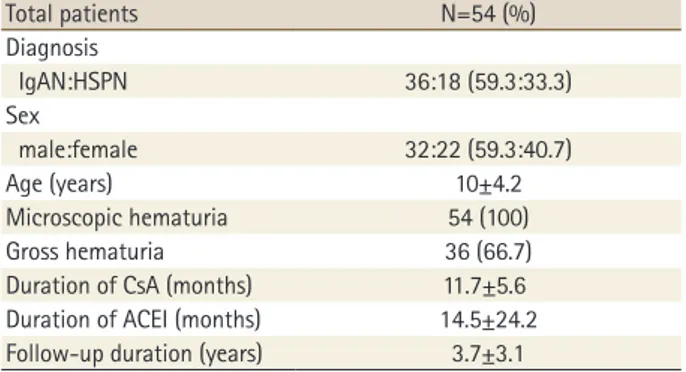
At the latest follow-up after the end of CsA treatment, Table 1. Clinical Characteristics of Children with Immunoglo- bulin A Nephritis and Henoch-Schönlein Purpura Nephritis
Total patients N=54 (%)
Diagnosis
IgAN:HSPN 36:18 (59.3:33.3)
Sex
male:female 32:22 (59.3:40.7)
Age (years) 10±4.2
Microscopic hematuria 54 (100)
Gross hematuria 36 (66.7)
Duration of CsA (months) 11.7±5.6 Duration of ACEI (months) 14.5±24.2 Follow-up duration (years) 3.7±3.1
Abbreviations: IgAN, Immunoglobulin A nephropathy; HSPN, Henoch- Schönlein purpura nephritis; CsA, cyclosporine A; ACEI, Angiotensin converting enzyme inhibitor
Fig. 1. Elementary renal lesions observed in patients with IgA nephropathy are shown repre sentatively according to the Oxford classification: a) segmental sclerosis (S1); b) mesangial hypercellularity (M1); and c) tubular atrophy/interstitial fibrosis >50% (T2).
Table 2. Comparison of Laboratory Data before and after the Treatment of Cyclosporin A
Variables Pre-treatment Post-treatment P value Total protein (g/dL) 5.5±0.8 7.1±0.4 <0.001
Albumin (g/dL) 2.8±0.6 4.3±0.2 <0.001
Na (mMol/L) 139.5±2.7 140.2±1.7 0.040
K (mMol/L) 4.2±0.4 4.4±0.4 0.111
Cl (mMol/L) 104.4±3.3 104.8±2.0 0.691
BUN (mg/dL) 14.0±4.2 14.4±4.8 0.851
Creatinine (mg/dL) 0.6±0.2 0.7±0.2 0.005
Cholesterol (mg/dL) 205.9±77.9 166.0±44.0 <0.001
Uric acid (mg/dL) 4.8±1.3 5.3±1.6 0.004
24 patients returned to normal urinalysis and maintained normal renal function (state A), 8 patients had microscopic hematuria without proteinuria (state B1), and 17 patients had proteinuria of <40 mg/m2/hr (state B2). The proportion of children with active renal disease with proteinuria of
>40 mg/m2/hr (state C) was decreased from 30 (55.6%) to 5 (9.3%). Side effects of CsA treatment, including gingival hypertrophy and trichosis, were minimal, and no one withdrew from CsA treatment because of the absence of severe side effects or complications. The
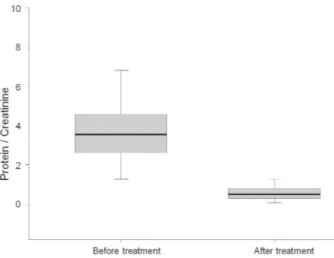
protein to creatinine ratio significantly decreased from 3.7±1.5 to 0.6±0.4 after CsA treatment (P<0.001) (Fig. 3).
Fig. 4 exhibits that if the basal level of proteinuria was set to 1, a 50% reduction in proteinuria was achieved 2.6 months after treatment with CsA. CsA treatment was well tolerated in all patients, and no patients required drug discontinuation or had any serious adverse effects that warranted hospitalization.
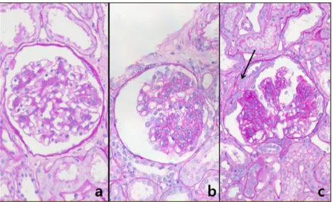
Fig. 2. Elementary renal lesions observed in patients with HSP nephritis are shown represen tatively according to the ISKDC classification: a) minimal glomerular abnor- mality; b) pure mesangial proliferation; and c) mesangial proliferation with crescent formation (arrow).
Table 3. Follow-up Histological Data in 6 HSPN and 6 IgA Patients Treated with Cyclosporin A
HSPN case 1st Biopsy 2nd Biopsy
Glomeruli (n) ISKDC AI CI TI Glomeruli (n) ISKDC AI CI TI
1 32 II 2/0 0 0 39 II 1/0 0 0
2 25 IIIb 3/0 0 0 30 II 2/0 1 1
3 64 IIIa 1/0 1 0 45 IIIb 3/1 0 0
4 43 IIIb 3/1 0 0 56 II 1/0 1 2
5 29 IIIb 2/1 0 0 39 II 2/0 0 1
6 47 III 2/0 1 1 37 II 2/0 1 1
IgAN case 1st Biopsy 2nd Biopsy
CIF CTA Oxford CS CIF CTA Oxford CS
1 0 0 M1E0S1T0 0 1 M1E0S1T0
2 0 0 M1E1S0T0 0 0 M1E1S0T0
3 0 0 M0E0S0T0 0 0 M0E0S0T0
4 0 0 M1E0S0T0 0 0 M1E0S0T0
5 0 0 M1E0S0T0 0 0 M1E0S0T0
6 0 0 M0E0S0T0 0 0 M0E0S0T0
Abbreviations: Bx, biopsy; ISKDC, International Study of Kidney Disease in Childhood; AI, activity index; ChI, chronicity index; TI, tubulointerstitial change; CIF, cortical interstitial fibrosis; CTA, cortical tubular atrophy; CS, classification system
Discussion
As previously suggested [16, 17], IgAN and HSPN repre- sent a spectrum of clinical and pathologic presentations of the same or a similar disorder, probably sharing a common pathogenesis. The features supporting this hypothesis include reports on the occurrence of these two diseases in the same family [18, 19], patients presenting with the combination of symptoms of HSP and IgAN [20, 21], and the indistinguishable nature of the renal histopathologic lesion [22]. However, according to our pathologic experiences, the two diseases are distingui- shable not only in clinical presentations but also in pa- thology. Particularly, electron microscopy reveals that much larger one or two electron dense deposits are
located in paramesangium and near the capillary wall in IgAN, whereas characteristically small and scattered electron dense deposits are often within and throughout the mesangial regions in HSPN [23]. This pathognomonic feature gives us a clue to pathologically differentiate and diagnose the two different diseases of IgA deposits.
Although the majority of patients with IgAN have good prognosis, 20-30% or sometimes up to 40% of patients will progress to ESRD over the course of 10-20 years [1, 2, 24]. These patients who are at high risk of progressive renal disease need to take a more aggressive therapeutic approach. Both clinical and histologic features at first biopsy should thoroughly be evaluated to determine whether the patients with IgAN are at high risk of pro- gressive renal disease. While HSP is often a benign and a self-limited condition, approximately 30-90% of pe- diatric patients develop nephritis within 4 to 6 weeks of the initial presentation [8]. HSP is the most common cause of crescentic glomerulonephritis in children, which often manifests hematuria and proteinuria of variable intensity [25].
The treatment of severe IgAN and HSPN is still con- troversial, and recommendations are based on small, often uncontrolled series. Some studies have reported favorable results of treatments including intravenous administration of methylprednisolone followed by oral treatment with corticosteroids, corticosteroids in combi- nation with azathioprine, cyclophosphamide with/with- out anticoagulants, plasmapheresis, and mycophenolate mofetil [26-28]. Nevertheless, there have been only a few studies of CsA treatment for IgAN or HSPN, respec- tively. Thus, the main goal of our study was to demon- strate the clinical efficacy of CsA in glomerular diseases characterized by IgA deposits and particularly to determine whether there were any severe glomerular histologic changes after CsA use according to the ISKDC and the Oxford classification.
Our study showed that CsA treatment has antiprote- inuric effect in patients with glomerular injury by IgA deposits. In our study, there was a patient (HSPN, patient 3) who showed histological changes from ISKDC IIIa to IIIb, although he achieved clinical remission in nephritic- range proteinuria. In the remaining patients, 3 patients Fig. 3. Protein to creatinine ratio before and after cyclosporine
treatment. Protein to creatinine ratio decreased from 3.7±1.5 to 0.6±0.4 (P<0.001).
Fig. 4. Proteinuria was reduced to the half of its pretreatment level after 2.6 months of CsA treatment (P<0.001).
(HSPN, patients 2, 4, and 5) showed improvements in histological grades (ISKDC IIIb to II in 3 patients) and 2 patients (HSPN, patients 1 and 6) showed the same histological grades (ISKDC II to II in 2 patients) with clinical improvements. In IgAN cases, all patients showed same histological grades with clinical improve ments. These results are in accordance with those of the previous studies. According to a study by Shin et al. [13], nine patients achieved stable remission after treatment with CsA during a mean follow-up of 4.6 years in 14 pediatric IgAN patients with nephrotic-range proteinuria. In addi tion, the laboratory findings exhibited a decline in the rate of 24-hr urinary protein excretion and a signi- ficant increase in serum albumin after CsA treatment.
Furthermore, immunofluorescent analysis at the first and second biopsies showed a significant reduction of mesangial IgA deposits in seven (50%) of the 14 patients.
A controlled study with a small number of patients by Lai et al. [29]. also demonstrated a >50% reduction in proteinuria in 83% of 12 cyclosporine-treated IgAN patients and decreased IgA levels in 80% as well as increased serum albumin. Ronkainen et al. also reported that in seven pediatric HSPN patients with nephrotic-range proteinuria, four patients achieved stable remission after treatment with CsA during a mean follow-up of 6.0 years [9]. Consistent with these previous studies, the protein to creatinine ratio and urinary protein excretion in 24-h collection significantly decreased after CsA treat- ment in our study. However, our participants were different from those of previous studies in that our patients, who did not respond to prednisolone, did not receive any other immunosuppressive agents such as cyclophosphamide or azathioprine before CsA treatment.
The mechanism of CsA immunosuppressive action in glomerular diseases is still elusive. One possible ex- planation for the immunological basis of CsA efficacy is related to its ability to inhibit the secretion of cytokines by infiltrating T cells and macrophages [30]. In addition, CsA also stabilizes the podocyte actin cytoskeleton, resulting in reducing massive proteinuria [31].
Although CsA is a very effective immunosuppressive agent for steroid-dependent and steroid-resistant nephritic syndrome [32], nephrotoxicity is one adverse effect that
limits its long-term use. Significantly increased creatinine level might result from CsA treatment in our study [33].
Notably, CsA-induced toxicity is related to the duration of treatment (>24 or 36 months) and dose of CsA [34, 35]. All of our patients were carefully observed and monitored during the treatment period, and the trough level of CsA was maintained between 100-200 ng/
mL during the entire period. Due to relatively low CsA trough levels, there were no significant side effects that warranted hospitalization or discontinuation of treatment.
The ideal duration of CsA treatment for patients with renal disease characterized by IgA deposits is not known. Our results indicated that treatment duration of 2.6 months was necessary to achieve a 50% reduction of proteinuria from levels at the initial diagnosis. Thus, based on the result, we suggest that CsA should be treated for at least three months or more to achieve the goal of reducing massive proteinuria in patients with glomerular injury by IgA deposits.
The present study has some limitations: 1) this was a retrospective study, and the sample size was small; 2) the study had relatively short duration of CsA treatment and follow-up period; and 3) selection bias may have existed in that only mild patients were recruited at renal biopsy. 4) we were not able to analyze the date of control group receiving ACEi alone due to unavailability of date.
All our patients received an ACEi, which is known to provide antiproteinuria and renoprotective effect against deterioration in renal function in various renal diseases [36]. Despite these limitations, the current data clearly show that patients with nephrotic-range proteinuria can effectively and safely be treated with CsA for less than one year. Also, the current pathologic results reveal that no patients treated with CsA for less than one year progress to renal deterioration and aggravation in the glomerular diseases. In other words, CsA is not inferior to other immunosuppressive agents in the treatment of patient with IgA deposits without causing renal cytoto- xicity in one-year usage.
In conclusion, our study indicates that CsA can be used to reduce massive proteinuria without any pathologic deterioration within one year. Additionally, our findings suggest that CsA treatment should be maintained for
at least three months or more to obtain a satisfactory effect on decreasing massive proteinuria in glomerular injury associated with IgA deposits, such as IgAN and HSPN.
Conflict of interest
The authors have no financial conflicts of interest.
한글요약
목적: IgA 신병증, HSP 신병증은 사구체의 메산지움에 IgA가 침착되는 대표적인 질환이다. 본 연구는 소아에서, 이 두 가지 질환에 대한 Cyclosporin A 의 임상적 및 병리 학적 효과를 평가하기 위하여 시행되었다.
방법: 총 54명의 환자(IgA 신병증: Henoch-Schönlein purpura 신병증=36:18)를 대상으로 후향적으로 연구를 진 행하였다. CsA는 5mg/kg/day 으로 투여하였으며, 투여 전, 후로 단백뇨의 양을 측정, 병리학적 변화를 조사하기 위 해 신생검을 시행하였다. HSP 신병증 및 IgA 신병증의 신생 검은 병리학적으로 각각 ISKDC 분류법, Oxford 분류체계 (2009)로 구분하였다.
결과: 혈청 단백/크레아티닌 비는 치료 전후로 3.7±1.5 에서 0.6±0.4으로 호전되었고(P<0.001), 총 54명 중 32명의 환자(59.2%)에서 CsA 치료 1년 후 단백뇨의 관해를 보였 다. 신생검의 병리학적 소견은 호전되거나, 또는 치료 전후 로 유지되는 양상을 보였으며, CsA로 인한 합병증은 없었 다.
결론: CsA 는 IgA의 사구체 침착을 특징으로 하는 IgA 신병증, HSP 신병증 환자에서 단백뇨 감소효과 및 말기신 부전으로의 진행을 예방하는 데에 효과적인 것으로 사료 된다.
References
1) Donadio JV, Grande JP. IgA nephropathy. N Engl J Med 2002;
347:73848.
2) D'Amico G. Natural history of idiopathic IgA nephropathy: role of clinical and histological prognostic factors. Am J Kidney Dis 2000;36:22737.
3) Katafuchi R, Vamvakas E, Neelakantappa K, Baldwin DS, Gallo GR. Microvascular disease and the progression of IgA ne
phropathy. Am J Kidney Dis 1990;15:729.
4) Ozen S, Ruperto N, Dillon MJ, Bagga A, Barron K, Davin JC, et al. EULAR/PReS endorsed consensus criteria for the classi
fication of childhood vasculitides. Ann Rheum Dis 2006;65:
93641.
5) Koutkia P, Mylonakis E, Rounds S, Erickson A. Leucocytoclastic vasculitis: an update for the clinician. Scand J Rheumatol 2001;30:31522.
6) Davin JC, Weening JJ. HenochSchönlein purpura nephritis:
an update. Eur J Pediatr 2001;160:68995.
7) Lee TH, Lee EY, Cho YS, Yoo B, Moon HB, Lee CK. Concurrent occurrence of chylothorax and chylous ascites in a patient with HenochSchönlein purpura. Scand J Rheumatol 2003;
32:3789.
8) Kaku Y, Nohara K, Honda S. Renal involvement in Henoch
Schönlein purpura: a multivariate analysis of prognostic factors. Kidney Int 1998;53:17559.
9) Ronkainen J, AlaHouhala M, Huttunen NP, Jahnukainen T, Koskimies O, Ormälä T, et al. Outcome of HenochSchoenlein nephritis with nephroticrange proteinuria. Clin Nephrol 2003;60:804.
10) Counahan R, Winterborn MH, White RHR, Heaton JM, Meadow SR, Bluett NH, et al. Prognosis of HenochSchönlein nephritis in children. Br Med J 1977;2:114
11) Working Group of the International IgA Nephropathy Network and the Renal Pathology Society, Cattran DC, Coppo R, Cook HT, Feehally J, Roberts IS, et al. The Oxford classification of IgA nephropathy: rationale, clinicopathological correlations, and classification. Kidney Int 2009;76:53445.
12) Working Group of the International IgA Nephropathy Network and the Renal Pathology Society, Roberts IS, Cook HT, Troyanov S, Alpers CE, Amore A, et al. The Oxford classification of IgA nephropathy: pathology definitions, correlations, and repro
ducibility. Kidney Int 2009;76:54656.
13) Shin JI, Lim BJ, Kim PK, Lee JS, Jeong HJ, Kim JH. Effects of cyclosporin A therapy combined with steroids and angio
tensin converting enzyme inhibitors on childhood IgA ne
phropathy. J Korean Med Sci 2010;25:7237.
14) Park JM, Won SC, Shin JI, Yim H, Pai KS. Cyclosporin A therapy for HenochSchönlein nephritis with nephroticrange pro
teinuria. Pediatr Nephrol 2011;26:4117.
15) Shin JI, Park JM, Shin YH, Kim JH, Lee JS, Jeong HJ. Henoch
Schönlein purpura nephritis with nephroticrange protei
nuria: histological regression possibly associated with cyclosporin A and steroid treatment. Scand J Rheumatol 2005;34:3925.
16) Egido J, Sancho J, Mampaso F, Lopez Trascasa M, Sanchez Crespo M, Blasco R, et al. A possible common pathogenesis of the mesangial IgA glomerulonephritis in patients with
Berger's disease and SchonleinHenoch syndrome. Proc Eur Dial Transplant Assoc 1980;17:6606.
17) Waldo FB. Is HenochSchonlein purpura the systemic form of IgA nephropathy? Am J Kidney Dis. 1988;12:3737.
18) Levy M. Familial cases of Berger's disease and anaphylactoid purpura: more frequent than previously thought. Am J Med 1989;87:2468.
19) Montoliu J, Lens XM, Torras A, Revert L. HenochSchonlein purpura and IgA nephropathy in father and son. Nephron 1990;54:779.
20) Thorner PS, Farine M, Arbus GS, Poucell S, Baumal R. IgA ne
phropathy: HenochSchonlein purpura and Berger's disease in one patient. Int J Pediatr Nephrol 1986;7:1316.
21) Chishiki M, Kawasaki Y, Kaneko M, Ushijima Y, Ohara S, Abe Y, et al. A 10yearold girl with IgA nephropathy who 5 years later developed the characteristic features of HenochSchonlein purpura nephritis. Fukushima J Med Sci 2010;56: 15761.
22) Meadow SR, Scott DG. Berger disease: HenochSchonlein syndrome without the rash. J Pediatr 1985;106:2732.
23) Rai A, Nast C, Adler S. HenochSchonlein purpura nephritis. J Am Soc Nephrol 1999;10:263744.
24) D'Amico G. Natural history of idiopathic IgA nephropathy and factors predictive of disease outcome. Semin Nephrol 2004;24:17996.
25) Jardim HM, Leake J, Risdon RA, Barratt TM, Dillon MJ. Cres
centic glomerulonephritis in children. Pediatr Nephrol 1992;
6:2315.
26) Niaudet P, Habib R. Methylprednisolone pulse therapy in the treatment of severe forms of SchönleinHenoch purpura nephritis. Pediatr Nephrol 1998;12:23843.
27) Shin JI, Park JM, Shin YH, Kim JH, Lee JS, Kim PK, et al. Can azathioprine and steroids alter the progression of severe HenochSchönlein nephritis in children? Pediatr Nephrol
2005;20:108792.
28) Yoshikawa N, Ito H, Sakai T, Takekoshi Y, Honda M, Awazu M, et al. A controlled trial of combined therapy for newly diagnosed severe childhood IgA nephropathy. The Japanese Pediatric IgA Nephropathy Treatment Study Group. J Am Soc Nephrol 1999;10:1019.
29) Lai KN, MacMoune Lai F, VallanceOwen J. A shortterm con
trolled trial of cyclosporine A in IgA nephropathy. Transplant Proc. 1988;20(3 Suppl 4):297303.
30) Borel JF. Pharmacology of cyclosporine (sandimmune). IV.
Pharmacological properties in vivo. Pharmacol Rev 1990;41:
259371.
31) Wiegmann TB, Sharma R, Diederich DA, Savin VJ. In vitro ef
fects of cyclosporine on glomerular function. Am J Med Sci 1990;299:14952.
32) Faul C, Donnelly M, MerscherGomez S, Chang YH, Franz S, Delfgaauw J, et al. The actin cytoskeleton of kidney podo
cytes is a direct target of the antiproteinuric effect of cyclo
sporine A. Nat Med 2008;14:9318.
33) Seikaly MG, Prashner H, NoldeHurlbert B, Browne R. Long
term clinical and pathological effects of cyclosporin in children with nephrosis. Pediatr Nephrol 2000;14:2147.
34) Iijima K, Hamahira K, Tanaka R, Kobayashi A, Nozu K, Naka mura H, et al. Risk factors for cyclosporineinduced tubulointerstitial lesions in children with minimal change nephritic syndrome.
Kidney Int 2002;61:18015.
35) KengneWafo S, Massella L, DiomediCamassei F, Emma F.
Idiopathic membranous nephropathy associated with poly
cystic kidney disease. Pediatr Nephrol 2010;25:9613.
36) Hausberg M, Barenbrock M, Hohage H, Müller S, Heidenreich S, Rahn KH. ACE inhibitor versus betablocker for the treatment of hypertension in renal allograft recipients. Hypertension 1999;33:8628.