희수나무 캘러스로부터 기관분화에 의한 식물체 재분화
배대호*·박화식·황성진*·황 백*†
*전남대학교 생물학과, 전라남도 산림자원연구소
Plant Regeneration through Organogenesis from Callus of Camptotheca acuminata Decaisne
Dae Ho Bae*, Whoa-Shig Park, Sung Jin Hwang*, and Baik Hwang*†
*
Department of Biology and Institute of Plant Resources, Chonnam National Univ., Kwangju 500-757, Korea.
Forest Resources Research Institute, JeollaNamdo, Korea.
ABSTRACT : Camptotheca acuminata , a native of South China is a well known natural source of monoterpene-indole alka- loid camptothecin(CPT), one of the most promising anti-tumoural compounds. This study was conducted to optimize plant growth regulators and culture conditions on plantlets regeneration through organogenesis from callus of Camptotheca acu- minta. Callus were induced from various explants of in vitro germinated plantlets of C. acuminta using WPM medium con- taining 0.2
㎎/L 2,4-D. Hypocotyl segments were exhibited higher embryogenic callus than the other explants. Shoot buds formation from embryogenic callus was affected by plant growth regulators, pre-treated dark condition and liquid culture.
Organogenesis was optimal in WPM liquid medium containing 0.5
㎎/L BA. The dark pre-treatment for 2 weeks before the solid culture was effective for organogenesis. The regenerated shoots were rooted in WPM medium with 0.2
㎎/L NAA and successfully acclimated in green-house conditions.
Key Words : Camptotheca acuminata , Camptothecin, Organogenesis, Plant regeneration
서 언
희수나무
( Camptotheca acuminata Decaisne)
는 중국 남쪽 지방에서 자생하고있는 니사나무속의 낙엽고목이다.
희수나 무는중국에서주로암치료를위한한약재로사용되고있으며 뿌리,
줄기,
잎,
과실에서의ethly alcohol
추출물은각종암세포에 대한 항균작용을 나타내고 있다
(Perdue et al ., 1970).
특히
,
희수나무의줄기와 수피에는 여러 종류의alkaloid
계통 화합물이있는것으로밝혀져있으며,
알려진이차대사산물로 는venoterpine, 3,3',4-tri-o-methlyellagic, sitosterol, campto- thecin, hydroxycamptothecin, methoxycamptothecin, deoxyca- mptothecin, betulic acid, vincoside-lactam
등이 있다.
그 중camptothecin (CPT)
은가장 잘알려진 항암물질이며(Hengel et al ., 1992),
이물질을이용한 많은항암유도체가 개발되어있다
.
CPT
계열의항암제는 독성을줄이고 수용성을높이기위한여러 연구를 거처
1994
년 대장암에 효과가 있는irinotecan (Camptosar, Japan)
과1995
년에난소암,
소세포폐암에효과가있는
topotecan (Hycamtin, UK)
등이 개발되었다(Chao and Joseph, 2001).
또한국내에서는난소암,
자궁경부암에효과가 있는belotecan (camptobel, Korea)
과irinotecan
의항암효과을 유지하면서설사등의부작용을개선한SK 2134(SK
케미칼, Korea)
등이 상품으로 개발되었다(Klaus and Peter, 2004).
그외에도
Glaxo-Wallcome, Daiichi
등의 기업에서개발하고 있는약10
개의유도체들이임상실험중에 있으며(Choi and Byun, 2000),
앞으로도CPT
유도체는 간단한 반합성 과정을 통해서CPT
으로부터 쉽게 만들어질 수있기 때문에 더욱더 많은상품이개발될것으로보인다.
CPT
는항암제연구에서점점더활용도가높아지고있으며세계시장뿐만아니라국내시장에서항암제상품생산을위 한 원료로써이용도가 높아지고 있는 추세다
(Maliepaard et al ., 2001).
그러나야생의희수나무로부터CPT
생산은생산지마다현저한차이가있고같은지역에서도일정한
CPT
생산을 조절하기가어렵다.
그예로,
희수나무에는잎의 반점과뿌리 의썩음을일으키는주요균들이있으며,
이로인하여희수나무의재배에 어려움을줄뿐만아니라
CPT
함량도감소시킨†
Corresponding author: (Phone) +82-62-530-0790 (E-mail) [email protected]
Received 2009 March 10 / Revised 2009 May 12 / Accepted 2009 June 8
다는 보고가 있다
(Li et al ., 2005).
이 같은 문제 때문에CPT
의높은요구에도불구하고원료확보에어려움을가지고있다
.
희수나무의한정된자생지를극복하고주요균으로부터 피해를막기위한방법뿐만아니라천연자원식물의보존과안 정적인원료공급을위하여희수나무의외부환경조건을적절 히조절할수있고좁은공간에서도대량배양이가능한이점 이 있는 기내배양 및 대량증식을 통한 연구가 필요하다(Tuskan et al ., 1990).
희수나무의조직배양에 관한 연구는
shoot buds
와axillary buds
을 이용한 식물체의 대량화에 관한 보고(Liu and Li., 2001; Wang et al ., 2005; Lu et al ., 2004)
와 잎절편으로부 터 기관형성을통한 식물의 재분화에 관한 보고(Zu et al. , 2005)
가있으며,
최근에는temporary immersion system (TIS)
에서
somatic embryogenesis
을통한 재분화 식물체(Sankar- Thomas et al ., 2008)
와Agrobacterium
을이용한형질전환식물체가얻어졌다고보고된바있다
(Wang and Zu, 2007).
희수나무의대량증식체계와형질전환에관한연구는외국 에서상당한수준에도달하고있으나국내에서는아직도희수 나무의대량증식과형질전환에관한 연구가체계적으로이루 어지지못하고있는실정이다
.
따라서본연구에서는기내배 양된희수나무의캘러스로부터신초 기관형성을통한 식물체 재생에 적합한 조건을 구명하기 위하여신초 재생에 미치는 생장조절제,
암조건,
액체배양등의영향을조사하여최적조건 을구명함으로서국내에서희수나무의대량증식뿐만 아니라 이를 자료로 하여 형질전환 식물체의 유도를 위한 연구에도 기초자료를제공하는데그목적이있다.
재료 및 방법
1. 식물재료
본연구에사용된희수종자는
2007
년11
월에전남산림자원 연구소에서분양 받았다.
종자는 휴면타파를위해 흐르는물 에5
일동안담가놓은후사용하였고,
종자의표면살균은과 피와외종피를먼저제거한다음70% ethanol
에서2
분, 0.3%
sodium hypochloride
용액에서8
분 동안 처리 후 멸균수로3~4
회 세척하였다.
사용된 배지는3% sucrose, 0.3%
phytagel (Sigma, USA)
이첨가된MS
기본배지(Murashige and Skoog, 1962)
를이용하였으며, pH
는멸균하기전에5.7
로적 정한후멸균하여사용하였다.
배양조건은광50
µ㏖m
−2s
−1,
광주기
16/8 h,
온도25
±1
℃으로하였다.
2. 캘러스 유도를 위한 식물생장조절제 처리와 최적 배지 선발 캘러스유도를위해기내발아후
3
주동안기내에서배양된식물체를자엽
,
배축,
뿌리를절취하여캘러스유도에이용하였다
.
캘러스 유도배지는3% sucrose, 0.3% phytagel
이첨가된
MS
기본배지에IAA, NAA, 2,4-D
를 각각0.2, 1.0, 3.0, 10.0
㎎처리한것을 사용하였다.
배지별캘러스성장률을보기위하여식물 절편으로부터유도된캘러스를
5 × 5
㎜크기 로 분할하여3% sucrose, 0.3% phytagel
과2,4-D 0.2
㎎/L
을 함유한MS, B
5(Gamborg et al ., 1968), WPM (Lloyd and McCown, 1980), SH (Schenk and Hildebrandt, 1972)
배지 에캘러스를치상하였다.
치상된 절편은2
주후동일 조성의 새로운배지에 계대배양하였고, 4
주후식물체절편에 따른캘러스 유도
,
식물생장조절제에따른 캘러스유도 및배지별캘러스 생장률을 조사하였다
.
배양조건은25
±1
℃,
암조건에 서배양하였다.
3. 신초 기관형성 유도 및 식물체 재분화
신초기관형성을위해기내에서
4
주동안증식시킨캘러스 를이용하였고이를 기관형성유도 배지에옮겼다.
기관형성유도배지는
3% sucrose, 0.3% phytagel
이 첨가된WPM
배지를 기본 배지로 사용하였으며
,
여기에 시토키닌, NAA
와BA
의조합처리,
암배양,
액체배양를실시하였다.
시토키닌류 는BA, TDZ, Zeatin
을각각0.5, 1.0, 2.0
㎎/L
첨가하였으며, NAA
와BA
는 각각1.0, 5.0, 10.0
㎎/L
와0.5, 1.0, 2.0
㎎/L
를조합첨가한배지를이용하였다
.
암처리는무처리와1~4
주 간암처리한후명조건상태로옮겼으며,
액체배양은phytagel
이첨가되지 않은
WPM
배지에BA 0.5, 1.0, 2.0
㎎/L
를첨 가한 배지를이용하였다.
배양조건은광50
µ㏖m
−2s
−1,
광주 기16/8 h, 25
±1
℃에서 배양하였으며,
치상된 캘러스는2
주 간격으로동일 조성의 새로운배지에 계대배양하였고,
배양 기간이4
주가되었을때,
신초재분화를조사하였다.
뿌리 유도를 위해
3% sucrose, 0.3% phytagel
이 첨가된WPM
배지에NAA
를0.2
㎎/L
첨가한 배지를사용하였으며, 50
µ㏖m
−2s
−1,
광주기16/8 h, 25
±1
℃에서 배양하였다.
뿌리가발생된식물체는배지를완전하게씻어낸다음
,
식물체의잎을
1/2
정도제거하고버뮤큘라이트가담긴화분으로이식한후에 랩으로 덮어 상대습도
85%
로유지하면서 약2
주간 순화시켰다.
결과 및 고찰
1. 캘러스 유도
발아 후
3
주동안 기내에서 배양된식물체로부터 얻은 각 식물체절편 부위와식물생장조절제에따른캘러스유도를알 아 본결과(Table 1),
식물체 절편별 캘러스유도는 치상 후1
주후부터 절단 부위에서캘러스가 형성되기시작하였으며,
배축 절편에서가장 많은 캘러스가생성되었다
.
그러나 뿌리절편에서는캘러스가형성 되지 않았고갈변화 되었다
.
식물생장조절제에 따른 캘러스 유도는
0.2
㎎/L 2,4-D
에서 가장활발하게나타났으며
,
다음으로1.0
㎎/L NAA, 1.0
㎎/L 2,4- D, 3.0
㎎/L NAA
순으로캘러스의 유도가일어났다.
유도된캘러스는반투명한색을띄고있었으며쉽게부셔졌고수분을 많이 포함한부드러운 세포괴를형성하였다
.
반면IAA
와고 농도의10.0
㎎/L NAA, 3.0
㎎/L 2,4-D
와10.0
㎎/L 2,4-D
에 서는 캘러스가 형성되지 않고 갈변화 상태로 변하였으며, 0.2
㎎/L NAA
배축에서는부정근이발생하였다.
배축절편으 로부터 유도된 캘러스를0.2
㎎/L
의2,4-D
를 첨가한 배지에 치상하였을 때,
캘러스의 성장량(Fig. 1)
은WPM
배지에서0.870 g
으로 가장 높았고, B
5 배지(0.850 g)
와MS
배지(0.799 g)
는 비교적 양호한 성장을 보였다.
반면SH
배지는0.042 g
정도로 가장 낮았다.
또한 캘러스의 건중량에서도WPM
배지와B
5 배지에서 각각0.107 g
과0.106 g
로 많았고SH
배지에서는0.057 g
로가장저조하였다.
이결과로희수나 무의 캘러스유도최적배지는0.2
㎎/L 2,4-D
가함유된WPM
배지로판단되었고이후이를실험에반영하여
WPM
배지를 희수나무기본배지로사용하였다.
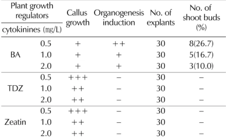
2. 신초 기관형성 유도를 위한 시토키닌류의 효과
배양중인캘러스로부터신초기관형성유도하기위해
BA, TDZ, Zeatin
이포함된고체배지에치상하여명조건에서4
주간 배양한 결과는Table 2
와 같다. BA
처리구에서는 캘러스의 생장은비교적낮은편이었지만치상2
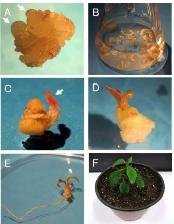
주후에배발생캘러스 를 볼 수 있었다(Fig. 2A).
그 중0.5
㎎/L BA
처리구에서26.7%
로가장양호한shoot buds
를유도되었으며BA
의농도가 증가함에 따라 그 수는 감소하였다
.
초기 발생된 배발생캘러스는대부분반투명한구형형태였으며
,
점차성장함에따 라빨간색소가생성되어붉은색을띄었다(
데이터미제시).
반면
TDZ
와Zeatin
에서는 캘러스생장만 하였고shoot buds
로는 유도 되지 않았다
. Gultai
와Jaiwal (1992)
은Vigna
radiata
의잎절편배양에있어서시토키닌이단독으로첨가되었을경우캘러스의형성과신초의재분화율이좋았으나
NAA
와
BA
가조합된배지에서는신초의증식과뿌리의분화가일어나지 않는다고 보고하였고
, Eun
등(1994)
은Pelargonium citrosa
의잎절편체배양에있어서2,4-D
와BA
를이용한배 지조성에서BA
가신초의분화에효과적이라고보고한바있 다.
본연구에서BA
가배발생 캘러스유도에영향을 미치는 사실을알수있었지만이캘러스는정상적인신초로발달하 지못하였다.
3. 신초 재분화 유도를 위한 BA와 NAA의 조합 효과 캘러스로부터신초재분화유도에미치는
BA
와NAA
의조 Table 1.Effect of plant growth regulators on callus induction from
various explants types of C. acuminata after 4 weeks culture.
Auxins (
㎎/L) Callus formation Cotyledon
segments Hypocotyl
segments Root segments IAA
0.2
− − −1.0
− − −3.0
− − −10.0
− − −NAA
0.2 + +(AR) -
1.0 ++ +++ +
3.0 + +
−10.0
− − −2,4-D
0.2 +++ +++ +
1.0 + + +
3.0
− − −10.0
− − −† MS basal medium containing 3% sucrose, 0.3% phytagel. (AR):
Adventitious root formation. Callus formation, +: poor, ++:
moderate, +++: good,
−: not-detected.
Fig. 1.
Effects of various media on callus growth of C. acuminata after 4 weeks culture. All media contain 0.2
㎎/L 2,4-D.
Table 2.
Effects of cytokinines on shoot buds formation from embryogenic callus of C. acuminata after 4 weeks culture in light condition.
Plant growth
regulators Callus
growth Organogenesis induction No. of
explants No. of shoot buds cytokinines (
㎎/L) (%)
BA 0.5 + ++ 30 8(26.7)
1.0 + + 30 5(16.7)
2.0 + + 30 3(10.0)
TDZ 0.5 +++
−30
−1.0 ++
−30
−2.0 ++
−30
−Zeatin 0.5 +++
−30
−1.0 ++
−30
−2.0 ++
−30
−† WPM basal medium containing 3% sucrose, 0.3% phytagel. Callus
formation, +: poor, ++: moderate, +++: good,
−: not-detected.
합처리의 영향을 알아 본 결과
(Table 3), shoot buds
는BA 0.5
㎎/L
와NAA 5.0
㎎/L
에서30.0%
로 가장 높게 나타났으 며, BA 0.5
㎎/L
이함유된 조합배지에서대체로양호하게형 성되었고BA
농도가BA 0.5
㎎/L
보다 증가함에따라shoot buds
수는 감소되는 경향을 나타냈다.
이결과는Wang
등(2006)
의의해 연구된 잎절편으로부터 희수나무의재분화에관한연구에서보여준결과와유사하였으며
,
이농도의BA
가희수나무의
shoot buds
에최적의농도라고보고된바있다.
또한NAA 1.0
㎎/L
의농도에서는캘러스의생장이더높게나타나는모습을볼수있었다
.
이는Oryza sativa
에서1.0
㎎/L
농 도의옥신류가캘러스분화및발달에유리하고신초의분화 에는옥신에비해시토키닌이요구된다는연구와유사한결과 를 보였다(Chen et al ., 1985).
그러나BA
와NAA
의 조합처리에서는기관형성에영향을미쳤지만완전한신초로발달 은보이지않았다
.
이러한이유로Konan
등(1997)
은세포배 양을통한 기관발생이나체세포배발생에서먼저기관이탈분 화되기때문에세포가갖는특성상여러가지 변이체가나타날수있는가능성이있고계대배양기간이길어질수록부정 아형성능력이감소할뿐만아니라배수체
,
이수체등의유전 적으로비정상적인세포의발생빈도가높다고보고한바있 으며, Kim
등(1997)
은높은농도의옥신활성으로인한 캘러 스화가신초의발달및생장을저해한다고보고한결과와유 사한결과를얻었다.
4. 신초 재분화 유도를 위한 암배양 처리 효과
암배양조건에서신초재분화유도를알아보기위하여
Table
4
와같이4
주동안 처리하여 관찰하였다.
그결과암배양2
주후이상에서비교적 높은
shoot buds
유도율를확인할수 있었다.
암배양2~4
주 처리에서50.0%
로 가장 높은shoot buds
을보였으며,
암배양처리1
주에서는33.3%,
무처리에서는23.3%
로낮은유도율을보였다. Dufour (1990)
은사과잎에서유도된캘러스로부터
2
주간암배양시가장많은재분화신초 을얻었다고보고했으며또한Lee
와Bae (1999)
는배양초기단계에 암처리 시
Gypsophia puniculata
의 잎절편으로부터Fig. 2.
Plantlets regeneration from embryogenic callus of C.
acuminata . (A) Shoot bud formation from pre-embryo
-genic callus, (B) Embryogenic callus in WPM liquid medium containing 0.5
㎎/L BA, (C) Adventitious shoot from pre-embryogenic callus, (D) Shoot regeneration from embryogenic callus after 6 weeks, (E) Rooted plantlets from regenerated shoots of C. acuminata after 3 weeks.
(F) Acclimatized plant of C. acuminata after 6 weeks.
Table 3.
Effects of auxins and cytokinines on plantlet formation via organogenesis from embryoenic callus of C. acuminata after 4 weeks culture in light condition.
Plant growth
regulators (
㎎/L) No. of
explants Callus
growth No. of shoot buds (%)
BA NAA
0.5 1.0 30 ++ 7(23.3)
5.0 30 ++ 9(30.0)
10.0 30 + 5(16.7)
1.0 1.0 30 ++ 4(13.3)
5.0 30 + 1(3.3)
10.0 30
− −2.0 1.0 30
− −5.0 30
− −10.0 30
− −† WPM basal medium containing BA 0.5 mg/L, 3% sucrose, 0.3%
phytagel. Callus formation, +: poor, ++: moderate, +++: good,
−: not-detected.
Table 4.
Effects of dark pre-treatments on plantlet formation via organogenesis from embryoenic callus of C. acuminata after 4 weeks culture.
Dark treatment
(weeks) No. of
explants No. of
shoot buds (%)
0 30 7(23.3)
1 30 10(33.3)
2 30 15(50.0)
3 30 14(46.7)
4 30 15(50.0)
† WPM basal medium containing BA 0.5 mg/L, 3% sucrose, 0.3%
phytagel.
75%
정도로재분화율이증가했다고보고했다.
본실험에서암 처리효과2
주이상처리했을때shoot buds
유도율이무처 리보다약2.1
배정도높은효과가있음을확인했으며,
시토키 닌류의BA 0.5
㎎/L
에서shoot buds
유도율과비교했을때에 도1.9
배정도 높은효과를 알수있었다.
하지만 암처리조 건에서도완전한신초재분화는유도되지않았다.
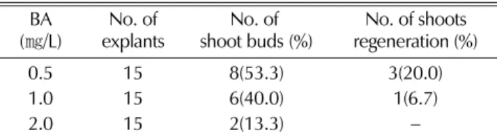
5. 신초 재분화 유도를 위한 액체배양 효과
액체배양에서캘러스로부터식물체의재분화유도를알아보
기위해
BA
를 농도별로처리한WPM
액체배지에배양하여Table 5
과 같은 결과를 얻었다. Shoot buds
유도는BA 0.5
㎎/L
에서53.3%
로 가장 높았으며, BA 1.0
㎎/L
에서40.0%, BA 2.0
㎎/L
에서는13.3%
정도로 관찰되어BA
와NAA
의조합처리와암처리보다액체배양에서높은shoot bud
가유도된다는것을확인할수있었다
(Fig. 2B).
액체배양에 서배양2
주후신초의 성장을보였고(Fig. 2C), 6
주후정 상적인신초의재분화를관찰하였다(Fig. 2D).
또한0.5
㎎/L BA
에서20.0%
와1.0
㎎/L BA
에서6.7%
로신초 재분화율을 보였다(Table 5).
이결과는Cheng
등(2007)
의Schisandra
chinensis
를재료로 하여체세포배발생과 식물의재분화에관한연구에서
2,4-D
를첨가한MS
액체배지를통한 액체배양시 배발생세포가 빠르게 성장하였고 분화하였다고 보고와
Mythili
등(1999)
의Sorghum dimidiatm
의배발생세포를액체 배양하여신초로재분화하였다는보고와유사한결과로희수 나무의캘러스로부터액체배양이신초재분화에가장 적합한 처리라고사료된다.
또한재분화된신초의뿌리유도는WPM
기본배지에
NAA
를0.2
㎎/L
의 농도로 첨가하여 치상12
일후부정근을확인했으며
(Fig. 2E),
뿌리가 발생된식물체는 버미큐라이트가담긴화분으로이식한후랩(wrap)
으로덮어 상대습도를85%
로유지시키면서약6
주간순화시켰을때,
완 전히활착한희수나무를관찰하였다(Fig. 2F).
감사의 글
본연구는 전남대연구비
(
관리번호: 2007-0647)
지원에 의 해수행된것으로이에감사드립니다.
LITERATURE CITED
Chao HH and Joseph T. (2001). New Advances in Long Cancer Chemotherapy: Topotecan and the Role of Topoisomerase I Inhibitors. Oncology. 61:14-24.
Chen TH, Lam L and Chen SC. (1985). Somatic embryogenesis and plant regeneration from cultured inflorescence of
Oryza sativaL. Plant Cell, Tissue and Organ Culture. 4:51-54.
Cheng HL, Niu Y, Zhao B, Ghimire BK, Kil HY, Heo K, Kim MJ, Eom SH, Cho DH and Yu CY. (2007). Somatic embryogenesis and plant regeneration from embryogenic cell suspension cultures of
Schisandra chinensisBaill. Korean Journal of Medicinal Crop Science. 15(5):346-351.
Choi H and Byun SY. (2000). Enhanced production of antlcancer agent camptothecin by double ellcitors in suspension cultures of
Camptotheca acuminata. Korean Journal of Biotechnology and Bioengineering. 15(5):428-433.
Dufour M. (1990). Improving yeild of adventitious shoots in apple. Acta Horticulturae. 280:51-61.
Eun JS, Ko JA, Kim YS and Kim MJ. (1994). Micropro- pagation by leaf and meristem cultures of
Pelargonium citrosaVan leenen. Korean Journal of Plant Tissue Culture. 21:247- Gamberg OL, Miller RA and Ojima K. 252. (1968). Natural requirenments of suspension cultures of soybean root cells.
Experimental Cell Research. 50:148-151.
Gultai A and Jaiwal PK. (1992).
In vitroinduction of multiple shoots and plant regeneration from shoot tips of mung bean (
Vigna radiata(L.) Wilczek). Plant Cell, Tissue and Organ Culture. 29:199-205.
Hengel AJ, Harkes MP, Wichers HJ, Hesselink PGM and Buitelaar RM. (1992). Characterization of callus and camptothecin production by cell lines of
camptotheca acuminata. Plant Cell, Tissue and Organ Culture. 28:11-18.
Kim KS, Seung NS, Kim MW, Pyo BS, Hwang B. (1997).
Micropropagation of
Achyranthes japonicathrough axillary buds culture. Koeran Journal of Plant Tissue Culture. 24:357- Klaus MD and Peter HK. 360. (2004). Death receptors in
chemotherapy and cancer. Oncogene. 23:2960-2966.
Konan NK, Schopke C, Carcamo R and Beachy RN. (1997).
An efficient mass propagation system for cassava (
Manihot esculentaCrantz) based on nodal explants and axillary bud- derived meristens. Plant Cell Reports. 16:444-449.
Lee SW and Bae JJ. (1999).
In vitroregeneration using leaf segment in
Gypsophila paniculataL. 'Bristol Fairy'. Korean Journal of Horticultural Science and Technology. 17:765-767.
Li S, Zhang Z, Cain A, Wang B, Long M and Taylor J. (2005).
Antifungal activity of camptothecin, trifolin, and hypiroside isolated from
Camptotheca acuminata. Journal of Agricultural and Food Chemistry. 53:32-37.
Liu, Z and Li Z. (2001). Micropropagation of
Camptotheca acuminataDecaisne from axillary buds, shoot tips, and seed embryos in a tissue culture system. In Vitro Cellular and Developmental Biology- Plant. 37:84-88.
Lloyd GB and McCown BH. (1980). Commercially feasible micropropagation of mountain laurel, Kalmia latifolia, by the
Table 5.