Journal of Trauma and Injury Vol. 26, No. 4, December, 2013
[ J Trauma Inj 2013;26:286-290 ]
� Address for Correspondence : Hee-Gon Park
Department of Orthopedic Surgery, Dankook University, School of Medicine 16-5 Anseo-dong, Cheonan, Choongnam 330-715, Korea
Tel : 82-41-550-6579, Fax : 82-41-559-7897, E-mail : [email protected]
Submitted : November 6, 2013 Revised : December 19, 2013 Accepted : December 19, 2013
원위 대퇴골 골절에 대한 고령 환자군의 치료 결과 비교 -임상적, 방사선학적 비교-
단국대학교 의과대학 단국대학교병원 정형외과 박희곤, 김연준, 장호성
- Abstract -
Clinical and Radiological Outcome of Distal Femoral Fracture in Elderly Patient Group
Hee-Gon Park, M.D., Yeon-Jun Kim, M.D., Ho-Seong Jang, M.D.
Department of Orthopedic Surgery, Dankook University, School of Medicine, Chonan
Purpose: To report the postoperative Clinical and Radiological outcomes following distal femoral fractures in
elderly patients compared with young patients.Methods: From March, 1996 to March, 2012, 83 patients who received surgical treatment for fractures of the
distal femur were enrolled in this retrospective study. Ages more than 65 was named group A. Group A was 49 cases and mean age is 72.5 year(65~91year). Group B was 49 cases and mean age is 45.7 year(16~61 year).Surgical methods are retrograde IM nail, locking compression plate, cannulated screw and postoperative reha- bilitation is no difference between two groups. Clinical results were evaluated using Neer scores, radiographic results and the presence of clinical complications.
Results: The mean union period was 18.4(12-40) weeks in group A and 17(10-24) weeks in group B. Neer
functional scores are no significant statistical difference between two groups. There are 5 cases metal breakage in group A and 1case in group B. There are 3 cases nonunion in group A and 1 case in group B.Conclusion: In the case of fractures of the distal femur in elderly patients, locking plate using minimally inva-
sive percutaneous periosteal osteosynthesis (MIPPO) technique may be one of the most effective methods and preoperative bone stock evaluation in important.Key Words: Distal Femur Fracture, Elderly, Retrograde, Locking plate
I. 서 론
대퇴골 원위부 골절은 모든 골절의 0.4%를, 대퇴골 골절 의 3~7% 정도를 차지하는 흔하지 않은골절이며, 그 중 50%
가량은 고령에서 발생하며, 슬관절을 포함하고 있어 치료 후 관절 구축 등의 후유증이 흔히 발생한다.(1) 젊은 환자의 경 우 대부분 고에너지 손상에 의해서 발생하며, 고령 환자의 경우 저에너지 손상에서도 발생이 가능하나,(2,3) 골다공증 이 동반되어 있기 때문에 분쇄 골절 및 주위 연부 조직 손상 으로 인한 불유합, 지연유합, 부정유합, 관절강직, 감염 등의 합병증이 흔히 발생할 수 있는 치료가 어려운 골절로 알려져 있다. 또한, 관절면을 침범한 경우가 흔해 수술적 접근을 통 한 관절면의 정확한 정복이 요구되며, 수술 방법으로는 금속 판, 교합성 골수강내 금속정 등을 이용한 내고정술이 있으 며, 고령 환자에서는 수술에 따른 관절구축뿐만 아니라, 근 위축, 침상안정에 따른 심부 정맥 혈전증과 같은 결과를 초 래할 수 있어 최소침습적 금속판 고정술 등이 고안되게 되었 다.(4) 이러한 고정방법에 대한 생역학적 연구들이 보고되어 있으나, 모두 골다공증이 없는 성인 대퇴골을 이용한 연구이 기에 골다공증 환자에서 발생하는 골절의 치료와는 부합하지 않는다. 또한 Schatzker 등(5)은 골다공증성 노인 골절의 경 우 술자의 술기와는 상관없이 좋지 않은 결과를 보일 수 있 다고 하였다. 고령 환자군에서 다양한 방법의 내고정술이 시 도 되었으나, 젊은 연령층의 환자군과 골절의 양상 및 수상
기전, 동반손상 여부, 술 후 합병증 및 그 예후에 대한 전반 적인 비교결과가 부족하다고 생각되어 저자들은 원위 대퇴골 골절 환자를 65세를 기준으로 두 군을 나누어 연령에 따라 비교하여 고찰해 보기로 하였다.
II. 대상 및 방법
1996년 3월부터 2012년 3월까지 원위 대퇴골 골절로 수 술적 치료받은 환자 중, 12개월 이상 추시가 가능했던 83명 을 대상으로 후향적 조사하였다(Fig. 1). 65세 이상을 고령 환자군으로 분류 하여 A군이라 하였으며 남자 16례, 여자 33례로 모두 49례였으며, 65세 미만의 환자군은 B군으로 남 자 26명, 여자 8명으로 모두 34례였다. 수상 당시 평균 연령 은 A군은 72.5세(65~91세)이었고, B군은 45.7세(18~62세) 이었다. AO/OTA 분류를 사용하였으며, A군에서 관절외 골 절(TypeA)이 29례, 관절면을 침범한 골절(TypeC)은 12례이 었으며, B군에서는 관절외 골절(TypeA)이 9례, 관절면을 침 범한 골절(TypeC)은 22례였다. 개방성 골절은 A군에서 6례 (Gustillo-Anderson type I: 1례, II: 2례, III: 3례)이었고, B군에서는 9례(Gustillo-Anderson type I: 1례, II: 5례, III: 3례)였다. 모든 환자에서 수술적 치료를 시행하였으며, 수술방법으로는 A군에서는 후향적 골수강내 고정술(25례), 삽관 나사못(5례), 외측 잠김 압박 금속판(19례)를 사용하였 고, B군에서 후향적 골수강내 고정술(1례), 삽관 나사못(0
Fig. 1. Preoperative AP (A) and lateral (B) radiographs of a distal femur fracture.
A B
례), 외측 잠김 압박 금속판(3례)를 사용하였다. 최소침습적 수술법을 사용한 군은 A군 6례, B군 8례로 총 14례였으며, 슬개 외측 주변 절개를 통해 금속판을 근육 밑으로 밀어 넣 어 고정하여 골절부의 혈행을 최대한 보존하며 연부조직 손 상을 최소화 하였다. 골수강내 고정법을 시행한 경우 A군 2 례에서 골유합의 지연으로 인해 역동화 수술을 하였으며, 골 이식은 A군에서 13례, B군에서는 18례에서 시행하였고, 추 가적인 수술적 처치를 필요로 하는 감염은 A군에서 3례, B 군에서 1례 발생하였다(Table 1).
술 후 재활로는 A군 및 B군에서 차이점을 두지 않았다. 술 후 3일째 야간 부목을 적용하여 지속수동운동 및 대퇴사두근 강화 운동을 시작하였으며, 추시 방사선 검사상 가골이 형성 되는 시기부터 부분체중 부하를 시행하였고, 전후면과 측면 사진 모두 가골 형성 시 전 체중 부하를 시행하였다.
추시시 환측 슬관절의 운동범위 및 방사선학적 검사를 하 였으며 최종 추시시 운동범위 및 기능적 평가를 통해 환자의 만족도를 측정하였다. 기능적 평가로는 Neer의 기능적 평가 척도를 사용하였으며, 1. 우수(85점 이상), 2. 만족(70~84 점), 3. 불만족(55~69), 4. 실패(55점 미만)의 네 단계로 나
누어 비교하였다. 통계학적 분석은 SPSS version 19.0 (SPSS Inc., Chicago, II, USA)을 사용하였으며, T-test 및 Chi-square test 방법을 사용하였다.
III. 결 과
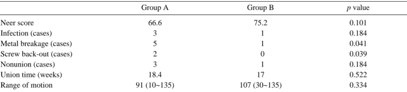
수상 기전으로 A군에서 단순 낙상(서있는 상태에서 넘어 짐을 포함)이 24례로 B군(9례)에 비해 의미 있게 많았으며 (p=0.012), 동반손상으로는 A군에서 동측 대퇴골 간부(2례) 및 경부 골절(0례), 동측 경골 골절(7례), 슬개골 골절(1례), 전완부 골절(2례) 있었으며, B군에서는 동측 대퇴골 간부(5 례) 및 경부 골절(3례), 동측 경골 골절(3례), 슬개골 골절(6 례), 전완부 골절(2례)가 관찰되었다. A군에서 3례, B군에서 1례 총 4례를 제외하고 모두 유합을 얻었으며, 평균 유합 기 간은 A군에서 18.4주(12~40주), B군에서 17주(10~24주)였 으며, 최종 추시시 Neer 기능적 평가는 A군(우수: 1례, 만 족: 18례, 불만족: 15례)이 B군(우수: 5례, 만족: 18례, 불만 족: 9례)에 비해 좋지 않은 결과를 보여주었고, 만족 이상이 B군과 비교하여 유의하게 적었다(p=0.037). 내고정물 파단
Table 1. Demographic data of two groups
Group A Group B p value
Age (Years) 72.5 (65~91) 45.7 (16~62)
Gender (male:famale) 16:33 26:8 0.057
Acconmapined injury (cases) 11 13 0.119
Follow up period (months) 22.1 24.9 0.452
Injury to operation time (days) 04.6 02.7 0.084
Bone graft (cases) 13 18 0.157
BMI (kg/m
2) 23.7 22.8 0.116
Fig. 2. Postoperatively 22 month metal breakage (A) and postoperatively 2 month Screw loosening (B).
A B
은 B군에서 1례가 관찰된 반면, A군에서는 5례가 관찰되었 고(p=0.041) (Fig. 2), 불유합은 A군에서 3례, B군에서 1례 가 관찰되었다(p=0.184). 최종 추시시 슬관절의 평균 운동범 위는 A군에서 91도(10~135도), B군에서 107도(30~135도) 로 통계적 유의한 차이는 없었으며(p=0.334) 인공관절로의 치환한 경우는 1례 있었다(Table 2).
A군에서 골밀도 검사를 시행한 례는 20례로 T-score는 고관절부 평균 -2.424, 척추부 평균 -3.136로 측정되었으 며, 15례에서 T-score -2.5미만의 골다공증 소견을 보였다.
IV. 고 찰
고령환자에서의 골절은 골다공증이 흔히 동반되며, 조기 보행을 요하므로 골절의 안정적 고정이 중요시된다. 또한 당 뇨 등의 기저 질환으로 전신상태의 면밀한 검사를 요하며 수 술 후 조기 거동 및 재활이 매우 중요하므로 평소 거동여부 및 활동성, 근육량 등이 치료 후 결과에 많은 영향을 미친다.
원위 대퇴골 골절의 경우, 체중부하의 역학적 관점에서 많은 연구가 행해졌고, 역행성 골수강내 고정술, 잠김 금속판 등 이 사용되고 있으며. 또한 골절부의 혈행을 유지하기 위해 최소침습적 금속판 고정술 등이 소개되었다. Stromsoe(6)는 골다공증 환자에서 골절의 유합능력은 보존되나 주로 고정의 유지에 문제가 발생한다고 보고하였다. 잠김 금속판을 사용 할 경우, Pull-out에 대한 저항력이 강하며 Screw의 Core diameter의 증가로 축성 압박력에 저항하는 힘이 커져 골다 공증 환자에서 좋은 고정물로 생각되고 있다.(7) 역행성 골수 강내 고정술의 경우 역학적으로 외측 금속판 고정술에 비해 유리하다고 생각되어지며, 절개의 양이 크지 않아 많이 사용 되고 있으나,(8) AO/OTA Type C와 같은 관절을 침범한 골 절의 경우 골편의 정복에 어려움이 있고, 불가피하게 관절 내부를 노출시켜야 하는 단점이 있다.(10) 본 연구에서는 잠 김 금속판을 사용한 경우에는 고정물 파단이나, 고정의 소실 이 총 5례에서 발생하였으며, 원인으로 불유합이 3례, 감염 이 2례였다.
Dirk Wahnert 등은 폴리우레탄 폼으로 만들어진 인공 원 위 대퇴골을 이용한 역학적 연구에서, 원위 대퇴골이 해면골
로 이루어져 있어 골절 중 원위부 나사못의 고정(Screw anchorage)이 정복의 유지에 가장 중요한 점으로 생각하였 으며, 역행성 골수강내 고정술의 경우 축성 압박력에 강하 고, 금속판의 경우 torsional stability에 강점이 있다고 하 였다.(11-16) Schandelmaier P. 등(17)은 안정적인 고정이 유지될수록 좋은 임상적 결과를 가져온다고 하였다.
Schutz M 등(18)에 의하면 일차적인 골이식은 수술 시간 의 연장 및 공여부 문제의 가능성이 있어 골절부의 혈행이 손상되지 않을 경우 필요하지 않다고 하였으며, 본 연구에서 도 골이식을 시행한 례가 A군에서 13례, B군에서 18례로 총 21례였으며, A군 5례와 B군 1례 총 6례를 제외한 77례에서 골유합을 얻을 수 있었다.
Nieves 등은 원위 대퇴골 골절 환자에서 당뇨, 심혈관 질 환 등으로 인해 최소한 한가지 이상의 약물을 복용하는 환자 가 32%이상으로 보고하고 있으며,(19) 이러한 기저 질환으 로 인해 환자의 사망률을 대퇴골 경부 골절 환자의 사망률과 비슷하게 보고되기도 하였다. 본원에서 입원 도중 사망한 례 는 없었으나, 기저 질환을 가진 경우가 54%(A군 67%, B군 31%)였으며, 고령 환자의 경우 전신적인 주의를 요할 것으로 생각된다. 본 연구에서는 최종 추시 이후의 사망여부 및 수 상 후 사망까지의 기간은 알 수 없었다.
본 연구는 후향적으로 시행되었으며 , 적은 환자의 증례수 및 골절의 분류 따른 비교와 수술적 방법에 따른 비교에 대 해 교차 분석은 시행하지 않은점이 한계점으로 생각된다.
V. 결 론
고령의 원위 대퇴골 골절 환자에서 골절의 양상이 상이함 을 인지 하여야 하며, 최소 침습적 술기를 통해 잠김금속판 을 이용한 수술을 권장할 수 있으며, 평가에 있어서 내고정 물의 고정력과 implant cutout 발생률에 관계를 고려 할 때 모든 환자에 있어서 개별적인 골질에 대한 평가가 선행되어 야 한다고 생각된다.
Table 2. Clinical and radiological outcomes of two groups
Group A Group B p value
Neer score 66.6 75.2 0.101
Infection (cases) 3 01 0.184
Metal breakage (cases) 5 01 0.041
Screw back-out (cases) 2 00 0.039
Nonunion (cases) 3 01 0.184
Union time (weeks) 18.4 17 0.522
Range of motion 91 (10~135) 107 (30~135) 0.334
REFERENCES