Vol. 29, No. 3, September, 2016 http://dx.doi.org/10.20408/jti.2016.29.3.76
� Address for Correspondence : Hyung Keun Song, M.D.
Department of Orthopeadic Surgery, Ajou University School of Medicine, 164 Worldcup-ro, Yeongtong-gu, Suwon, Korea
Tel : 82-31-219-5220, Fax : 82-31-219-5229, E-mail : [email protected]
Submitted : August 18, 2016 Revised : September 2, 2016 Accepted : September 12, 2016
Analysis of Radiological and Clinical Results in Treatment of Open Segmented Tibia Fractures: A Comparison between Intramedullary Nailing and Minimal Invasive Plate Osteosynthesis
Ji Wan Kim, M.D., Hyung Keun Song, M.D.
1Department of Orthopaedic Surgery, Haeundae Paik Hospital, Inje University, Busan, Korea
1Department of Orthopaedic Surgery, Ajou University School of Medicine, Suwon, Korea
Purpose: The purpose of this study was to compare the data comprehensively including not only the clinical and radiographic outcomes but some parameters related to operation between the minimally invasive plate osteosynthesis (MIPO) technique and intramedullary nailing (IMN) for treatment of segmental tibia shaft fractures.
Methods: We conducted a retrospective study of 31 patients (mean age, 49.3 years, range, 27-74 years), with a mean follow-up of 14.1 months (range, 12-19 months) with acute segmental tibial fractures (AO 42-C2) who underwent either surgical treatment of MIPO or IMN. In accordance with the Gustilo-Anderson classification, 11 were type I, 5 were type II, and 15 were type III. Initial compartment syndrome was confirmed in 2 cases.
Results: There were no statistically significant differences in terms of the patient demographic data between the two groups. The time to definitive fixation was longer in the MIPO group (mean 13.7±10.9 days; range, 2-27) than in the intramedullary group (mean 5.4±9.6 days; range, 0-35) with statistically significant difference (p=0.002). Bony union was observed in most of cases but except 5 cases of nonunion were diagnosed (3 in the MIPO vs 2 in IMN, p=0.188).
The average bone healing time was 27.1 weeks (10 to 56 weeks) in MIPO group and 23.2 weeks (13 to 66 weeks) in IMN group, respectively (p=0.056). Overall complications were 5 cases in MIPO group and 2 cases in the IMN group.
Difference in LEFS was not statistically significant between both groups (p=0.824).
Conclusion: This study showed that segmental tibia shaft fractures treated with both MIPO and intramadullary nail- ing was challenging with relatively high complication rate. A well planned sequential strategy with keeping the soft tis- sue and personality of fracture in mind is utmost significant as much as the choice of surgical modalities. [ J Trauma Inj 2016; 29: 76-81 ]
Key Words: Segmental tibia fracture, MIPO, Intramedullary nail
I. Introduction
The segmental tibia shaft fracture has a distinc- tive personality of fracture with two different level of one tibia and inevitably accompanied with mod- erate or severe soft tissue problem caused by high energy injury. The high potential for complications
is much to do with the precarious blood supply of the intermediate segment of tibia and vulnerable soft tissue envelope.(1) High complication rates are reported for segmental tibial fractures including infection, nonunion, malunion and amputation.(2-5) These worrisome problems are the reason there is no clinical consensus about the best way to treat
these complicated fracture,(6) although traditionally several treatment methods have been reported to manage the segmental tibia fracture with non-oper- atively and surgically.(3,6-9) The intramedullary nailing (IMN) is commonly and widely used surgical modality due to its biomechanical property.(3,7,10) As widely known, however, the intramedullary nail- ing is a technically demanding and frequently needs additional procedure, such as a blocking screw, an additional plate and position of bone clamp so as to prevent the malalignment of entire tibia.(11) Moreover, the complications of the segmental frac- ture dealt with the intramedullary nailing were not within the surgeon’s sphere of influence because of inherent characteristics of the segmental fracture itself.
Recently, the minimally invasive plate osteosyn- thesis (MIPO) technique has been applied to lower extremities and even more extended into the seg- mental tibia shaft fracture as well.(12,13) Although they recommended the MIPO technique for these difficult fracture with good functional outcomes, we wondered whether the MIPO technique is an alter- native and as effective and safe as the outcomes of the intramedullary nailing. To our knowledge, how- ever, there was no clinical study on comparative data with the MIPO and intramedullary nailing for the segmental tibia shaft fracture.
The purpose of this study was to compare the data comprehensively including not only the clinical and radiographic outcomes but some parameters related to operation between the MIPO technique and intramedullary nailing for treatment of segmental tibia shaft fractures. Our null hypothesis was that there were no significant differences in all compar- ative data between two surgical techniques.
II. Materials and Methods
During the period from October 2010 to April 2015, the authors have gone through prospectively col- lected orthopedic trauma databases in two hospitals (level I trauma center), and identified consecutive series of 31 adult patients with acute segmental tib- ial fractures (AO 42-C2) who underwent either sur- gical treatment of MIPO or IMN that were eligible for inclusion in our retrospective case series.
Inclusion criteria were patients who are skeletally mature (age≥18 years); those with a displaced sepa- rate proximal and distal tibial shaft fractures (each fracture is not involved into the rule of the square);
those who were underwent operation with either MIPO or IMN; those who were followed up during a minimum 1-year. Exclusion criteria were as follows:
prior amputation, Gustilo-Anderson type IIIc open fracture, fracture with bone loss, spinal cord deficit,
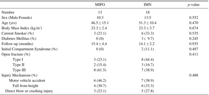
Table 1. Demographic data between MIPO and intramedullary nail groups
MIPO IMN p-value
Number 13 18
Sex (Male:Female) 10:3 13:5 0.552
Age (yrs) 46.5±15.1 51.3±10.4 0.470
Body Mass Index (kg/m2) 23.3±2.40 23.3±3.70 0.674
Current Smoker (%) 3 (23.1) 6 (33.3) 0.535
Diabetes Mellitus (%) 0 (0) 3 (09.7) 0.245
Follow-up (months) 15.4±4.40 14.1±2.20 0.935
Initial Compartment Syndrome (%) 0 (0) 2 (11.1) 0.497
Open fracture (%) 0.411
Type I 3 (23.1) 8 (44.4)
Type II 2 (15.4) 3 (16.7)
Type III 8 (61.5) 7 (38.9)
Injury Mechanism (%) 0.488
Motor vehicle accident 6 (46.2) 7 (38.9)
Fall from height 4 (30.7) 6 (33.3)
Direct blow or crushing injury 3 (23.1) 5 (27.8)
* statistically significant difference (p<0.05).
significant brain injury, and pathologic fractures.
Total consecutive 31 patients were included in this study, which was conducted after obtaining approval from the institutional review board at our hospital.
This study included 23 men (74.2%) and 8 women (25.8%), with mean age of 49.3 years (range, 27-74 years). Detailed history of enrolled patients, includ- ing smoking, diabetic status, and body mass index, were reviewed. All other demographics in regard with fractures such as fracture classification of proximal and distal site by AO/OTA classification, Gustilo-Anderson classification of open fracture, presence of initial compartment syndrome, and injury mechanism are described in Table 1.
The electronic medical records and radiographs of each patient were reviewed. Data recorded included the age, gender, mechanism of injury, fracture AO/OTA type and Gustilo-Anderson grade, treat- ment information, complications, and time to radi- ographic fracture healing. The time to surgery was defined as the time between the injury and definitive surgical treatment. Routine follow up radiographs were obtained every 4 weeks until solid continuous callus formation was observed; callus formation on 3/4 of the cortices and radiographic evidence of frac- ture line fading were considered signs of fracture union. Limb rotation and alignment were assessed at all follow-up visits. Malalignment (or malunion) was defined as angulation deformity of 10 degree or more, compared to the uninjured leg. Rotational malalignment was checked with thigh foot angle compared to the uninjured leg. Final clinical out- comes were evaluated using the Lower Extremity Functional Scale(14) (LEFS; 0, unable to perform any activity to 80, excellent function) by a physician who was unaware of the patients’ information.
Complications were recorded as union-related or soft tissue-related. SPSS version 18.0 (SPSS Inc. Chicago, IL, USA) was used for the statistical analyses.
All open fractures were managed with early, thor- ough debridement and irrigation, followed by addi- tional debridement when indicated. Injuries with a significant deformity and/or soft tissues that were deemed unsafe for primary definitive fixation were treated with temporary spanning external fixation.
Definitive surgical treatment was performed when
the status of the soft tissue was sufficiently stabi- lized for soft tissue reconstruction and there was no evidence of infection. Patients were encouraged to start active and passive range of motion exercises at the knee and the ankle as soon as possible. The majority of patients were encouraged to partially weight bear at 2 or 3 weeks after surgery. Patients were allowed to fully weight bear whte there was no pain at the fracture site and radiological evidence of bone union.
III. Results
The open injury was graded as Gusilo-Anderson type I in 11 patients (35.5%), type II in 5 patients (16.1%), and type III in 15 patients (48.4%). Two patients had an impending compartment syndrome that required double incision fasciotomy. The mean follow-up period was 14.6 months (range, 12?25 months). The initial stabilization was intramedullary (IMN) nailing in 13 patients (41.9%), percutaneous plating (MIPO) in 2 patients (6.5%), and provisional external fixation in 16 patients (51.6%). In those 16 patients who had undergone provisional external fixation, definitive surgery was performed with either an IMN in 5 patients or a MIPO in 11 patients.
Definitive surgery after provisional external fixation was performed at a mean of 16.2 days after injury (range, 2-35 days).
In IMN group, the conventional patella tendon split technique (knee flexed) was used in five patients and the semi-extended nail insertion tech- nique (3 medial parapatella, 3 lateral parapatella, 7 suprapatella) was used in 13 patients. In MIPO group, proximal lateral periarticuluar tibial plate was inserted in 4 patients, distal medial periarticu- lar tibial plate in 3 patients, and combination with proximal lateral and distal medial plate was in 6 patients. Soft tissue reconstruction was performed in 15 patients (six split thickness skin graft and nine free flap). All free flaps were elevated anterolateral thigh fasciocutaneous free flaps.
No significant differences were identified with respect to patient age (p=0.470), sex (p=0.552), body mass index (p=0.674), patient’s number of smoking (p=0.535), and open fracture grade (p=0.411) between
MIPO group and IMN group (Table 1). The mean time injury to definitive fixation was 13.7 days in MIPO group and 5.4 days in IMN group, respectively (p=0.002).
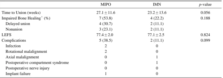
Bony union was observed in most of cases but except 6 cases of nonunion were diagnosed (3 in the MIPO vs 3 in IMN, p=0.188). All nonunion were treated with autogenous bone graft (1 proximal and 2 distal in MIPO group, and 3 proximal in IMN group) (p=0.625). The average bone healing time was 27.1 weeks (range 10-56 weeks) in MIPO group and 23.2 weeks (range 13-66 weeks) in IMN group, respectively (p=0.056). Differences in LEFS were not statistically significant between each group (p=0.824) (Table 2).
Complications were noted in 7 patients (22.6%):
superficial infection, 1 case; deep infection, 1 case;
rotational malunion, 2 cases; angulation, 1 case;
postoperatively compartment syndrome, 1 case;
implant failure, 1 case and implant failure, 1 case. In the deep infection case, the fracture was healed when the infection occurred, so treatment involved surgical debridement, implant removal, and antibi- otic therapy (Table 3).
IV. Discussion
This study compared clinical and radiographic results and surgery-related parameters of two dif- ferent surgical techniques. Although there was no statistical difference in regards to pre- and post- operative parameters according to surgical modali- ties, the time to definitive surgery was statistically significantly longer in MIPO group than in IMN group. This outcome appears to be attributable to the fact that plate fixation is more affected by the
Table 2. Comparison of parameters related to operation between 2 groups
MIPO IMN p value
Initial External Fixation 11 (84.6) 5 (27.8) 0.002*
Injury to definitive fixation (days) 13.7±10.9 5.4±9.6 0.002*
Soft tissue reconstruction (%) *8 (61.5) 7 (38.9)
STSG *1 (*7.7) 5 (27.8) 0.213*
Free flap *7 (53.8) 2 (11.1)
Bone graft (%) *3 (23.1) 2 (11.1) 0.625*
Operation time (minutes) 86.5±10.2 79.1±8.60 0.389*
* statistically significant difference (p<0.05).
STSG: split-thickness skin graft
Table 3. Clinical and radiologcal outcomes between two groups
MIPO IMN p-value
Time to Union (weeks) 27.1±11.6 23.2±13.6 0.056
Impaired Bone Healing�(%) 7 (53.8) 4 (22.2) 0.188
Delayed union 4 (30.7) 2 (11.1)
Nonunion 3 (23.1) 2 (11.1)
LEFS 77.4±2.00 77.1±2.50 0.824
Complications 5 (38.5) 2 (11.1) 0.099
Infection 2 0
Rotational malalignment 2 0
Axial malalignment 0 1
Postoperative compartment syndrome 0 1
Postoperative nerve injury 0 0
Implant failure 1 0
* statistical significant difference.
�delayed union and nonunion taken together.
LEFS: lower extremity functional score
condition of adjacent soft tissues in determining the timing of surgical intervention than intramedullary nailing.
IMN is the most commonly used surgical tech- nique. This is because that IMN has distinct mechanical and biological advantages compared to other fixation techniques.(3,7,8,10) Despite these benefits, this modality requires the surgeon’s expe- rience and skills because of challenging surgical procedures and is more prone to malalignment including valgus deformity, anterior angulation and others.(3,4) Segmental tibial fractures accompany precarious blood supply to the intermediate segment of tibia and are at risk of secondary injury due to intermediate fragment rotation when reaming.(15)
Open segmental fractures are difficult to be man- aged with conventional open reduction and compres- sion due to massive soft tissue damage caused by high energy injury. Recent studies have achieved favorable results that MIPO can be used as an alter- native technique to replace IMN.(12,13) MIPO is more ideal for minimizing periosteal and soft tissue injury by submuscular insertion compared to conventional open reduction and compression. Moreover, instead of intramedullary nail insertion, this technique does not damage blood supply to the intramedullary canal by preserving the intramedullary environment after reaming.
Several investigators have reported high compli- cation rates in segmental tibia fractures, and asso- ciated complications include problems with bone union, infection, malalignment, amputation and others.(3,5,10,15-17)
In this study, complications occurred in 7 cases (22.7%), and no statistical difference was found between the two groups. Even though infection did not occur in patients with IMN, there were two cases with infection in MIPO group. This outcome was comparable to a previous study that conducted plate fixation.(12,13) McMahon et al. have addressed that malunion, one of the common complications, devel- oped at a lower rate in the group with open plate fixation compared to the group with IMN.(6)
In the present study, malunion occurred in both groups that underwent plate osteosynthesis or IMN.
Rotational deformity commonly occurred in MIPO
group, while axial malalignment (valgus deformity) chiefly developed in IMN group. Rotational malalign- ment occurred in a patient who received MIPO using proximal lateral and distal medial plates concurrently.
Malalignment of the proximal and intermediate frag- ments developed as intermediate and distal fragments were anatomically reduced. Rotational deformity has not been reported in other previous studies on inter- nal fixation.(12,13) Malalignments in the proximal tibia occurred in the group with IMN, these are valgus deformity and anterior angulation that are frequently seen in proximal tibia fractures. Anterior angulation can be corrected with insertion with knee in flexion to some degree. On the contrary, valgus deformity can be overcome by the use of multiple techniques includ- ing blocking screw insertion, insertion of additional plates, intramedullary nail insertion after reduction using reduction forceps and others.
Bone graft was performed in 5 cases, 3 with plate osteosynthesis and 2 with IMN. This procedure was done to treat nonunion at the bone defect site of the medial tibia due to valgus malalignment of the proximal fragment in the group with IMN, and at the bone defect site of the lateral tibia due to rota- tional deformity of the distal fragment in 2 out of 3 cases in the group with MIPO. The other case had fracture gap caused by displacement of butterfly fragments at the distal fractured site and nonunion was treated with autogenous bone graft.
No statistical difference was found in age, gender, BMI, severity of open fractures and other baseline characteristics between IMN and MIPO groups.
Debridement and external fixation were carried out as an initial treatment in cases of requiring damage con- trol surgery due to severe initial or associated injuries.
Since the fracture was reduced and alignment was restored to some degree through external fixation, plate osteosynthesis was primarily performed in definitive surgery while maintaining reduction with external fixation, instead of conducting IMN that requires removal of an external fixator (p=0.002). For this reason, the time to definitive surgery after the initial injury was longer in MIPO group than in IMN group (p=0.002). There was no statistical difference with respect to time to bone healing, clinical scores, and incidence of complications.
This retrospective study was limited by the rela- tively small sample size. In addition, the two surgi- cal interventions, MIPO and IMN, were not random- ly performed in patients. Despite these limitations, the present study was meaningful in that it com- pared open segmental fractures of the tibia, a rare fracture pattern, through a multicenter study. More prospective, multicenter, randomized studies with a larger sample size are warranted in the future.
V. Conclusion
This study has identified that segmental tibia shaft fractures treated with both MIPO and IMN are technically challenging and have a relatively high complication rate. Since the two surgical techniques had no statistical difference in pre- and post-oper- ative variables, these interventions are anticipated to be appropriate to manage segmental tibial frac- tures. S Regardless of surgical modalities, satisfac- tory outcomes are expected by preserving soft tis- sues and achieving anatomical alignment through a well-planned sequential strategy.
REFERENCES
01) Arastu MH, Sheehan B, Paolucci EO, Buckley RE. Does it really spin? Intra-medullary nailing of segmental tibial frac- tures--a cadaveric study. Injury 2015; 46: 643-8.
02) Foster PA, Barton SB, Jones SC, Morrison RJ, Britten S. The treatment of complex tibial shaft fractures by the Ilizarov method. J Bone Joint Surg Br 2012; 94: 1678-83.
03) Huang CK, Chen WM, Chen TH, Lo WH. Segmental tibial fractures treated with interlocking nails. A retrospective study of 33 cases. Acta Orthop Scand 1997; 68: 563-6.
04) Kakar S, Tornetta P, 3rd. Segmental tibia fractures: a prospec- tive evaluation. Clin Orthop Relat Res 2007; 460: 196-201.
05) Teraa M, Blokhuis TJ, Tang L, Leenen LP. Segmental tibial fractures: an infrequent but demanding injury. Clin Orthop Relat Res 2013; 471: 2790-6.
06) McMahon SE, Little ZE, Smith TO, Trompeter A, Hing CB.
The management of segmental tibial shaft fractures: A sys- tematic review. Injury 2016; 47: 568-73.
07) Kim KC, Lee JK, Hwang DS, Yang JY, Kim YM. Tibial unreamed intramedullary nailing using schanz screws in dis- placed diaphyseal segmental fractures. Orthopedics 2007; 30:
906-8.
08) Ozturkmen Y, Karamehmetoglu M, Karadeniz H, Azboy I, Caniklioglu M. Acute treatment of segmental tibial fractures with the Ilizarov method. Injury 2009; 40: 321-6.
09) Sarmiento A, Latta LL. Functional treatment of closed seg- mental fractures of the tibia. Acta Chir Orthop Traumatol Cech 2008; 75: 325-31.
10) Wu CC, Shih CH. Segmental tibial shaft fractures treated with interlocking nailing. J Orthop Trauma 1993; 7: 468-72.
11) Yoon RS, Bible J, Marcus MS, Donegan DJ, Bergmann KA, Siebler JC, et al. Outcomes following combined intramedullary nail and plate fixation for complex tibia fractures: A multi- centre study. Injury 2015; 46: 1097-101.
12) Ma CH, Tu YK, Yeh JH, Yang SC, Wu CH. Using external and internal locking plates in a two-stage protocol for treat- ment of segmental tibial fractures. J Trauma 2011; 71: 614-9.
13) Reynders P. Open acute segmental tibial fracture fixation using the Less Invasive Stabilisation System (LISS): study of 23 consecutive cases. Injury 2009; 40: 449-54.
14) Binkley JM, Stratford PW, Lott SA, Riddle DL. The Lower Extremity Functional Scale (LEFS): scale development, mea- surement properties, and clinical application. North American Orthopaedic Rehabilitation Research Network. Phys Ther 1999; 79: 371-83.
15) Rommens PM, Coosemans W, Broos PL. The difficult heal- ing of segmental fractures of the tibial shaft. Arch Orthop Trauma Surg 1989; 108: 238-42.
16) Giannoudis PV, Hinsche AF, Cohen A, Macdonald DA, Matthews SJ, Smith RM. Segmental tibial fractures: an assess- ment of procedures in 27 cases. Injury 2003; 34: 756-62.
17) Woll TS, Duwelius PJ. The segmental tibial fracture. Clin Orthop Relat Res 1992: 204-7.