pISSN 1738-3544 eISSN 2288-1662
The Correlation between Toxin Genotype and Antibiotic Resistance in Methicillin Resistant Staphylococcus aureus Isolated from Clinical Specimen of Intensive Care Unit
Chul Park 1 , Chi Nam Seong 2
1
Department of Clinical Laboratory Science, Gwangyang Health Science University, Gwangyang 57764, Korea
2
Department of Biology, College of Life Science and Natural Resources, Sunchon National University, Suncheon 57922, Korea
중환자실의 임상검체로부터 분리된 Methicillin 내성
Staphylococcus aureus 의 독소유전자형과 항생제내성의 상관관계
박 철 1 , 성치남 2
1
광양보건대학교 임상병리과,
2순천대학교 생물학과
This study is aimed to determine the correlation between the toxin gene types and antibiotic resistance from MRSA (methicillin-resistant Staphylococcus aureus ). Fifty-two strains of MRSA, between January 2014, and December 2014, were isolated from clinical specimens obtained from 2,664 cases in the intensive care unit of a hospital in Suncheon, Jeonnam, Korea. Genes encoding mec A, enterotoxin (SE), toxic shock syndrome toxin-1 (TSST-1), exfoliative toxin (ET), and Panton-Valentine leukocidin (PVL) were detected by multiplex PCR-mediated amplification using specific primers. Toxin genes ( seg and sei ) were present in 40 strains (76.9%), followed by tst in 34 strains (65.4%). Other genes ( eta , etb , sea , sed , see , seh , sej , and pvl ) were not detected. Forty strains (76.9%) of MRSA had 2 or more toxin genes simultaneously; 5 coexistent toxin-genes ( seb , sec , seg , sei , tst ) were the most common in 28 strains (53.8%), and 6 strains (11.5%) had seg and sei genes. The coexistence of genes were 72.5∼100%, showing a high correlation among genes ( seb , sec , seg , sei and tst ). As strains ( seb , sec , tst ) that had particular toxin genes ( seb , sec , seg , sei , tst ) in multiple showed 100% resistance to ciprofloxacin, clindamycin, erythromycin, we were able to find that seb , sec , and tst genes have a close relationship to the aforementioned antibiotics.
It showed a higher resistance to ciprofloxacin, clindamycin, erythromycin, and tetracycline compared with strains that had toxin genes independent from multiple toxin genes.
Key words: Methicillin-resistant Staphylococcus aureus , Antibiotic resistance, Intensive care unit, Toxin gene
Corresponding author: Chul Park Department of Clinical Laboratory Science, Gwangyang Health Science University, Gwangyang 57764, Korea
Tel: 82-61-760-1443 Fax: 82-61-760-9009 E-mail: [email protected]
This is an Open Access article distributed under the terms of the Creative Commons Attribution Non-Commercial License (http://creativecommons.org/licenses/by-nc/4.0) which permits unrestricted non-commercial use, distribution, and reproduction in any medium, provided the original work is properly cited.
Copyright © 2016 The Korean Society for Clinical Laboratory Science. All rights reserved.
Received: May 9, 2016
Revised 1
st: June 9, 2016
Revised 2
nd: June 11, 2016
Revised 3
rd: July 1, 2016
Revised 4
th: July 3, 2016
Accepted: July 3, 2016
서 론
MRSA (methicillin resistant Staphylococcus aureus )는 일반 적으로 모든 penicilli계, cephalosporin계, carbapenem계 및
-lactam 혼합제에 대하여 내성을 나타내어 항생제 감수성검사 결 과가 감수성으로 나올지라도 모두 내성으로 나타나기 때문에 사용 가능한 항생제는 극히 제한적이다. 1961년 MRSA가 처음으로 보 고된 후[1] 꾸준히 발생율이 증가하고 있으며, 실제로 미국 중심정 맥관 관련 혈류 감염(central line-associated bloodstream infections, CLABSI) 연구에 따르면, 미국 중환자실에서 MRSA 관 련 감염율은 1997년 48%에서 2007년 65%로 증가했음을 확인할 수 있다[2]. 국내에서도 1980년 3차병원에서 분리된 S . aureus 의 25%를 차지하고, 1990년에는 점차적으로 증가하여 54%, 2000년 에는 70%를 보였으며[3] 2005년과 2007년에 한국 내성ㆍ세균조 사단(Korean Nationwide Surveillance of Antimicrobial Resis- tance, KONSAR)의 발표에 의하면 전국적으로 임상 분리균주의 MRSA 비율 또한 이차, 삼차병원 모두에서 70% 전후의 결과를 보 여주고 있다[4]. 1998년 전국 15개 종합병원의 의료관련감염률을 조사한 연구에서는 일반병동 분리주의 68.4%가 MRSA인데 비해 1990년대 이미 중환자실(intensive care unit, ICU)의 MRSA비율 은 90%이상 이었으며 2007∼2008년 전국병원감시체계(Korean Nosocomial Infection Surveillance System, KONIS) 자료에서 도 적극적인 감염관리활동으로 점차 감소하였으나 89.7% 분리율 로 여전히 세계 최고 수준의 내성율을 보이고 있다[5,6]. 또한 항생 제 감수성검사에서도 -lactam제재 이외의 다른 약제에도 다양한 내성을 획득함과 동시에 균주에 따라 병원성을 나타내는 독소유전 자를 다양하게 갖고 있다[7]. 즉, 발열, 발적, 혈압 저하로 인한 장기 에 장애등을 일으켜 toxic shock syndrome과 같은 치명적인 질환 을 유발하는 독성 쇼크 증상독소-1 (toxic shock syndrome toxin-1, TSST-1) [8,9]와 백혈구를 파괴하는 백혈구 용해 독소 (Panton-Valentine leukocidin, PVL) 및 피부박탈효소인 ex- foliative toxin A와 B ( eta , etb )가 있다. 또한 위장관성 독소로 작 용할 뿐 아니라 알러지 및 자가 면역성 질환에 관여하는 장독소 (enterotoxin, SE)가 있으며 항원성 차이에 의해 5종류의 주요 혈 청형(SEA, SEB, SEC, SED, SEE)을 포함하여 SEG, SEH, SEI, SEJ, SEK, SEL, SEM, SEN, SEO 등 19종류가 밝혀져 있다[10-12]. 이러 한 균주들이 중환자실에서 감염 전파로 원내 폐렴의 발병률이 높게 나타남으로서 높은 사망율과 심각한 합병증을 나타낼 수 있어 중환 자실 내에서의 MRSA에 대한 효과적인 병원 감염관리는 매우 중요 하다 할 수 있다[13]. 본 연구의 목적은 S . aureus 의 다양한 독소유 전자중 toxic shock syndrome toxin-1 (TSST-1), Panton-Va-
lentine leukocidin (PVL), exfoliative toxin A와 B ( eta , etb ), 및 Enterotoxin ( se )인 sea , seb , sec , sed , see , seg , seh , sei , sej 를 포 함하여 모두 13종류의 독소 유전자에 대한 specific primer를 이용 해 Multiplex PCR을 실시하여 독소유전자의 다양성과 항생제 감 수성검사 결과와의 연관성을 조사 하고자 하였다.
재료 및 방법
1. 연구대상
2014년 1월부터 12월까지 전라남도 순천의 일개 종합병원 중환 자실에서 의뢰된 2,664건의 환자 가검물로부터 MRSA 균주를 분 리하였다. Muller-Hinton agar (MHA)에서 oxacillin 항생제 디스 크 주위에 형성된 억제대의 직경이 10 mm 이하인 균주를 선별하 는 디스크 확산법으로 CLSI M100-S19 지침[14]에 따라 MRSA를 1차 판정하였으며 자동화 기기인 Vitek automated system (bioMérieux, Hazelwood, MO, USA)의 AST-P601 카드를 이용 한 최소억제 농도(MIC)가 4 ug/mL 이상임을 확인 하였으며 이때 동일 환자로부터 반복 분리된 MRSA 균주는 제외하였다. 분리된 균 주의 계대배양은 trypticase soy agar (TSA, Becton Dickinson) 배지를 이용하였으며 균주의 보존은 20% glycerol을 이용하여 − 20 o C에 냉동 보관하였다.
2. 독소 유전자 유형 분석을 위한 Genomic DNA 추출
TSA 배지에 순수배양에서 자란 단일 집락 1 loop 2 mg을 lysis buffer [10 mM Tis-HCl (pH 8.0), 1 mM EDTA, 10 mM NaCl, 2%
SDS] 100 L와 2 small spoon (0.5 g)의 glass bead (size: 0.4 mm) 혼합체에 넣고 10분간 TOMY mixer (Tomy, Seiko, Japan)에 혼합 하고, 1×TE buffer 200 L와 phenol:chloroform:isoamyl alcohol (25:24:1) 300 L를 넣고, 3분간 TOMY mixer에 다시 혼 합 한 후 원심분리(12,000 rpm, 4 min) 하였다. 상층액을 새로운 tube에 옮긴 후 RNase A 20 mg/mL 3 L을 넣고 37 o C에 30분간 배양하였고, 0.1 volume의 3 M sodium acetate (pH 5.2)와 2 volume의 100% ice ethanol을 넣고 DNA를 침전 시킨 후 원심분 리(12,000 rpm, 10 min, 4 o C) 하였다. 차가운 70% ethanol로 세척 한 후 건조하여 멸균된 증류수에 녹여 실험에 사용할 때까지 –20 o C 에 냉동 보관하였다[10].
3. Multiplex PCR을 이용한 mecA와 독소 유전자 분석
mec A와 독소 유전자를 증폭하기 위해 사용한 primer는 Table
1과 같다. Primer는 3 Set (A, B, C)로 구분하여 모두 동일한 조건으
로 multiplex PCR을 실시하였다. PCR 반응액은 dNTP (각 2.5
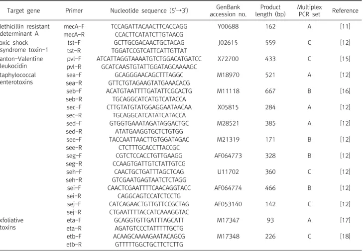
Table 1. Nucleotide sequences and anticipated sizes of PCR product for the Staphylococcus aureus gene-specific oligonucleotide primers used in this study
Target gene Primer Nucleotide sequence (5'→3') GenBank accession no.
Product length (bp)
Multiplex
PCR set Reference Methicillin resistant
determinant A
mecA-F TCCAGATTACAACTTCACCAGG Y00688 162 A [11]
mecA-R CCACTTCATATCTTGTAACG
Toxic shock syndrome toxin-1
tst-F GCTTGCGACAACTGCTACAG J02615 559 C [12]
tst-R TGGATCCGTCATTCATTGTTAT Panton-Valentine
leukocidin
pvl-F ATCATTAGGTAAAATGTCTGGACATGATCC X72700 433 C [15]
pvl-R GCATCAASTGTATTGGATAGCAAAAGC Staphylococcal
enterotoxins
sea-F GCAGGGAACAGCTTTAGGC M18970 521 A [12]
sea-R GTTCTGTAGAAGTATGAAACACG
seb-F ACATGTAATTTTGATATTCGCACTG M11118 667 B [16]
seb-R TGCAGGCATCATGTCATACCA
sec-F CTTGTATGTATGGAGGAATAACAA X05815 284 A [12]
sec-R TGCAGGCATCATATCATACCA
sed-F GTGGTGAAATAGATAGGACTGC M28521 385 A [12]
sed-R ATATGAAGGTGCTCTGTGG
see-F TACCAATTAACTTGTGGATAGAC M21319 171 B [12]
see-R CTCTTTGCACCTTACCGC
seg-F CGTCTCCACCTGTTGAAGG AF064773 328 B [12]
seg-R CCAAGTGATTGTCTATTGTCG
seh-F CAACTGCTGATTTAGCTCAG U11702 360 C [12]
seh-R GTCGAATGAGTAATCTCTAGG
sei-F CAACTCGAATTTTCAACAGGTACC AF064774 466 B [12]
sei-R CAGGCAGTCCATCTCCTG
sej-F CATCAGAACTGTTGTTCCGCTAG AF053140 142 C [12]
sej-R CTGAATTTTACCATCAAAGGTAC Exfoliative
toxins
eta-F GCAGGTGTTGATTTAGCATT M17347 93 A [17]
eta-R AGATGTCCCTATTTTTGCTG
etb-F ACAAGCAAAAGAATACAGCG M17348 226 C [18]
etb-R GTTTTTGGCTGCTTCTCTTG
Abbreviation: F, forward; R, reverse.
Table 2. Sources of methicillin-resistant S . aureus obtained from intensive care unit
Sources Sputum Pus Urine Blood Central venous
catheter tip Body fluid wound Total
Number of isolates 20 11 8 4 4 3 2 52
mM), MgCl 2 2 mM, primer 각 20 pmol, Taq DNA polymerase (Bioneer, Daejeon, Korea) 0.5 U, genomic DNA 100 ng 및 반응 완충용액(10 mM Tris-HCl, 40 mM KCl, 1.5 mM MgCl 2 )에 총 부 피가 50 L가 되도록 증류수를 첨가하였다. PCR 반응은 PCR thermal cycler TP 600 (Takara Bio, Foster City, CA, USA)을 이 용하였으며 PCR 반응 조건은 95 o C에서 5분간 초기 denaturation 시키고, 95 o C에서 30초, 59 o C에서 30초, 72 o C에서 40초의 cycle 을 30회 반복한 후 마지막으로 72 o C에서 10분 동안 반응 시켰다.
PCR 산물은 2% agarose gel에 전기영동한 후 특이 밴드를 확인하 였다.
4. 항생제 감수성 검사
분리균의 항생제 감수성 시험은 CLSI M100-S19 지침[14]의 방
법에 따라 디스크 확산법(disc diffusion method)으로 각각의 항
생제가 함유된 디스크 6 mm를 사용하여 실시하였다. 혈액배지에
배양한 균을 생리식염수로 McFarland 0.5관에 맞춘 세균현탁액을
멸균된 면봉에 충분히 적신 후 MHA 배지에 접종한 후 항생제 디스
크를 올려놓고 37 o C incubator 에서 16∼24시간 배양한 후 디스
크 주위에 형성된 억제대의 직경을 mm단위로 측정하여 판정 기준
표와 비교하여 감수성, 중간내성, 저항성으로 판정하였다[14]. 매
시험마다 대조 균주로 S . aureus ATCC 25923를 사용하였다. 최소
억제 농도(MIC)는 자동화 기기인 Vitek automated system
Fig. 1. Agarose gel electrophoresis of the multiplex PCR amplification pro- ducts from methicillin-resistant Sta- phylococcus aureus intensive care unit isolates. Lanes M, 1 kb plus DNA ladder; lanes 31∼34, primer set A;
lanes 46∼50, primer set B; lanes 47∼51, primer set C. Lane 31 ( mec A and sec ); lane 46 ( seb , sei and seg );
lane 47 ( tst ).
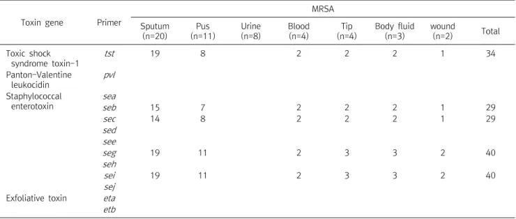
Table 3. Frequency of toxin genes detected in the methicillin-resistant Staphylococcus aureus
Toxin gene Primer
MRSA Sputum
(n=20)
Pus (n=11)
Urine (n=8)
Blood (n=4)
Tip (n=4)
Body fluid (n=3)
wound
(n=2) Total
Toxic shock syndrome toxin-1
tst 19 8 2 2 2 1 34
Panton-Valentine leukocidin
pvl
Staphylococcal enterotoxin
sea
seb 15 7 2 2 2 1 29
sec 14 8 2 2 2 1 29
sed see
seg 19 11 2 3 3 2 40
seh
sei 19 11 2 3 3 2 40
sej Exfoliative toxin eta etb
Abbreviation: Tip, Central venous catheter tip.
(bioMérieux, USA)의 AST-P601 카드를 이용 하였다.
결 과
1. 대상 균주의 검체별 분리원
2,664건의 환자 검체에서 균이 분리된 검체는 객담에서 20균주, 농 11균주, 소변 8균주, 혈액 4균주, 중심정맥관 팁 4 균주, 체액 3 균주, 창상 2균주로 총 52균주 이었다(Table 2).
2. 독소 유전자 유형 분석
독소유전자의 특이적인 primer를 사용하여 3 Set (A, B, C)로 구 분하여 multiplex PCR을 실시하였다(Fig. 1). 장독소 중 seg 와 sei 유전자는 각각 40 균주(76.9%)에서 확인되었고 seb 와 sec 는 각각 29균주(55.8%)가 보유하고 있었으며 sea 와 sed , see , sej , seh , see 유전자들은 검출되지 않았으며 피부 박탈성 독소 유전자 eta 와
etb , leukocidin 독소 유전자인 pvl 또한 검출되지 않았다. 독성 쇼
크 증상독소-1 유전자 tst 를 보유한 균주는 34균주(65.4%)로 확인
되었으며 검체별 객담에서 가장 많이 분리된 독소유전자는 tst ,
seg , sei 를 각각 19균주(95.0%)에서 보유하고 있었으며 농에서는
seg , sei 를 각각 11균주(100%) 모두에서 보유하고 있었다(Table
3). 2개 혹은 그 이상의 독소 유전자를 동시에 보유한 균주들의 분포
는 Table 4에 나타내었다. 40균주(76.9%)가 2개 이상의 독소 유전
자를 보유하고 있었으며, 6균주(11.5%)가 2개의 독소유전자( seg ,
sei )를, 5균주(9.6%)가 3개의 독소유전자( seg , sei , tst )를, 1균주
(1.9%)가 4개의 독소유전자( seb , seg , sei , tst )를, 28균주(53.8%)
가 5개의 독소유전자( seb , sec , seg , sei , tst )를 동시에 보유하고 있
었다. 독소 유전자들 간의 동시 유전자 보유율은 tst 유전자가 seg ,
sei 유전자들을 100% 동시 보유하였고 seb 가 tst , seg , sei 와 sec 가
tst , seg , sei 와 seg 가 sei 와 sei 가 seg 유전자를 100% 동시 보유하였
다. seb , sec , seg , sei 와 tst 유전자들 사이의 동시보유율은 seg 또
Table 4. Combination of toxin genes detected in methicillin-resistant Staphylococcus aureus isolated from intensive care unit Specimen
Toxin gene
Sputum (n=20)
Pus (n=11)
Urine (n=8)
Blood (n=4)
Tip (n=4)
Body fluid (n=3)
wound (n=2)
Total (n=52)
seb , sec , seg , sei , tst 14 7 2 2 2 1 28
seb , seg , sei , tst 1 1
seg , sei , tst 4 1 5
seg , sei 3 1 1 1 6
None 1 8 2 1 12
See Table 3.
Table 5. Coexistance of toxin genes in methicillin-resistant Staphylococcus aureus isolated from intensive care unit
Toxin genes tst seb sec seg sei
tst - 29/29 (1.000)* 29/29 (1.000) 34/40 (0.850) 34/40 (0.850)
seb 29/34 (0.853)
†- 28/29 (0.966) 29/40 (0.725) 29/40 (0.725)
sec 29/34 (0.853) 28/29 (0.966) - 29/40 (0.725) 29/40 (0.725)
seg 34/34 (1.000) 29/29 (1.000) 29/29 (1.000) - 40/40 (1.000)
sei 34/34 (1.000) 29/29 (1.000) 29/29 (1.000) 40/40 (1.000) -
*The rate of existence of a gene of row ( tst ) in the strains harboring a gene of column ( seb ).
†
The rate of existence of a gene of column ( seb ) in the strains harboring a gene of row ( tst ).
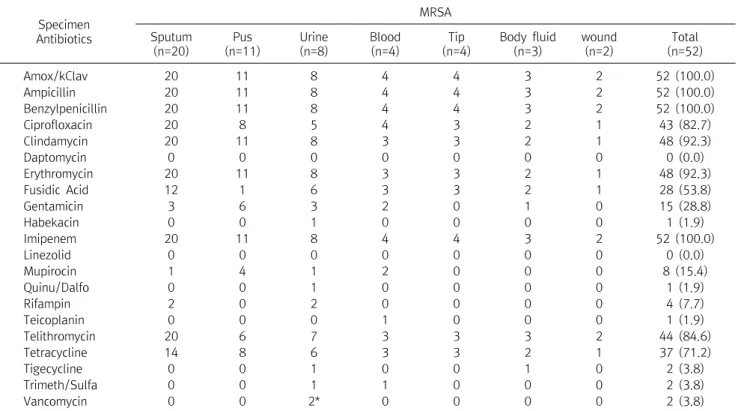
Table 6. Antibiotic resistance of methicillin resistant Staphylococcus aureus isolated from intensive care unit
Specimen Antibiotics
MRSA Sputum
(n=20)
Pus (n=11)
Urine (n=8)
Blood (n=4)
Tip (n=4)
Body fluid (n=3)
wound (n=2)
Total (n=52)
Amox/kClav 20 11 8 4 4 3 2 52 (100.0)
Ampicillin 20 11 8 4 4 3 2 52 (100.0)
Benzylpenicillin 20 11 8 4 4 3 2 52 (100.0)
Ciprofloxacin 20 8 5 4 3 2 1 43 (82.7)
Clindamycin 20 11 8 3 3 2 1 48 (92.3)
Daptomycin 0 0 0 0 0 0 0 0 (0.0)
Erythromycin 20 11 8 3 3 2 1 48 (92.3)
Fusidic Acid 12 1 6 3 3 2 1 28 (53.8)
Gentamicin 3 6 3 2 0 1 0 15 (28.8)
Habekacin 0 0 1 0 0 0 0 1 (1.9)
Imipenem 20 11 8 4 4 3 2 52 (100.0)
Linezolid 0 0 0 0 0 0 0 0 (0.0)
Mupirocin 1 4 1 2 0 0 0 8 (15.4)
Quinu/Dalfo 0 0 1 0 0 0 0 1 (1.9)
Rifampin 2 0 2 0 0 0 0 4 (7.7)
Teicoplanin 0 0 0 1 0 0 0 1 (1.9)
Telithromycin 20 6 7 3 3 3 2 44 (84.6)
Tetracycline 14 8 6 3 3 2 1 37 (71.2)
Tigecycline 0 0 1 0 0 1 0 2 (3.8)
Trimeth/Sulfa 0 0 1 1 0 0 0 2 (3.8)
Vancomycin 0 0 2* 0 0 0 0 2 (3.8)
Abbreviation: Amox/kClav, amoxicillin/clavulanic acid; Quinu/Dalfo, quinupristin/ dalfopristin; Trimeth/Sulfa, trimethoprim/sulfamethoxazole;
Tip, Central venous catheter tip.
*vancomycin resistance is intermediate. Number of resistant strains is expressed.
는 sei 보유균주가 seb 또는 sec 를 동시에 보유한 비율인 72.5%에서
sei 보유균주가 seg 를 동시에 보유한 비율인 100%에 달했다(Table 5). 3. 항생제 감수성
분리된 MRSA 52균주의 항생제 내성 양상은 amoxicil-
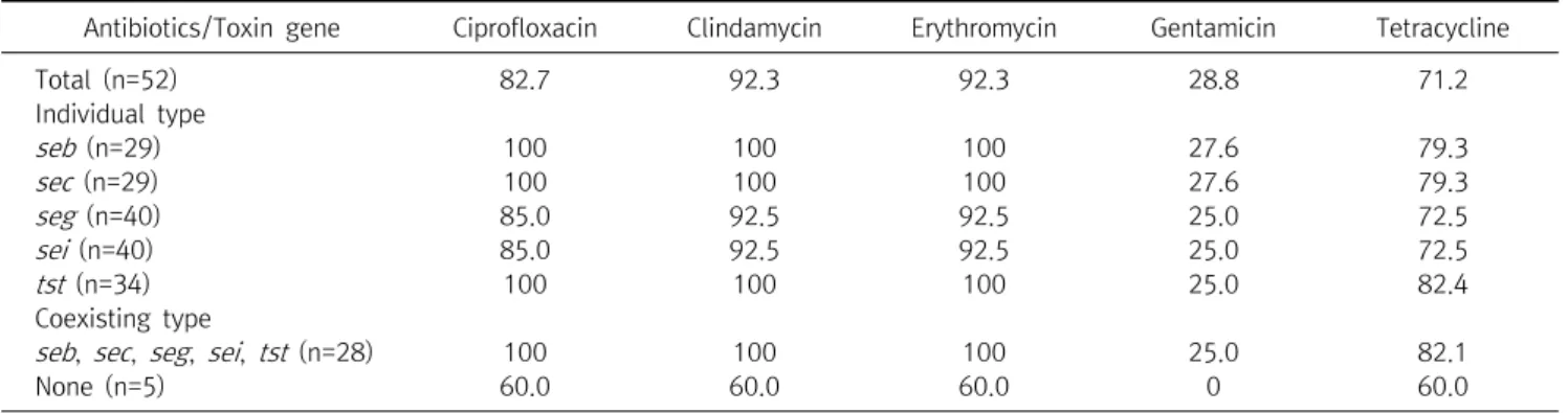
Table 7. Correlationship between toxin genes and antibiotic resistance in methicillin resistant Staphylococcus aureus isolated from intensive care unit
Antibiotics/Toxin gene Ciprofloxacin Clindamycin Erythromycin Gentamicin Tetracycline
Total (n=52) 82.7 92.3 92.3 28.8 71.2
Individual type
seb (n=29) 100 100 100 27.6 79.3
sec (n=29) 100 100 100 27.6 79.3
seg (n=40) 85.0 92.5 92.5 25.0 72.5
sei (n=40) 85.0 92.5 92.5 25.0 72.5
tst (n=34) 100 100 100 25.0 82.4
Coexisting type
seb , sec , seg , sei , tst (n=28) 100 100 100 25.0 82.1
None (n=5) 60.0 60.0 60.0 0 60.0
lin-clavulanic acid, benzylpenicillin, ampicillin, imipenem은 100% 내성을 보였고 erythromycin, clindamycin 항생제에 대한 내성율은 똑같이 92.3%로 나타났다. 또한 telithromycin, ci- profloxacin, tetracycline은 각각 84.6%, 82.7%, 71.2% 내성율을 나타냈다. Daptomycin, linezolid, quinupristin-dalfopristin, teicoplanin, tigecycline, vancomycin에 대한 내성은 없었다 (Table 6).
4. 독소 유전자형 MRSA의 항생제 내성 분석
독소유전자형에 따른 MRSA의 항생제 내성 양상은 seb , sec 와 tst 유전자 보유 균주들은 ciprofloxacin, clindamycin과 ery- thromycin에 대한 항생제 내성율이 100%를 보였고, seg , sei 를 보 유한 균주들 또한 ciprofloxacin, clindamycin, erythromycin, tetracycline 항생제에 각각 85.0%, 92.5%, 92.5%, 72.5%로 내성 을 보였다. 그리고, 5개의 독소유전자( seb , sec , seg , sei , tst )를 동 시에 보유한 균주들도 ciprofloxacin, clindamycin, erythromycin 에 100% 내성율을 보였고 tetracycline, gentamicin은 각각 82.1%, 25.0%의 내성율을 보였다(Table 7).
고 찰
MRSA로 인한 유행적 발생은 대부분 중환자실과 화상병동 등과 같이 면역상태가 저하되거나 병원 치료력이나 항생제 사용력이 길 고 각종 침습적 치료를 실시하는 환자들이 모여 있는 특수병동에서 주로 발생하는 것으로 보고되고 있다[19].
분리된 MRSA 균주들 중 식중독을 유발한다고 알려진 5개의 독 소 유전자( sea , seb , sec , sed , see ) 외의 seg 와 sei 유전자가 각각 40 균주(76.9%)로 가장 많은 보유율을 나타냈으며 다음으로 독성 쇼 크증상 독소 1 유전자 tst 는 34균주(65.4%), 장독소 seb , sec 는 각 각 29균주(55.8%) 순으로 검출 되었으며 검체별 객담과 농으로부
터 분리된 MRSA 균주가 혈액과 소변으로부터 분리된 MRSA에 비 해 독소유전자 보유 빈도가 높음을 알 수 있었고 특이하게 소변에 서 분리된 균주들은 독소유전자를 한 균주도 갖고 있지 않음을 확 인하였다. 한편 피부 박탈성 독소(exfoliative toxin) 유전자 eta 와 etb 그리고 장독소 sea , sed , see , seh , sej 와 주로 피부감염이나 병 원감염보다는 지역감염 환자로부터 분리되는 leukocidin 독소 pvl 유전자는 검출 되지 않았다. 이러한 결과는 한국의 다른 병원에서 분리한 MRSA의 독소 유전자 보유율과 비교 해보았을때 sea 보유 율은 낮았지만, seb 와 sec 보유율은 높은 결과였으며 독성 쇼크 증 상 독소-1 유전자 tst의 보유율은 65.4%로 Kim 등[20]의 결과 43.2%보다 높은 검출율을 보였고, 또 다른 보고[21]에서 보다 seb , sec , seg , sei , tst 각각의 유전자 보유율이 20∼30% 이상 독소 유전 자수가 더 늘어 증가하고 있음을 확인하였다. 또한 최근 국외 논문 [22]과의 독소 유전자 보유율을 비교해 보았을 때 seb , sec 와 tst 유 전자 보유율 또한 본 연구에서 결과가 높게 분포했지만 검출되지 않았던 sea , eta , etb 유전자는 Sabouni 등[22]의 결과에서는 40.6%, 11.3%, 9%로 각각 검출되어 본 연구에서의 결과와 국내ㆍ 외의 독소유전자 분포 양상에 차이가 있어 독소형은 지역과 시간의 흐름에 따라 분포가 다양한 것으로 생각된다.
분리된 균주들중 독소유전자를 2개 이상 동시에 보유한 조합의 MRSA는 40균주(76.9%)였으며 분포도를 살펴보면 seb , sec , seg , sei , tst 의 5개 유전자를 동시 보유한 조합은 28균주(53.8%)를 보였 으며 다음으로 seg , sei 유전자 동시 보유와 seg, sei, tst 유전자 동 시 보유 조합은 각각 6균주(11.5%), 5균주(9.6%)를 보였다(Table 4). 이것은 여러 다른 보고에 의한 것과 마찬가지로 대부분의 MRSA 균주가 2∼4개의 독소유전자를 동시에 보유한다[23]는 결과와 일 치 하였으며, 오히려 본 연구에서는 다른 보고에 의한 것 보다 더 많 은 5개( seb , sec , seg , sei , tst )까지의 독소유전자를 동시 보유하고 있음을 확인 할 수 있었다.
독소 유전자들 간의 동시 유전자 보유율은 tst 유전자가 seg , sei
유전자들을 100% 동시 보유하였으며 뿐만 아니라 seb 가 tst , seg , sei 를 sec 가 tst , seg , sei 를 seg 가 sei 를 sei 가 seg 유전자들을 100%
동시 보유하고 있었다. 본 연구 결과는 특정한 독소 유전자 seb , sec , seg , sei 와 tst 유전자간의 상관성이 높음을 말해준다. 다른 연 구자들의 보고 자료에 의한 S . aureus 가 tst 와 sec 유전자를 동시에 보유한다[24]는 사실을 본 연구에서도 확인 할 수 있었다.
한편, 분리된 MRSA 52균주의 항생제 내성율은 amoxicil- lin-clavulanic acid, benzylpenicillin, ampicillin, imipenem은 100% 내성을 보였고 erythromycin, clindamycin 내성율은 각각 92.3%로 같은 내성율을 보였다. 또한 telithromycin, ciprofloxacin, tetracycline은 각각 84.6%, 82.7%, 71.2%의 내성을 보였다. 이와 같은 항생제 내성의 결과는 다른 연구 결과[5]와도 거의 일치함을 확인 할 수 있었다.
특정 독소유전자 보유에 대한 항생제 내성과의 연관성을 분석한 결과 5개 독소유전자( seb , sec , seg , sei , tst )를 동시에 보유한 균주 들과 개별적으로 독소유전자 seb , sec , seg , sei 와 tst 를 각각 1개씩 만을 보유한 균주들은 ciprofloxacin, clindamycin, erythromycin, tetracycline에 대한 내성율의 차이는 크지는 않았지만 독소유전 자를 하나도 갖지 않은 균주들 보다는 내성율이 높게 나타났다. 그 러나 gentamicin은 다른 항생제와 비교해 볼때 내성율은 훨신 낮 았으며 또한 독소유전자를 보유한 균주들은 보유하지 않은 균주들 보다 내성율은 높았지만 전체 평균 내성율과는 큰 차이가 없는 결 과를 보였다. 결론적으로 독소유전자를 하나 또는 5개 독소유전자 ( seb , sec , seg , sei , tst )를 동시에 보유한 균주들과의 항생제 내성율 의 차이는 독소유전자 보유 갯수와는 상관 없이 모두 내성율이 높 았음을 확인하였다. 그렇지만 독소유전자를 보유하지 않은 균주들 보다는 내성율이 높게 나타났다. 그리고 5개의 독소유전자( seb , sec , seg , sei , tst )를 보유한 균주와 개별적 독소유전자 seb , sec , tst 유전자를 각각 보유한 균주의 조합에서 ciprofloxacin, clindamycin, erythromycin 항생제에 100% 내성을 보임으로서 공통적으로 포 함된 seb , sec , tst 유전자와 이 항생제의 내성과는 연관이 있음을 확인 할 수 있었다. 이렇듯 독소유전자를 보유한 MRSA 균주들이 중환자실 환자들에게 감염이 된다면 효과적인 항생제 치료가 제한 적이므로 내성균의 확산 방지를 위한 철저한 감염관리가 더욱 필요 하다고 생각한다.
요 약
본 연구는 methicillin-resistant Staphylococcus aureus (MRSA)로부터, 독소 유전자형과 항생제 내성의 상관 관계를 결정 하는 것을 목표로 하였다. 2014년 1월∼12월까지 전남 순천의 한
병원 중환자실의 임상검체 2,664건에서 얻어진 MRSA 52균주를 분리하였다. 유전자들이 암호화하고 있는 mecA , 장독소(staphy- lococcal enterotoxins; sea , seb , sec , seg , seh , sei , sej ), 독성 쇼 크 증상독소-1 (toxic shock syndrome toxin-1; tst - 1 ), 표피박탈 성독소(exfoliative toxin; eta , etb ), 백혈구 용해 독소(Pan- ton-Valentine leukocidin; pvl )를 특이적 프라이머를 이용한 multiplex PCR로 증폭 검출 하였다. 독소 유전자 seg 와 sei 유전자 가 각각 40균주(76.9%)로 가장 많은 보유율을 나타냈으며 다음으 로 tst 34균주(65.4%) 순으로 검출 되었으며 eta , etb , sea , sed , see , seh , sej 와 pvl 유전자들은 검출 되지 않았다. 2개 이상의 독소 유전자를 동시에 보유한 조합의 MRSA는 40균주(76.9%) 였는데 5 개 유전자( seb , sec , seg , sei , tst )를 동시 보유한 조합이 28균주 (53.8%)로 가장 많은 분포를 보였으며 다음으로 seg , sei 유전자 동 시 보유 조합으로 6균주(11.5%)에서 나타났다. 유전자들 간의 동 시 보유율은 72.5∼100%로서 특정한 독소 유전자 seb , sec , seg , sei와 tst 유전자간의 상관성이 높게 나타났다. 특정 다수의 독소유 전자( seb , sec , seg , sei , tst )를 동시에 보유한 균주들이 개별적 독소 유전자를 보유한 균주( seb , sec , tst )와의 항생제 내성의 상관성은 ciprofloxacin, clindamycin, erythromycin 항생제에 100% 내 성을 보임으로서 공통적으로 포함된 seb , sec , tst 유전자와 이 항생 제의 내성과는 밀접한 연관이 있음을 알았다.
Acknowledgements: None Funding: None
Conflict of interest: None
References