pISSN 1738-3544 eISSN 2288-1662
Antioxidant and Antimicrobial Activities of Fruiting Bodies of Phellinus gilvus Collected in Korea
Ki-Nam Yoon, Hyung Seok Jang
Department of Clinical Laboratory Science, Ansan University, Ansan 15328, Korea
국내에서 수집된 마른진흙버섯 자실체의 항산화 및 항균 효과
윤기남, 장형석
안산대학교 임상병리과
This study was initiated to evaluate the antioxidant and antimicrobial activities of methanol extract (ME) and hot water extract (HWE) obtained from the fruiting bodies of medicinal mushroom, Phellinus gilvus. The free radical scavenging activity of ME from P. gilvus on 1,1-diphenyl-2-pi- crylhydrazyl (DPPH) were 93.65% at 2 mg/mL, which was comparable with the positive control, butylated hydroxytoluene (BHT, 96.97%) at the same concentration. The ferrous ion-chelating ability of ME and HWE was significantly higher than that of BHT at all concentration levels. The antimicrobial assay of ME was performed against six bacteria and one species of fungus. ME exhibited antibacterial activity against 5 out of 6 bacteria: Staphylococcus aureus, Streptococcus mutans, Bacillus subtilis, Escherichia coli, and Pseudomonas aeruginosa; whereas, ME did not show antimicrobial activity against gram-negative bacterium Vibrio vulnificus and fungal species Candida albicans. The minimum inhibitory concentration (MIC) of ME against 5 strains of bacteria, such as S. aureus, S. mutans, B. subtilis, E. coli, and P. aeruginosa, was 100, 100, 50, 100, 200 mg/mL, respectively. The results suggest that good antioxidant and microbial activities of P. gilvus fruiting bodies might be used for natural antioxidant and antimicrobial agents.
Key words: Antimicrobial, Antioxidant, Medicinal mushroom, Phellinus gilvus
Corresponding author: Hyung Seok Jang Department of Clinical Laboratory Science, Ansan University, 155 Ansandaehak-ro, Sangnok-gu, Ansan 15328, Korea Tel: 82-31-400-6935 Fax: 82-31-363-7702 E-mail: [email protected]
This is an Open Access article distributed under the terms of the Creative Commons Attribution Non-Commercial License (http://creativecommons.org/licenses/by-nc/4.0) which permits unrestricted non-commercial use, distribution, and reproduction in any medium, provided the original work is properly cited.
Copyright © 2016 The Korean Society for Clinical Laboratory Science. All rights reserved.
Received: October 5, 2016 Revised 1st: November 20, 2016 Revised 2nd: November 25, 2016 Revised 3rd: November 29, 2016 Revised 4th: November 29, 2016 Accepted: November 29, 2016
Introduction
Mushrooms have become attractive as a functional food because of high contents of protein, crude fiber, minerals and vitamins [1] and also contain physiologically beneficial bioactive substances that promote good health [2]. The secondary metabolites produced from mushrooms possess antibacterial, antifungal, antioxidant, antiviral, antitumor, antiallergic, antiatherogenic, hypoglycemic, anti-inflammatory
and hepatoprotective activities [3,4].
Reactive oxygen species (ROS) were produced by a result
of normal cellular metabolism in a living organism. At low to
moderate concentrations, ROS function in physiological cell
processes, but at high concentrations, they provoke adverse
effects to cell components, such as lipids, proteins, and DNA
and caused degenerative diseases including cancer, cardio-
vascular disease, neural disorders, and diabetes. Protection
against free radicals can be enhanced by intake of dietary
antioxidant. Experimental evidence suggested that foods containing antioxidant nutrients may be of major importance in disease prevention possibly caused by free radicals. The intake of natural antioxidant sources such as vegetables, fruits, and mushrooms may be effective over the long term for health. Therefore, antioxidant from natural products may be of great benefit for improving the quality of life by preventing or postponing the onset of degenerative diseases [5].
Antibiotic resistance has become global problems in recent years. Even though a number of natural and synthetic antimicrobial agents have been isolated and synthesized against pathogenic microorganisms, infectious diseases remain one of the major threats to human health [6]. The increasing antibiotic resistance of pathogenic microorganisms caused by overuse of newly developed antibiotics has led to the screening of novel sources for potential antimicrobial activity [7]. Although mushrooms belong to Basidiomycota have not been fully studied on antimicrobial activity, a variety of antimicrobial compounds produced by the fungi suggested that they may be a new and useful sources to combat against pathogenic microorganisms [8]. In the natural environment, the fungi including mushrooms need defense chemical compounds to fight against bacterial and fungal invasion.
Therefore, antimicrobial compounds produced by many mushroom species could be useful to develop new antibiotics.
Although many antimicrobial compounds have been isolated and identified from mushrooms, the antimicrobial compounds prescribed in the hospital are still originated from microscopic fungi and actinomycetes [9].
P . gilvus , a white rot fungus, belongs to family Hymeno- chaetaceae of Basidiomycota and known to as 'Sanghwang' and consumed for medicinal purposes in Korea, China, and Japan for thousand years [10]. The functional and physiological activities of the P . gilvus have been reported by several researchers. The hot water extract of fruiting bodies from P . gilvus has been reported to contain inhibitory effect on pulmonary inflammation [11]. The polysaccharide extracted from fruiting bodies of P . glivus exhibited inhibitory effect on melanoma of rats [12]. The polysaccharide extracted from P . glivus also showed wound healing effect on diabetic rats [13].
Hence, the aim of this study was to evaluate the antioxidant
and antimicrobial activities of ME and HWE from fruiting bodies of P . gilvus . In addition to this, profiles of phenols and flavonoid compounds concentrations of the mushroom were also analyzed.
Materials and Methods
1. Mushroom sample
Fruiting bodies of P . gilvus were collected from Bukhan mountain of Seoul city in the summer of 2015 and identified by Dr. Kyung-Rim Lee (mycologist). The voucher specimens were deposited in the “Culture Collection of Mushrooms”, Incheon National University. The fruiting bodies were fully air dried at 45
oC and ground into a fine powder.
2. Preparation of the extracts
To obtain the ME, 20 g of the mushroom powder was extracted with 200 mL of 80% methanol at orbital shaker (150 rpm) for 24 hr at 25
oC. Then, the mixture was filtered through Whatman no. 4 filter paper, and the remaining residue was extracted with 200 mL of methanol twice as described above.
To obtain the HWE, 20 g of the mushroom powder was boiled for 3 hr in 200 mL of distilled water, cooled to 25
oC and filtered with filter paper. Then, the remaining residue was extracted with 200 mL of 80% methanol or distilled water twice as described above, respectively. The ME and HWE were evaporated to dryness at 50
oC under reduced pressure in a rotary evaporator, and remaining solvent was removed with a freeze-drier [14]. Extracts were kept in the dark at −80
oC for prior to use.
3. Scavenging effect on DPPH free radicals
The hydrogen atoms or electron donation ability of the ME
and HWE was measured by the method bleaching of the
purple colored DPPH methanol solution [15]. One mL of 0.1
mM DPPH radical solution in methanol was mixed with 1 mL
aliquots of varying mushroom extract concentrations (0.125,
0.25, 0.5, 1.0 and 2.0 mg/mL). The reaction was carried out by
shaking vigorously and then allowing it to stand for 30 min in
the dark before measuring the absorbance at 517 nm using a
spectrophotometer. Inhibition of the DPPH free radical in
percent (I%) was calculated as:
I%=[(A
control−A
sample)/A
control]×100,
where A
controlis the absorbance of the control reaction (containing all reagents except the test compound), and A
sampleis the absorbance of the test compound. BHT was used as the positive control.
4. Chelating effects on ferrous ions
The chelating effect of the ME and HWE was determined according to the method of Dinis et al . [16]. Briefly, various concentrations (0.063∼1.0 mg/mL, 2 mL) of the extracts in methanol were added to a solution of 2 mM FeCl
2(0.05 mL).
The reaction was initiated by adding 5 mM ferrozine (0.2 mL).
Total volume was adjusted to 5 mL with methanol, and the mixture was shaken vigorously and left at room temperature for 10 min. The absorbance of the solution was measured spectrophotometrically at 562 nm. The inhibition percentage of the ferrozine-Fe
2+complex formation was calculated using the following formula:
Ferrous ion chelating effect (%)=[(A
control−A
sample)/A
control]
×100,
where A
controlis the absorbance of the control (control contained FeCl
2and ferrozine; complex formation molecules), and A
sampleis the absorbance of the test compound. BHT was used as the positive control.
5. Reducing power assay
The reducing power of the ME and HWE was determined according to the method of Gulcin et al . [17]. Each extract (1∼8 mg/mL) in methanol (2.5 mL) was mixed with 200 mM sodium phosphate buffer (pH 6.6, 2.5 mL) and 1% potassium ferricyanide (2.5 mL), and the mixture was incubated at 50
oC for 20 min. Then, 10% trichloroacetic acid (2.5 mL) was added, and the mixture was centrifuged at 200 × g (6K 15;
Sigma, Munich, Germany) for 10 min. The upper layer (2.5 mL) was mixed with deionized water (2.5 mL) and 0.1% ferric chloride (0.5 mL). Finally, the absorbance was measured at
700 nm against a blank. BHT was used as the positive control.
6. Determination of bioactive components
Phenolic compound concentration in the ME and HWE of the mushroom was determined by a colorimetric assay, based on procedures described by Singleton and Rossi [18] with minor modifications. Briefly, 1 mL of sample was mixed with 1 mL of Folin and Ciocalteu’s phenol reagent. After 3 min, 1 mL of saturated sodium carbonate solution was added to the mixture and adjusted to 10 mL with distilled water. The reaction was kept in the dark for 90 min, after which the absorbance was measured spectrophotometrically at 765 nm.
Gallic acid was used to calculate the standard curve. The results were mean value±standard deviation (SD) and expressed as g of gallic acid equivalents (GAEs) per mg of extract.
Flavonoid concentration of the ME and HWE was determined by a colorimetric method described by Jia et al . [19] with minor modifications. The ME and HWE solution (1 mL) of the mushrooms was diluted with 4.3 mL of 80%
aqueous ethanol, and 0.1 mL of 10% aluminum nitrate and 0.1 mL of 1 M aqueous potassium acetate were added. After 40 min at 25
oC, the solution was mixed well, and the intensity of the pink color was measured by spectrophotometer at 415 nm. Quercetin was used to calculate the standard curve. The results were mean value±standard deviation (SD) and expressed as g of quercetin equivalents (QEs) per mg of extract.
7. Determination of antimicrobial efficacy 1) Microbial test organisms
Three species of gram-positive bacterial strains such as S .
aureus KCTC 1621, S . mutans KCTC 3065, B . subtilis KCTC
1021, and 3 species of gram-negative bacterial strains
including E . coli KCTC 1045, P . aeruginosa KCTC 2513, and V .
vulnificus KCTC 2959, and one fungal species of C . albicans
KCTC 7965 were obtained from Korean Collection for Type
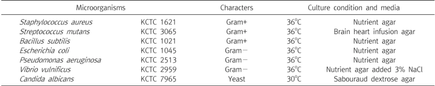
Cultures (KCTC). The characteristics, culture media, and
culture conditions of the microorganisms used in this study
were listed in the Table 1.
Table 1. List of microorganisms used for antimicrobial activity in this study
Microorganisms Characters Culture condition and media
Staphylococcus aureus KCTC 1621 Gram+ 36oC Nutrient agar
Streptococcus mutans KCTC 3065 Gram+ 36oC Brain heart infusion agar
Bacillus subtilis KCTC 1021 Gram+ 36oC Nutrient agar
Escherichia coli KCTC 1045 Gram− 36oC Nutrient agar
Pseudomonas aeruginosa KCTC 2513 Gram− 36oC Nutrient agar
Vibrio vulnificus KCTC 2959 Gram− 36oC Nutrient agar added 3% NaCl
Candida albicans KCTC 7965 Yeast 30oC Sabouraud dextrose agar
2) Antimicrobial assay
Antimicrobial activity of the ME of the mushroom was determined by the agar well diffusion method [20], with slight modification. Briefly, the ME and HWE were dissolved in 3%
dimethylsulfoxide (DMSO) to a final concentration of 200 mg/mL, and filter sterilized through 0.45-m membrane filter. Small wells (5 mm in diameter) were made in the agar plates by sterilize cork borer. One hundred L of the ME and HWE with 200 mg/mL concentration was loaded into the different wells. An overnight culture of each microbial isolate was emulsified with nutrient broth to a turbidity that was equivalent to 0.5 McFarland (10
5cfu/mL). In order to determine the antimicrobial efficacy of the extracts, aliquot of test culture (100 L) was evenly spread over the surface of the solidified agar. Bacteria were cultured on Mueller Hinton agar, and fungus on Sabouraud dextrose agar. Antibiotics such as streptomycin (30 g/well), tetracycline (30 g/well), and fluconazole (40 g/well) were used for positive controls for determining inhibition zone of selected bacteria and fungus, respectively. DMSO was used as negative control for testing selected microorganisms. All the plates previously loaded with respective extracts and test microorganisms were incubated for 24 hr at 37
oC for bacteria and 48 hr for fungus at 28
oC. After incubation, inhibition zone was measured in millimeters. All the tests were carried out in triplicate and their means and standard deviation (SD) were recorded.
3) Minimum inhibitory concentration (MIC)
The MIC of the mushroom extract was performed with ME since the HWE did not exhibit inhibition zone at 200 mg/mL concentration in the agar gel diffusion test. Therefore, antibacterial activity of MIC against 5 bacterial strains such as
S . aureus , S . mutans , B . subtilis , E . coli , and P . aeruginosa were further studied with ME by applying the broth dilution method [21] with minor modifications. The ME was diluted to give the final concentrations of 25∼400 mg/mL. One hundred
L of 10
5CFU/mL of the bacterial strains was inoculated in tubes with equal volume of nutrient broth and the ME. The tubes were incubated aerobically at 37
oC for 24∼48 hr. The lowest concentration (highest dilution) of the extract that produced no visible growth (no turbidity) in the first 24 hr when compared with the negative control tubes was considered as initial MIC. The dilutions that showed no turbidity were incubated further for 24 hr at 37
oC. The lowest concentration that produced no visible turbidity after a total incubation period of 48 hr was regarded as final MIC.
8. Statistical analysis
All experiments were carried out in triplicates. Data obtained were analyzed by one way analysis of variance and means were compared by Duncan’s multiple range tests (SPSS 11.5 version). Differences were considered significant at p <0.05.
Results
1. DPPH radical scavenging activities
The scavenging activities of the ME, and HWE from the fruiting bodies of P . gilvus on DPPH radicals increased with increasing concentration. At 0.125, 0.25, 0.5, 1.0, and 2.0 mg/mL concentration of the extracts, the scavenging abilities of the ME, and HWE on the DPPH radical ranged from 89.86%∼93.65%, and 88.18%∼91.45%, respectively (Fig. 1).
These results indicated that the ME and HWE possessed good
scavenging activity on DPPH radicals at the concentrations
Fig. 3. Reducing power levels of methanol and hot water extract from fruiting bodies of Phellinus gilvus at different concentrations.
Values are expressed as mean±SD (n=3). Fr. MeOH, fractions extracted with 80% methanol; Fr. HW, fractions extracted with hot water; BHT, butylatedhydroxytoluene. ***p≤0.001; **p≤0.01;
*p<0.05 vs. BHT group.
Fig. 1. Scavenging activity of methanol and hot water extract from fruiting bodies of Phellinus gilvus against 1,1-diphenyl-2-pi- crylhydrazyl. Values expressed as mean±SD (n=3). Fr. MeOH, fractions extracted with 80% methanol; Fr. HW, fractions extracted with hot water; BHT, butylatedhydroxytoluene. ***p≤0.001; **p≤ 0.01; *p<0.05 vs. BHT group.
Fig. 2. Chelating effect of methanol and hot water extract from fruiting bodies of Phellinus gilvus at different concentrations. Values are expressed as mean±SD (n=3). Fr. MeOH, fractions extracted with 80% methanol; Fr. HW, fractions extracted with hot water, BHT, butylatedhydroxytoluene. ***p≤0.001; **p≤0.01; *p<0.05 vs. BHT group.
tested. However, BHT, the positive control showed excellent scavenging abilities of 96.19∼96.97% at the concentration range of 0.125∼2.0 mg/mL, respectively.
2. Chelating activity of ferrous ion
The chelating activity of the ME and HWE from the fruiting bodies of P . gilvus toward ferrous ions was investigated at five different concentrations (0.125, 0.25, 0.50, 1.0, and 2.0 mg/mL). BHT was used as reference standard. As shown in Fig. 2, the chelating capacity of the ME and HWE increased with dose-dependent manner. The chelating effect of ME and HWE were 72.70% and 79.95% at 2.0 mg/mL, respectively, whereas chelating effect of BHT, the positive control was 68.87% at 2.0 mg/mL.
3. Reducing power assay
The reducing power of ME and HWE at five different concentrations (0.5, 1.0, 2.0, 4.0, and 6.0 mg/mL) ranged from 1.88 to 2.24, and 1.84 to 1.98, respectively, suggesting that reducing power of the ME, and HWE of P . gilvus fruiting bodies increased with increasing concentration of the extracts (Fig. 3). However, the reducing power of the reference antioxidants, BHT was much higher than those of the ME and HWE, indicating that reducing power of the ME, and HWE were moderate.
4. Bioactive compounds
Total phenolics and flavonoid contents of the ME and HWE from the fruiting bodies of P . gilvus were investigated. The contents of phenolic compound of the ME and HWE from fruiting bodies of the mushroom ranged from 31.17 and 12.83
g gallic acd equivalent (GAE)/mg, respectively. The flavonoid content of the ME and HWE ranged from 15.29 and 27.46 g quercetin equivalent (QE)/mg, respectively (Table 2).
5. Antimicrobial assay
1) Agar well diffusion method
Antimicrobial activity of ME and HWE of fruiting bodies of
P . gilvus against 6 bacteria such as S . aureus , S . mutans , B .
subtilis , E . coli , and P . aeruginosa , V . vulnificus and 1 species
of fungus, C . albicans was conducted using agar well diffusion
method. The ME of the fruiting bodies of P . gilvus exhibited
the various degrees of antimicrobial effects against 5 out of 7
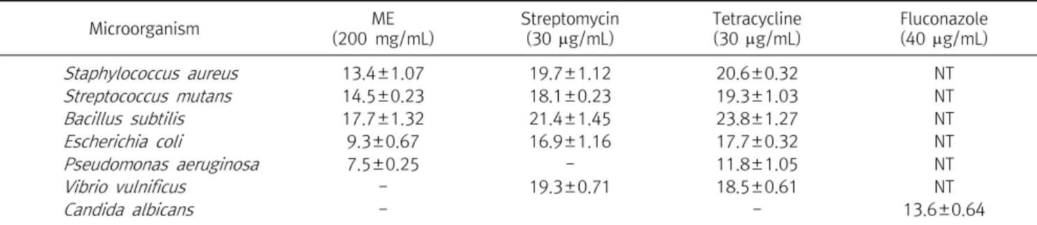
Table 3. Diameter of inhibition zone of methanol (ME) extract from Phellinus gilvus fruiting bodies against selected microorganisms
Microorganism ME
(200 mg/mL)
Streptomycin (30 g/mL)
Tetracycline (30 g/mL)
Fluconazole (40 g/mL)
Staphylococcus aureus 13.4±1.07 19.7±1.12 20.6±0.32 NT
Streptococcus mutans 14.5±0.23 18.1±0.23 19.3±1.03 NT
Bacillus subtilis 17.7±1.32 21.4±1.45 23.8±1.27 NT
Escherichia coli 9.3±0.67 16.9±1.16 17.7±0.32 NT
Pseudomonas aeruginosa 7.5±0.25 - 11.8±1.05 NT
Vibrio vulnificus - 19.3±0.71 18.5±0.61 NT
Candida albicans - - 13.6±0.64
Antimicrobial activity was determined by diameter (mm) of the inhibition zone surrounding well.
The data are expressed as means±standard deviation (SD) of triplicate determinations.
Values with different superscripts within same row are significantly different at p<0.05.
Abbreviation: -, Inhibition zone was not detected; NT, Not tested.
Table 2. Total Phenolic and flavonoids content in methanol and hot water extracts from fruiting bodies of Phellinus gilvus
Samples
Extraction yield
% (w/w)
Phenolic content (g GAEs/mg
extract)
Flavonoids content (g QEs/mg
extract) Methanol
extract
10.37 31.17±1.04 15.29±0.56
Hot water extract
7.62 12.83±0.51 27.46±1.87
The values are expressed as means±SD of three parallel measurements.
Abbreviation: GAEs, gallic acid equivalents; QEs, quercetin equivalents.
test microorganisms, whereas gram-negative bacterium V . vulnificus KCTC 2959 and fungal strain C . albicans KCTC 7965 did not show inhibition zone, indicating that the antimicrobial activity of the ME was not effective to all bacteria and the fungus tested. However, the HWE of P . gilvus did not exhibit inhibitory activity against 7 microbial strains such as six bacteria and one fungus. As summarized in Table 3, three gram-positive bacteria such as S . aureus KCTC 1621, S . mutans KCTC 3065, and B . subtilis KCTC 1021 have shown susceptibility against the ME at 200 mg/mL with inhibition zone diameter of 13.4, 14.5, and 17.7 mm, respectively, whereas 2 out of 3 gram-negative bacteria such as E . coli , KCTC 1045, and P . aeruginosa KCTC 2513 showed susceptible reaction against the ME with the inhibition zone diameter of 9.3, and 7.5 mm at the same concentration tested.
These results suggest that the highest inhibitory activity was determined against B . subtilis KCTC 1021, and the weakest inhibitory activity was determined against P . aeruginosa KCTC 2513. However, the inhibition zone diameter of
streptomycin and tetracycline, the positive controls ranged from 16.9∼21.4 mm, and 11.8∼23.8 mm, respectively.
2) Minimum inhibitory concentration
The MIC of the ME of P . gilvus was tested using broth
dilution method. Since the HWE of the fruiting bodies of P .
gilvus did not show inhibition zone against all microbial
strains tested in the agar well diffusion method, HWE was
excluded for the MIC test. The ME did not show inhibition
zone against a bacterial strain V . vulnificus , and a fungal strain
C . albicans at 200 mg/mL, these two microbial strains were
excluded in the MIC test. The 5 bacterial strains, which
showed susceptible reaction to the ME were used for the MIC
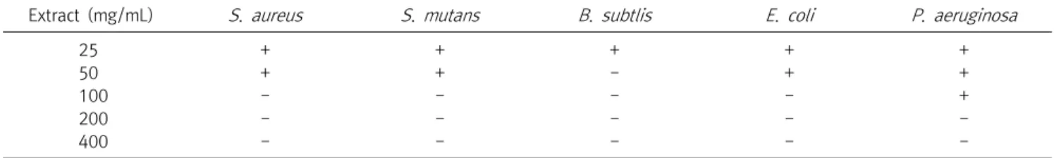
test. In the MIC test, S . aureus KCTC 1621 strain showed no
visible growth at 100 mg/mL of ME, whereas visible growth
was found at 25 and 50 mg/mL concentration. The S . mutans
KCTC 3065 strain showed visible growth at 50 mg/mL of the
ME, and no visible growth was found at 100 mg/mL. The B .
subtilis KCTC 1021 strain exhibited no visible growth at 50
mg/ml of ME and showed visible growth at 25 mg/mL. The E .
coli KCTC 1045 strain showed no visible growth at 100
mg/mL, whereas visible growth was observed at 50 mg/mL of
the ME. The P . aeruginosa KCTC 2513 strain exhibited visible
growth at 100 mg/mL of the ME, whereas no visible growth
was observed at 200 mg/mL (Table 4). Therefore, it is
concluded that the MIC values of the ME against 5 bacterial
strains such as S . aureus KCTC 1621, S . mutans KCTC 3065, B .
subtilis KCTC 1021, E . coli KCTC 1045, and P . aeruginosa
KCTC 2513 were 100, 100, 50, 100, 200 mg/mL, respectively.
Table 4. Minimum inhibitory concentration (MIC) of methanol extract of Phellinus gilvus fruiting bodies
Extract (mg/mL) S. aureus S. mutans B. subtlis E. coli P. aeruginosa
25 + + + + +
50 + + - + +
100 - - - - +
200 - - - - -
400 - - - - -
Abbreviation: -, Growth of microorganisms was not detected; +, Growth of microorganisms was detected.
Discussion
The antioxidant is capable of reducing the stable DPPH radical (purple) to the non-radical form DPPH-H (yellow).
The DPPH scavenging activities of antioxidant are known to as attributed to their hydrogen donating abilities to ROS. In the DPPH radical scavenging assay, the ME and HWE of the mushroom showed the good antioxidant activity, however the BHT, positive control showed excellent DPPH scavenging effect compared with the ME and HWE at the all concentration tested. The methanol extract of P . florida cultivated in India showed 38% DPPH scavenging ativity at 1.0 mg/mL [22], whereas the DPPH of the ME of P . gilvus was 92.76% at 1.0 mg/mL. Mau et al. [23] found that the radical scavenging abilities of methanol extracts from Grifola frondosa , Hericium erinaceus , Tricholoma giganteum , and Dictyophora indusiata ranged from 63.3%∼92.1% at a concentration of 6.4 mg/mL.
It seemed that the scavenging activity of ME and HWE of P . gilvus fruiting bodies was more effective than those mentioned mushrooms above.
Ferrous ions are regarded as the strongest pro-oxidant among various metal ions. Chelating activity of ferrous ions can prohibit free radical generation and protect cells from oxidative damage by removing iron, which may be involved in free radical generation [24]. Sarikurkcu et al. [25]
documented that chelating effect on ferrous ion by the methanol extract of Amanita caesarea , and Clitocybe geotropa were 74.1%, and 37.2% at 2.0 mg/mL, respectively. The chelating effects of methanol extracts from Auricularia fuscosuccinea , Auricularia polytricha , and Tremella fusiformis at 2.0 mg/mL were reported to be 91.28%, 88.04%, and 86.92%, respectively [26]. The ME and HWE determined herein exhibited significantly higher ferrous ion chelating
activity than that of the standard reference, BHT, at the 0.125–
2.0 mg/mL. However, the ME and HWE of P . gilvus demon- strated strong chelating abilities than those of all mentioned mushrooms above except C . geotropa . As ferrous ions are a potent pro-oxidant, the strong chelating effect found in P . gilvus fruiting bodies might be used as a natural antioxidant agent.
The reducing power assay was performed on ME and HWE of fruiting bodies from P . gilvus . The reducing power of the ME and HWE of the P . gilvus increased with increasing concentration of the extracts. The strongest reducing power (2.24) was observed at 6 mg/mL of ME, and the lowest reducing power inhibition (1.98) was exhibited by the same concentration of the HWE. The synthetic antioxidant BHT showed higher reducing power values of 2.92 at 6.0 mg/mL (Table 2). Menaga et al. [27] reported that methanol extract of P. florida fruiting bodies showed reducing power of 0.911 at 0.5 mg/mL, which was significantly lower than the reducing power of our methanol extract of 1.88 at 0.5 mg/mL. The reducing power of a hot water extracts of Hypsizygus marmoreus was 0.99 at 5.0 mg/mL [28], whereas those of Agaricus bisporus , Pleurotus eryngii , Pleurotus ferulae and Pleurotus ostreatus showed reducing powers of 0.76, 0.75, 0.70, and 0.61 at 2.0 mg/mL, respectively [29]. Yoon et al. [30]
demonstrated that reducing power of methanol extract of Agaricus brasiliensis fruiting bodies was 1.54 at 6.0 mg/mL, which was lower than that of methanol extract from P . gilvus fruiting bodies at the same concentration tested.
Our results showed that the reducing power of the both extracts from P . gilvus increased steadily as the extract concentrations increased, whereas the reducing power of BHT increased slowly as the concentration increased.
Reducing power components are generally associated with
the presence of reductones, which exert their antioxidant action by breaking the free radical chain and donating a hydrogen atom [30]. Therefore, it might be concluded that the good DPPH scavenging, ferrous ion chelating, and reducing power activities of the ME and HWE of P . gilvus fruiting body might be used for natural antioxidant sources.
All phenols and flavonoids are considered as compounds possessing the highest antioxidant activity because of their chemical structures. It is suggested that phenolic compounds known to have inhibitory effects on free radicals, and reduce the risk of heart disease and age related disorders by slowing the progression of atherosclerosis [31,32]. The total phenol content extracted with methanol (31.17 g GAE/mg) from fruiting bodies was higher than that of hot water extract (15.29 g GAE/mg), indicating that methanol is more efficient solvent extracting phenolics from fruiting bodies of P . gilvus . Mujic et al. [33] reported that the phenolic compounds extracted with 50% ethanol from Lentinula edodes , Hericium erinaceus , and Agrocybe aegerita were 11.70, 7.89, 23.07 g GAE/mg, respectively, whereas phenolics extracted by methanol from fruiting bodies of Ganoderma lucidum , and Morchell esculenta were 26.4, and 25.01 g GAE/mg, respectively [34]. The total phenolic compounds extracted with methanol from P . gilvus fruiting bodies was higher than those of mushrooms mentioned above, however phenolic compound contents extracted with hot water was lower than those of A . aegerita , G . lucidum , and M . esculenta mushrooms.
Therefore, the high antioxidant activity exhibited in the ME and HWE was likely due to relatively higher phenolic and flavonoid contents present in the P . gilvus fruiting bodies.
The results from this study showed that the HWE did not exhibit the antimicrobial activity against all seven selected microorganisms and the ME showed antimicrobial activity against 5 out of 7 microorganisms tested. However one gram-negative bacterium P . vulnificus , and a fungus C . albicans were resistant to the ME by exhibiting no inhibition zone in the agar gel diffusion assay. Therefore, it was considered that the ME of fruiting body from P . glivus has a relatively broad-spectrum antibacterial activity except fungus. Rahman et al. [35] documented antimicrobial study of ethanol extract of Pleurotus florida against 6 human
pathogenic bacteria. Among them, the highest antibacterial activity was recorded in both gram-positive bacteria S . marcescens and S . aureas , while the lowest antibacterial activity was observed in gram-negative bacteria P . aerugnosa , indicating that gram-negative bacterium was more resistant than gram-positive bacteria to antibacterial agents. Similar antibacterial potentials have been observed in the aqueous extract of Phellinus ignarius [36] and methanol extract fruiting bodies of Flammulina velutipes [37]. However, the mycelial culture of Lentinus edodes inhibited growth of both gram-positive and gram-negative bacteria [38]. Hong and Lee [39] demonstrated that ethanol extract of Houttuynia cordata exhibits antibacterial activity against 10 methicillin-resistant Staphylococcus aureus (MRSA) by exhibiting their IC
50values ranging from 2.05∼4.10 mg/mL. The IC
50values of H . cordata extract on MRSA were lower than those of P . gilvus methanol extract indicating that ethanol extract of H . cordata possessed strong antibacterial activity compared with P . glivus methanol extract.
It is also evident from our results that the gram-positive
bacteria such as S . aureus , S . mutans , and B . subtilis were
more sensitive to the ME of the P . gilvus , whereas
gram-negative bacteria such as E . coli , P . aeruginosa , and V .
vulnificus were more resistant to the ME. The results from our
study are in agreement with literature which showed that the
gram-positive bacteria were more sensitive to the
antimicrobial agents compared to gram-negative bacteria
[40]. It is well known that gram-negative bacteria possess
additional outer membrane and a periplasmic space, both of
which are absent from gram-positive bacteria. Our results of
antibacterial activity on ME may be explained by the
differences in the cell wall structure that can produce
differences in antibiotic resistant of the gram-positive and
gram-negative bacteria [40] This study has shown that the
antimicrobial activity of the P . gilvus fruiting bodies are
promising natural antimicrobial agents that can be used for
potential antibacterial agent. Since many phenolics and
flavonoids have been found to be responsible for antioxidant
and antimicrobial activities, it was expected that antimicrobial
activity of the ME against 5 bacterial species might be due to
in part by the phenols and flavonoid compounds present in
the fruiting bodies of P . gilvus . However, further extensive studies are recommended for identifying the bioactive components responsible for their antimicrobial activities from this mushroom.
요 약
본 연구에서는 우리나라에 자생하는 약용 버섯인 마른진흙버섯 의 자실체를 80%의 메탄올과 열수를 이용해 추출한 물질의 항산화 와 항균 효과에 대한 실험을 수행하였다. 마른진흙버섯 자실체의 메탄올 추출물은 2.0 mg/mL 농도에서 DPPH 라디칼 소거능 (93.65%)이 양성대조군인 BHT (96.97%)에 비해 낮았으나 상대적 인 효과는 높았다. 철이온 소거능 실험에서 추출물의 모든 농도 (0.125∼2.0 mg/mL) 범위에서 철이온 소거능 항산화 효과가 BHT 에 비해 월등하게 높은 것으로 나타났다. 또한 6종의 세균 S . aureus , S . mutans , B . subtilis , E . coli , P . aeruginosa , V . vulnificus 와 1종의 곰팡이 C . albicans 를 대상으로 200 mg/mL의 농도에서 메탄올 추출물을 이용해 agar well diffusion법으로 항균 효과를 측정한 결과, V . vulnificus 와 C . albicans 를 제외한 5종의 세균에서 inhibition zone을 보여 메탄올 추출물은 여러 종류의 그 람 양성세균과 그람 음성세균에 항균효과를 나타내는 것으로 나타 났다. 또한 메탄올 추출물에 inhibition zone을 나타낸 S . aureus , S . mutans , B . subts , E . coli , P . aeruginosa 등 5종의 세균을 대상 으로 최소저해농도 (MIC)를 측정한 결과 각각 100, 100, 50, 100, 200 mg/mL의 MIC를 나타내었다. 따라서 마른진흙버섯 자실체의 메탄올 추출물은 항산화 및 항균 효과가 높아서 이들 추출물은 천 연 항산화제와 항균제로 이용이 가능할 것으로 사료된다.
Acknowledgements: None Funding: None
Conflict of interest: None
References
1. Cheung PCK. Mini-review on edible mushrooms as source of dietary fiber: Preparation and health benefits. Food Sci Human Wellness. 2013;2:162-166.
2. Anke T. Basidiomycetes: A source for new bioactive secondary metabolites. Prog Ind Microbiol. 1989;27:51-66.
3. Wasser SP, Weis AL. Therapeutic effects of substances occur- ring in higher Basidiomycetes mushrooms: a modern perspective.
Crit Rev Immunol. 1999;19:65-96.
4. Lindequist U, Niedermeyer THJ, Julich WD. The pharmaco- logical potential of mushrooms. eCAM. 2006;2:285-299.
5. Valko M, Rhodes CJ, Moncol J, Izakovic M, Mazur M. Free radi- cals, metals and antioxidants in oxidative stress-induced cancer. Chem Biol Interact. 2006;160:1-40.
6. Nathan C, Cars O. Antibiotic resistance-problems, progress, and prospects. N Engl J Med. 2014;371:1761-1763.
7. Westh H, Zinn C.S, Rosahi VT. An international multicenter study of antimicrobial consumption and resistance in
Staphylo- coccus aureus
isolates from 15 hospitals in 14 countries.Microb Drug Resist. 2005;10:169-179.
8. Nwachukwu E, Uzoeto HO. Antimicrobial activity of some local mushrooms on pathogenic isolates. J Med Plants Res. 2010;
4(23):2460-2465.
9. Broadbent D. Antibiotics produced by fungi. Bot Rev. 1966;
32:219-242.
10. Park WH, Lee HD. Illustrated book of Korean medicinal mushrooms. 2nd ed. Seoul, Korea: KyoHak Publishing; 2003.
p213-p215.
11. Bae JS, Jang KH, Yim H, Jin HK. Polysaccharide isolated from
Phellinus gilvus
inhibit melanoma growth in mouse. Caner Lett.2005;218:43-52.
12. Bae JS, Jang KH, Jin HK. Polysaccharide isolated from
Phellinus gilvus
enhances dermal wound healing in streptozotocin-in- duced diabetic rats. J Vet Sci. 2005;6:161-164.13. Jang BS, Kim JC, Bae JS, Rhee MH, Jang KH. Song JC, et al.
Extracts of
Phellinus gilvus
andPhellinus baumii
inhibit pulmo- nary inflammation induced by lipopolysaccharide in rats.Biotechnol Lett. 2004;26:31-33.
14. Shim SM, Im KH, Kim JW, Shim MJ, Lee MW, Lee TS. Studies on immuno-modulatory and antitumor effects of crude poly- saccharides extracted from
Paecilomyces sinclairii
. Kor J Mycol.2003;31:155-160.
15. Galvez M, Martin-Cordero C, Houghton PJ, Ayuso MJ.
Antioxidant activity of
Plantago bellardii
All. Phytother Res.2005;19:1074-1076.
16. Dinis TCP, Madeira VMC, Almeida MLM. Action of phenolic de- rivates (acetoaminophen, salycilate and 5-aminosalycilate) as inhibitors of membrane lipid peroxidation and as peroxyl radi- cal scavengers. Arch Biochem Biophys. 1994;315:161-169.
17. Gulcin I, Buyukokuroglu ME, Oktay M, Kufrevioglu OI.
Antioxidant and analgesic activities of turpentine of
Pinus nigra
Arn. subsp.pallsiana
(Lamb.) Holmboe. J Ethnopharmacol.2003;86:51-58.
18. Singleton VL, Rossi JA. Colorimetry of total phenolics with phosphomolybdic-phosphotungstic acid reagents. Am J Enol Vitic. 1965;16:144-158.
19. Jia Z, Tang M, Wu J. The determination of flavonoid contents in mulberry and their scavenging effects on superoxide radicals.
Food Chem. 1999;64:555-559.
20. Oyetayo VO, Dong CH, Yao YJ. Antioxidant and antimicrobial properties of aqueous extract from
Dictyophora indusiata
. Open Myco J. 2009;3:20-26.21. Kosanic M, Rankovic B, Dasic M. Mushrooms as possible anti- oxidant and antimicrobial agents. Iran J Pharmaceut Res.
2012;11(4):1095-1102.
22. Prabu M, Kumuthakalavallia R. Antioxidant activity of oyster mushroom (
Pleurotus florida
[Momt.] Singer) and milky mush- room (Calocybe indica
P and C). Int J Curr Pharmaceut Res.2016;8(3):1-4.
23. Mau JL, Lin HC, Song SF. Antioxidant properties of several spe- cialty mushrooms. Food Res Int. 2002;35:519-526.
24. Singh N, Rajini PS. Free radical scavenging activity of an aque- ous extract of potato peel. Food Chem. 2004;85:611-616.
25. Sarikurkcu C, Tepe B, Semiz DK, Solak MH. Evaluation of metal concentration and antioxidant activity of three edible mush- rooms from Mugla, Turkey. Food Chem Toxicol. 2010;48:
1230-233.
26. Lin WY, Yang MJ, Hung LT, Lin LC. Antioxidant properties of methanol extract of a new commercial gelatinous mushrooms (white variety of
Auricularia fuscosuccinea
) of Taiwan. Afr J Biotechnol. 2013;12:6210-6221.27. Menaga D, Rajakumar S, Ayyasamy PM. Free radical scavenging activity of methanol extract of
Pleurotus florida
mushroom. Int J Pharm Pharmaceut Sci. 2013;5:601-606.28. Lee YL, Yen MT, Mau JL. Antioxidant properties of various ex- tracts from
Hypsizigus marmoreus
. Food Chem. 2007;104:1-7.29. Badu DR, Pandey M, Rao GN. Antioxidant and electrochemical properties of cultivated
Pleurotus spp.
and their sporeless/low sporing mutants. J Food Sci Technol. 2014;51(11):3317-3324.30. Yoon KN, Jang HS, Jin GH. Antioxidant, anti-diabetic, an- ti-cholinesterase, and nitric oxide inhibitory activities of fruit- ing bodies of
Agaricus brasiliensis
. Korean J Clin Lab Sci.2015;47:194-202.
31. Chukwuebuka E, Chinenye IJ. Biological functions and anti-nu- tritional effects of phytochemicals in living system. J Pharm Biol Sci. 2015;10:10-19.
32. Amjad L, Shafighi M. Evaluation of antioxidant activity, phe- nolic and flavonoid content in
Punica granatum
var.isfahan
Malas flowers. Int J Agri Crop Sci. 2013;5:1133-1139.33. Mujic I, Zekovic Z, Lepojevic Z, Vidovic S, Zivkovic J.
Antioxidant properties of selected edible mushroom species. J Cent Eur Agric. 2010;11(4):387-391.
34. Yildiz O, Can Z, Laghari AQ, Sahin H, Malkoc M. Wild edible mushrooms as a natural source of phenolics and antioxidants. J Food Biochem. 2014;39:148-154.
35. Rahman MM, Rahaman A, Nahar T, Uddin B. Basunia MA, Hossain S. Antioxidant and antimicrobial activity of
Pleurotus florida
cultivated in Bangladesh. J Med Plants Stu. 2013;1(3):166-175.
36. Sittiwet C, Puangpronpitag D. Anti-
Staphylococcus aureus
ac- tivity ofPhellinus igniarius
aqueous extract. Int J Pharmacol.2008;4(6):503-505.
37. Nedelkoska DN, Pancevska, NA, Amedi H, Veleska, D, Ivanova E, Karadelev M, et al. Screening of antibacterial and antifungal activities of selected Macedonian wild mushrooms. J Nat Sci Matica Srpska Novi Sad. 2013;124:333-340.
38. Komemushi S, Yamamoto Y, Fujita T. Purification and identi- fication of antimicrobial substances produced by
Lentinus edodes
. J Antibact Antifung Agents. 1996;24:21-25.39. Hong SB, Lee CH. Antimicrobial activity of
Houttuynia cordata
ethanol extract against major clinical resistant microorganisms.Kor J Clin Lab Sci. 2015;47:194-202.
40. Hugo WB, Russell AD. Pharmaceutical microbiology. 3rd ed.
Oxford: Blackwell Scientific; 1983.