Introduction
Kümmell’s disease has been known since 1891, when Kümmell reported 5 cases of post-traumatic vertebral col- lapse syndrome with no signs or symptoms for months, or even years, after a minor trauma. Intravertebral vacuum phenomenon (i.e., cleft sign, an intravertebral gas cleft) is
one of the common findings.8) The difference between Küm- mell’s disease and vertebral compression fracture is that Kümmell’s disease has progressive osteonecrosis of the an- terior vertebral body with new bone formation and fibro- sis around it.
Kaneda et al.6) reported that surgical treatment was need- ed to prevent delayed neurologic symptoms in Kümmell’s disease. Surgical treatment, including posterior fixation, has been preferred.9,10,15) However, there have been recent reports of the use of percutaneous vertebroplasty (PVP) or balloon vertebroplasty procedures rather than surgical treatment for Kümmell’s disease.1,2,14)
This study was designed to evaluate the radiologic and clinical results of PVP for Kümmell’s disease.
Kümmell’s Disease Treated with Percutaneous Vertebroplasty:
Minimum 1 Year Follow-Up
Jae Won Park, Jong-Hwa Park, Hong Jun Jeon, Jong Young Lee, Byung Moon Cho, and Se-Hyuck Park
Department of Neurosurgery, Hallym University Kangdong Sacred Heart Hospital, Hallym University College of Medicine, Seoul, Korea
Objective: To evaluate the radiographic and clinical outcomes of percutaneous vertebroplasty (PVP) in patients with Küm- mell’s disease.
Methods: A retrospective review was conducted for 19 vertebrae in 18 patients, between January 2012 and June 2016. A vi- sual analogue scale (VAS) score was used to determine each patient’s subjective level of pain (0=no pain to 10=severe pain) preoperative, immediately postoperative and at the last follow-up (at least 12 months after PVP).
Radiographic parameters such as regional and global kyphotic angle, lumbar lordosis (LL), thoracolumbar junction (TLJ) angle, vertebral height, cement leakage, refracture, and adjacent level fracture were evaluated by the clinician preopera- tive, immediate postoperative and at the last follow-up.
Results: The mean VAS score significantly decreased after PVP and the decrease was maintained through to the final fol- low-up (p<0.05). However, the regional and global kyphotic angle, LL, and TLJ angle were not improved. Cement leak- age was observed in 5 cases (26.3%): however, there were no cases of cement leakage into the spinal canal. No neurologi- cal deterioration was observed, even among patients with cement leakage. Adjacent level fractures were detected in 3 cases (15.8%).
Conclusion: PVP can be considered as an effective treatment option for pain relief and maintenance of sagittal balance in patients with Kümmell’s disease.
(Korean J Neurotrauma 2017;13(2):119-123) KEY WORDS: Bone cement ㆍKümmell’s disease ㆍVertebroplasty.
Received: August 30, 2017 / Revised: September 19, 2017 Accepted: September 20, 2017
Address for correspondence: Jong-Hwa Park
Department of Neurosurgery, Hallym University Kangdong Sa- cred Heart Hospital, Hallym University College of Medicine, 150 Seongan-ro, Gangdong-gu, Seoul 05355, Korea
Tel: +82-2-2224-2493, Fax: +82-2-473-7387 E-mail: [email protected]
cc This is an Open Access article distributed under the terms of Cre- ative Attributions Non-Commercial License (http://creativecommons.
org/licenses/by-nc/4.0/) which permits unrestricted noncommercial use, distribution, and reproduction in any medium, provided the original work is properly cited.
Korean J Neurotrauma 2017;13(2):119-123 https://doi.org/10.13004/kjnt.2017.13.2.119
Materials and Methods
From January 1, 2013 to June 30, 2016, 18 patients who underwent PVP for Kümmell’s disease were enrolled in this study. A total of 19 vertebrae were evaluated (there were two lesions in 1 patient).
The average age at the time of the procedure was 80.3 years (range, 69-88 years). The average follow-up period was 14.4 months (range, 12-45 months). The sites of the disease were in the following thoracic (T) or lumbar (L) vertebrae: T9 (1 case). T11 (3 cases), T12 (6 cases), L1 (4 cas- es), L2 (2 cases), and L3 (3 cases). Patients with decreased height of the vertebral body and the presence of a gas cleft identified by plain radiography were evaluated for Küm- mell’s disease using magnetic resonance imaging (MRI) (Figure 1). MRI criteria were as follow: collapse of the ver- tebral body, fracture line that appeared as a line of low sig- nal intensity on T1-weighted images (WI) or a band-like high signal intensity on T2-WI, and, confirmation of a gas-filled cleft.13) The diagnosis was confirmed if the radiologist’s reading was consistent with these criteria.
PVP was performed under local anesthesia. The patient’s position was fixed for maximal lordosis. Monitoring de- vices such as blood pressure, oxygen saturation, and elec- trocardiography were used. Antibiotics were administered 15 to 20 minutes before the procedure. A transpedicular ap- proach was used to access the vertebral body under C-arm
fluoroscopy. A Jamshidi needle was placed in the anterior one-third of the vertebral body and polymethyl methacry- late cement was injected through the needle. The mean amount of bone cement injected was 3.9 mL.
Each patient evaluated their level of pain using a visual an- alogue scale (VAS) ranging from 0=no pain to 10=severe pain. The clinician measured neurologic complications.
Radiologic parameters including regional kyphotic an- gle, global kyphotic angle, lumbar lordosis (LL), thoraco- lumbar junction (TLJ) angle, and the height of the anteri- or, middle, and posterior walls of the fractured vertebral bodies were measured. The presence of adjacent fractures and cement leakage were identified. LL was the Cobb’s an- gle between the lower endplate of T12 and the upper end- plate of the first sacral vertebra, and the TLJ angle was mea- sured between the lower endplate of T10 and the upper endplate of L2.
Statistical analysis was performed using SPSS version 19.0 (IBM Corp., Armonk, NY, USA) and the results were analyzed using a paired student t-test.
Results
Clinical outcomes
VAS scores are summarized in Table 1. Back pain, as mea- sured on the VAS, was statistically significantly reduced after PVP. Mean (± standard deviation) VAS scores preop-
FIGURE 1. Magnetic resonance image (MRI) of Kümmell’s disease. MRI of the lumbar spine shows a well-defined fluid signal in- tensity lesion surrounding dark rim with at L1 vertebral body. (A) Sagittal T2-weighted image (WI) showing band-like gas in the ver- tebral body. (B) Sagittal T1-WI showing a band-like low intensity signal. (C) Sagittal computed tomography image showing a recent compression fracture with retropulsion, and L1 intravertebral fluid and air-densities. (D) Sagittal X-ray image showing intravertebral gas cleft at L1.
D C
B A
erative, immediate postoperative, and final follow-up were 7.3±0.7, 3.3±0.8, and 2.5±1.1, respectively (p<0.05).
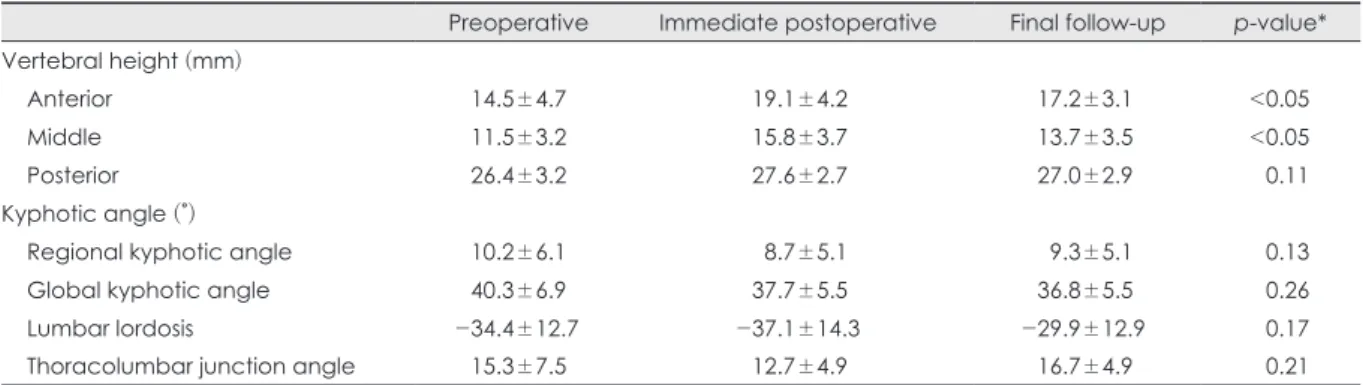
Radiographic outcomes
The results of the radiographic measurements are sum- marized in Table 2. The heights of the anterior and middle
vertebral bodies ware significantly increased after PVP (p
<0.05). There was no statistically significant change in the height of the posterior vertebral body (p=0.11). No statisti- cally significant changes were observed in the kyphotic an- gle measurements (p=0.13).
TABLE 1. Comparison of VAS scores for back pain
Preoperative Immediate postoperative Final follow-up p-value†
VAS* 7.3±0.7 3.3±0.8 2.5±1.1 <0.05
The data is presented as mean±standard deviation. Final follow-up occurred at 14 months on average, ranged from 12 to 45 months. *Scores ranged from 0 to 10, †statistically significant at p<0.05. VAS: Visual Analogue Scale
TABLE 2. Radiographic parameters: measurements of vertebral height and kyphotic angle
Preoperative Immediate postoperative Final follow-up p-value*
Vertebral height (mm)
Anterior 14.5±4.7 19.1±4.2 17.2±3.1 <0.05
Middle 11.5±3.2 15.8±3.7 13.7±3.5 <0.05
Posterior 26.4±3.2 27.6±2.7 27.0±2.9 0.11
Kyphotic angle (˚)
Regional kyphotic angle 10.2±6.1 8.7±5.1 9.3±5.1 0.13
Global kyphotic angle 40.3±6.9 37.7±5.5 36.8±5.5 0.26
Lumbar lordosis -34.4±12.7 -37.1±14.3 -29.9±12.9 0.17
Thoracolumbar junction angle 15.3±7.5 12.7±4.9 16.7±4.9 0.21
The data is presented as mean±standard deviation. Final follow-up occurred at 14 months on average, ranged from 12 to 45 months. *Statistically significant at p<0.05
FIGURE 2. A case of Kümmell’s disease treated with percutaneous vertebroplasty. (A) There was acute vertebral body collapse and diminished vertebral height at L1. An intravertebral vacuum cleft (intravertebral vacuum phenomenon) was seen on a preoper- ative radiograph as a horizontal linear radiolucency within the L1 vertebral body. (B) Immediate postoperative lateral standing plain radiography showed a cement mass in the L1 vertebral body. Bone cement was injected to fill the gap and stabilize the fractured site. (C) At the 1-year follow-up, a lateral standing plain radiography image showed no cement leakage and no adjacent vertebral body fracture. Bone cement was filled into the injection site.
A B C
Complications
Cement leakage occurred in 5 of 19 cases (26.3%). How- ever, there was no leakage into the spinal canal. No neuro- logic complications, such as paraparesis or paresthesia of the lower limbs, were reported.
Adjacent level fracture
Adjacent level compression fracture occurred in 3 cases (15.8%). Two cases occurred in patients with cement leak- age, and one case without cement leakage. The sites of the fractures were as follows: at L1 in a patient with Kümmell’s disease at L3, at L2 in a patient with Kümmell’s disease at L3, and in L1 in a patient with Kümmell’s disease at T11.
All were treated with additional PVP.
Discussion
In 1891, Dr. Herman Kümmell reported a disease in which patients developed severe back pain and angular kyphosis ranging from months to years after experiencing a trauma that had been asymptomatic. Schmorl and Junghanns11) demonstrated delayed post-traumatic vertebral fracture pathologically.
Kempinsky et al.7) attempted post decompression via to- tal laminectomy in Kümmell disease patient. Shikata et al.12) performed decompression with total laminectomy and removed the bony fragments that were projecting posteri- orly.2) Kaneda et al.6) reported that the lamina should be preserved because it is important for stabilization of sagittal balance. They reported that an anterior approach for de- compression and reconstruction was effective in osteopo- rotic patients, and there was a very low incidence of instru- mentation failure and very low morbidity.
In patients with Kümmell’s disease with neurologic com- plications, surgical treatment is more appropriate. Howev- er, most patients with Kümmell’s disease complain of back pain without neurologic symptoms. It has a risk for elderly patients to undergo surgery under general anesthesia. Con- servative treatments such as bed rest and analgesics could be another treatment option for these patients. However, the long-term immobilization might lead to secondary com- plications such as pneumonia, pulmonary embolism, and pressure sores.5)
Since Deramond et al.3) successfully treated vertebral hem- angioma using PVP and bone cement in 1984, PVP has been used to relieve pain by stabilizing the vertebral body. How- ever, because of the characteristic osteonecrosis and vacu- um phenomenon associate with Kümmell’s disease, it was generally considered that PVP would result in leakage af-
ter the cement injection and was therefore not an appropri- ate treatment.14) In spite of these concerns, many authors have described the efficacy of PVP for treating patients with Kümmell’s disease. Cho et al.2) reported that osteopo- rotic patients with Kümmell’s disease without neurologic complications showed improvement in pain after PVP. Ha et al.4) showed that PVP can effectively alleviate pain in pa- tients with. Back pain decreased after PVP in our study as well.
With regards to radiographic outcomes, vertebral height increased after PVP for both the anterior and middle verte- brae and this was maintained until the last follow-up eval- uation (p<0.05). For some patients, this was as long as 45 months after the procedure. The parameters associated with sagittal balance, such as regional kyphotic angle, global kyphotic angle, LL, and TLJ angle were not corrected. How- ever, there was no deterioration in the measurements and stasis was maintained at the follow-up assessment (Figure 2).
The results of this study show that PVP for Kümmell’s disease can alleviate back pain. In addition, the preoperative sagittal balance for these patients can be maintained, al- though it cannot be corrected.
The limitations of this study are as follows. First, the number of cases was small. Second, the follow up period was at least 1 year, but was not a long-term follow-up. Long- term follow-up is needed. Third, this study was a retrospec- tive analysis.
Conclusion
PVP can be considered as an effective treatment option for pain relief and maintenance of sagittal balance in patients with Kümmell’s disease.
■ The authors have no financial conflicts of interest.
REFERENCES
1) Chen L, Dong R, Gu Y, Feng Y. Comparison between balloon ky- phoplasty and short segmental fixation combined with vertebro- plasty in the treatment of Kümmell’s disease. Pain Physician 18:
373-381, 2015
2) Cho YS, Cho SD, Kim BS, Park TW, Lew S, Hwang SY. Küm- mell’s disease managed by percutaneous vertebroplasty. J Korean Soc Spine Surg 8:226-234, 2001
3) Deramond H, Depriester C, Galibert P, Le Gars D. Percutaneous vertebroplasty with polymethylmethacrylate. Technique, indica- tions, and results. Radiol Clin North Am 36:533-546, 1998 4) Ha KY, Lee JS, Kim YS, Chon JS, Seo JY. Comparison of verte-
broplasty between vertebral osteoporotic compression fracture and Kümmell’s disease. J Korean Orthop Assoc 40:458-464, 2005 5) Harper CM, Lyles YM. Physiology and complications of bed rest.
J Am Geriatr Soc 36:1047-1054, 1988
6) Kaneda K, Asano S, Hashimoto T, Satoh S, Fujiya M. The treat-
ment of osteoporotic-posttraumatic vertebral collapse using the Kaneda device and a bioactive ceramic vertebral prosthesis. Spine (Phila Pa 1976) 17:S295-S303, 1992
7) Kempinsky WH, Morgan PP, Boniface WR. Osteoporotic kypho- sis with paraplegia. Neurology 8:181-186, 1958
8) Kim HS, Heo DH. Percutaneous pedicle screw fixation with poly- methylmethacrylate augmentation for the treatment of thoracolum- bar intravertebral pseudoarthrosis associated with Kümmell’s osteonecrosis. Biomed Res Int 2016:3878063, 2016
9) Kim KT, Suk KS, Kim JM. Surgical treatment of Kümmell dis- ease with neurologic deficits: Posterolateral decompression and posterior reconstruction. J Korean Soc Spine Surg 8:136-142, 10) Kim SW, Chung YK. The surgical reconstruction of osteoporotic 2001
vertebral fractures. J Korean Soc Fract 14:30-36, 2001
11) Schmorl G, Junghanns H. The human spine in health and disease,
ed 2nd. New York, NY: Grune and Stratton, 1971
12) Shikata J, Yamamuro T, Iida H, Shimizu K, Yoshikawa J. Surgical treatment for paraplegia resulting from vertebral fractures in senile osteoporosis. Spine (Phila Pa 1976) 15:485-489, 1990
13) Yang DL, Yang SD, Chen Q, Shen Y, Ding WY. The treatment evaluation for osteoporotic Kümmell disease by modified poste- rior vertebral column resection: Minimum of one-year follow-up.
Med Sci Monit 23:606-612, 2017
14) Yi X, Lu H, Tian F, Wang Y, Li C, Liu H, et al. Recompression in new levels after percutaneous vertebroplasty and kyphoplasty compared with conservative treatment. Arch Orthop Trauma Surg 134:21-30, 2014
15) Zhang GQ, Gao YZ, Zheng J, Luo JP, Tang C, Chen SL, et al. Pos- terior decompression and short segmental pedicle screw fixation combined with vertebroplasty for Kummell’s disease with neuro- logical deficits. Exp Ther Med 5:517-522, 2013