Acute Osteomyelitis in the Proximal Humerus Caused by Pyogenic Glenohumeral Arthritis in an Elderly Patient - A Case Report
Yoon-Suk Hyun , Jae-Woo Kwon, Sung-Yup Hong, Kyeol Han
Department of Orthopaedic Surgery, Hallym University Kangdong Sacred Heart Hospital, Seoul, Korea
Reports of osteomyelitis in the proximal humerus with pyogenic glenohumeral arthritis of adjacent joints mostly involve pediatric pa- tients. Nowadays, osteomyelitis that is secondary to adjacent pyogenic glenohumeral arthritis is extremely rare, even more so in adults than in pediatrics. We report a rare case of the pyogenic glenohumeral arthritis followed by osteomyelitis of the proximal humerus in an elderly patient. Initially, we diagnosed a case of pyogenic glenohumeral arthritis only, which, despite arthroscopic synovectomy, did not resolve and severe pain continued. Subsequent radiological imaging, performed after our suspicion of a secondary involvement, allowed us to diagnose osteomyelitis combined with the pyogenic glenohumeral arthritis, which we had overlooked because of the extreme rar- ity of the condition in adults since the antibiotic era began.
(Clin Shoulder Elbow 2014;17(4):197-200) Key Words: Septic arthritis; Shoulder; Osteomyelitis
Osteomyelitis is an inflammation of the bone that is in gener- al caused by pyogenic arthritis, bacterial infections of the joints, and is mostly diagnosed in pediatric patients.1,2) Only one case of osteomyelitis has so far been diagnosed in adults after pyo- genic arthritis occurred in the patient’s joints during a reparative treatment for damaged rotator cuffs.3) To our best knowledge, we do not know of any cases of acute osteomyelitis caused by pyogenic arthritis that is not surgery-related in adults. However, in this case report, we describe our encounter with a case of pyogenic glenohumeral arthritis of non-surgery origin that led to osteomyelitis of adjacent bones in an elderly patient with no underlying disease.
Case Report
An 84-year-old female patient was referred to us for symp- toms, which began the night before, of severe pain and for focal swelling and fever over the affected bone in her right shoulder.
A week before referral, the patient had received intra-articular fluid injections for mild pain at a different hospital. At the time of
referral, the patient showed distinctive focal swelling and fever over the affected bone, severe pain, a limited range of motion of the joints indicated by passive flexion and abduction of below 20o, a body temperature of 39oC, and a generally sick physical appearance. At the outpatients’ ward, 20 ml of viscous yellowish synovial fluid was taken by arthrocentesis for synovial fluid tests.
Subsequent analysis showed a white blood cell count of 67,200 cells/mm3, of which 90% were neutrophil count. A Gram stain was positive for the presence of Gram-positive bacteria. Blood tests showed high levels of white blood cells, 13,110 cells/mm3, high levels of C-reactive protein, 209.7 mg/dl (normal range is below 3.0 mg/dl), and a high erythrocyte sedimentation rate (ESR) of 107 mm/h (normal range is below 26 mm/h).
Initially, we diagnosed pyogenic arthritis of the shoulder by physical examination and synovial fluid tests. Before surgery, we assessed the extent of the arthritis through magnetic resonance imaging (MRI) and found the following; a massive rotator cuff tear, severe subacromial bursitis, an abnormally high volume of synovial fluid, a thickened synovial capsule, which was en- hanced on the MRI image, and lastly, nothing to suspect an
Clinics in Shoulder and Elbow
CiSE
Copyright © 2014 Korean Shoulder and Elbow Society. All Rights Reserved.
This is an Open Access article distributed under the terms of the Creative Commons Attribution Non-Commercial License (http://creativecommons.org/licenses/by-nc/3.0) which permits unrestricted non-commercial use, distribution, and reproduction in any medium, provided the original work is properly cited.
pISSN 2383-8337 eISSN 2288-8721
CASE REPORT
Clinics in Shoulder and Elbow Vol. 17, No. 4, December, 2014 http://dx.doi.org/10.5397/cise.2014.17.4.197
Received August 21, 2014. Revised November 4, 2014. Accepted November 5, 2014.
Correspondence to: Yoon-Suk Hyun
Department of Orthopaedic Surgery, Hallym University Kangdong Sacred Heart Hospital, 150 Seongan-ro, Gangdong-gu, Seoul 134-701, Korea Tel: +82-2-2242-2230, Fax: +82-2-489-4391, E-mail: [email protected]
Financial support: None. Conflict of interests: None.
198
www.cisejournal.orgClinics in Shoulder and Elbow Vol. 17, No. 4, December, 2014
invasion of the infection to the adjacent bones (Fig. 1). For the emergency surgery, we carried out arthroscopic drainage, intra- articular lavage, synovectomy, debridement, and resectomy of the subacromial bursa. With the patient in lateral decubitus position, a posterior portal was used to insert the arthroscopy, and an anterior portal for surgical instruments. Synovial fluid was aspirated using an automated irrigation pump at a pressure of 30 mmHg. After the surgery, a Hemo-Vac drainage system was inserted through the posterior portal and 15 ml of blood was drained every day for 3 days. The bacterial culture of the drain- age fluid from the final day was positive for Methicillin-resistant Staphylococcus aureus (MRSA). During surgery, we observed hypertrophy of the synovial capsule and signs of acute infection, confirming pre-operative MRI findings. Further, we were able to confirm the massive rotator cuff tear and degenerative arthritis marked by partial damage of the articular cartilage of the humer- al head. After the surgery, following results from the Department of Infectious Diseases that the patient contracted MRSA after an intra-articular injection, we prescribed Teicoplanin for the pa- tient.4) The patient exhibited symptoms of intermittent fever of above 37.4oC even after surgery.
Despite initial signs of the patient’s symptoms resolving after the surgery, the symptoms gradually aggravated from the 5th postoperative day, and abnormal ranges of C-reactive protein (126.6 mg/dl) and ESR (107 mm/h) were seen on the 7th post- operative day. To assess the infection, we carried out further diagnostic MRI imaging and found bone marrow edema at the proximal humerus, and pus pockets and fluid collection at the axilla (Fig. 2). Arthrocentesis of 10 ml of the synovial fluid from the affected joint showed a viscous yellowish appearance. Sub-
sequent synovial fluid tests showed a white blood cell count of 104,000 cells/mm3, of which 90% were neutrophil count.
At the 8th postoperative day, we carried out a second sur- gery comprising an open intra-articular drainage of the proximal humerus and perforation and drainage of the distal humerus to remove the infection and the acute osteomyelitis. Using the anterior portal technique, we carried out a massive resectomy of the disordered granulation tissue of the shoulder joint. Then, we carried out perforation and drainage using Steinmann wire of 3.6 mm diameter. Through MRI imaging before the second sur- gery, we found that the fluid in the suspected pus pocket at the axilla was relatively transparent and colorless. After the second surgery, teicoplanin and clindamycin were co-prescribed after Gram staining and bacterial culture of the synovial fluid attained from the primary surgery showed presence of MRSA. The level of pain measured a day after the second surgery improved by about 60% of that measured a day after the primary surgery. Al- though we did not find any systemic or focal fever, we adminis- tered 3 weeks of intravenous antibiotics therapy to aid recovery of the elderly patient.
Susceptibility tests against various antibiotics of the bacte- rial culture from the bone marrow attained during the second surgery again showed the presence of MRSA. The resistance of this bacterium strain to many antibiotics led us to regard it as a community-acquired MRSA; thus, the patient was kept on intra- venous antibiotics for longer. We prescribed oral antibiotics, Cip- rofloxacin and Rifampin, for an additional 4 weeks until levels of ESR and C-reactive protein came down to normal ranges. At the final 6-month follow-up, we found normal ESR and C-reactive protein levels of 15.0 mm/h and 0.6 mg/dl, respectively, and an asymptomatic patient.
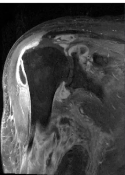
Fig. 1. An oblique coronal image of the initial magnetic resonance imaging (fat suppressed, contrast enhanced image) shows severe subacromial, subdeltoid bursitis, capsulitis with thick synovial enhancement, and a chronic massive rotator cuff tear without osteomyelitis in the proximal humerus.
Fig. 2. A magnetic resonance imaging before the second operation shows multiple pus pockets in the proximal humerus.
Acute Osteomyelitis in the Proximal Humerus after Pyogenic Arthritis - A Case Report Yoon-Suk Hyun, et al.
www.cisejournal.org
199 Discussion
The diagnosis of osteomyelitis of the shoulder proximal hu- merus secondary to pyogenic arthritis can be overlooked as it is extremely rare in adults. The use of antibiotics in the modern era has nearly wiped out osteomyelitis in adults, compounded by the fact in adults there is less synovial fluid within the joint cavity in adults than infants.5)
The pathophysiology of osteomyelitis with concomitant pyogenic arthritis in pediatrics is usually either through hema- togenous spread of an infection through the transepiphyseal or the epiphyseal blood vessels from adjacent joints, or a direct spread after a periosteum rupture induced by pressure from early stage edema and/or exudate.1) When osteomyelitis occurs alone, a secondary acute osteomyelitis of in adjacent joints may lead to a pyogenic arthritis.6) However, our case in this current report is peculiar in that primary pyogenic arthritis in a joint led to osteomyelitis of an adjacent bone. The putative pathophysiol- ogy of osteomyelitis secondary to a pyogenic arthritis is either a bacterial infection that spread due to the degenerative ero- sion of the humeral head cartilage, which we saw during open surgeries of the shoulder, or a direct epiphyseal exposure of the tendon attachment site, which formed the border between the joint capsule and the humeral head, and the infection wherein led to the humeral head. As Trueta7) found the epiphysis and the joint capsule receive blood from a common vascular supply, and thus through this medium the osteomyelitis may spread to nearby joints. According to animal studies by Alderson et al.,6) the growth of bacteria at the cartilage may destroy the cartilage, after which the infection is able to spread to adjacent structures as it gains access to the cartilagenous blood vessels. After the pri- mary surgery, we administered teicoplanin for 3 days by which the level of C-reactive protein began to decrease, but from the 4th day improvement stalled, and C-reactive protein levels re- mained at an abnormal range of over 100 mg/dl. We believe that the premature stopping of the Hemo-Vac drainage system on the 3rd postoperative day counteractively led to an increased intra-articular growth of bacteria, and subsequent erosion of the cartilage already partially eroded from arthritis, in our patient’s case. Then, the erosion of the cartilage may have led to the spread of the infection to the epiphysis, where a secondary os- teomyelitis formed. In support of this, we found that the strain of MRSA from the bone marrow of the humeral head and the joint cavity of the shoulder was the same.
Although the patient had a relatively healthy medical history, having only hypertension without other underlying diseases and a healthy lifestyle and nutritional level for an 84-year-old, the fact that the patient is an 84-year-old meant that her severity of infection may be as aggressive as that of a chronically diseased individual with immunosuppression. In adults or in the elderly, bacterial infections in the joint are mostly treated through simple
lavage, open or arthroscopic drainage and lavage, and, if neces- sary, a partial synovectomy.8,9) At the time of referral of the pa- tient, through synovial fluid tests, we diagnosed acute pyogenic arthritis and implemented arthroscopic lavage and drainage.
However, the infection may have spread to the humeral head due to the damage in the delicate cartilage, caused by the pres- sure we enforced during a synovial wash or by surgical tools, leading to a secondary proximal humeral osteomyelitis. To dem- onstrate the delicacy of pyogenic arthritis treatment, Abdel et al.8) found that a third of all arthroscopic treatments of primary pyogenic arthritis need further treatments. As well as this, Rhee et al.9) found that open surgeries give better results for injection- induced pyogenic arthritis than closed surgeries. We propose that for patients who have had a preoperative diagnosis of car- tilage erosion and/or massive rotator cuff tear, an open surgery from the start, rather than a closed surgery that stimulate spread of infection, may prevent acute osteomyelitis.
Taking an MRI image can help us to locate and predict the size of the pyogenic arthritis, and assist surgeons to devise posi- tions of arthroscopic portals and treatment methods. Impor- tantly, MRI images are important to discern possible invasion of the infection to the surrounding tissue. Bierry et al.10) have found that even after the treatment of pyogenic arthritis, post-treatment MRI image showed bone marrow edema or erosion of the adja- cent bones. However, as in this report, clinicians must be aware of the fact bone marrow edema, detected through an early MRI image, can progress to osteomyelitis.
Despite insufficient evidence to directly implicate the ero- sion of the articular cartilage or massive tear of the rotator cuffs as the causes of osteomyelitis secondary to pyogenic arthritis, this is a single report of a rare case. In this report, we found that pyogenic arthritis with underlying bone cartilage erosion and a massive rotator cuff tear, the bacterial infection may invade to adjacent bones, and in this case, to the glenohumerus in an elderly patient. If as in our case, concomitant treatment of drain- age and antibiotics therapy for pyogenic arthritis does not mark- edly alleviate pain nor resolve the patient’s symptoms, but rather increase in C-reactive proteins, we advise another diagnostic MRI to be performed to catch a secondary osteomyelitis at an early stage.
References
1. Goergens ED, McEvoy A, Watson M, Barrett IR. Acute osteo- myelitis and septic arthritis in children. J Paediatr Child Health.
2005;41(1-2):59-62.
2. Perlman MH, Patzakis MJ, Kumar PJ, Holtom P. The incidence of joint involvement with adjacent osteomyelitis in pediatric patients. J Pediatr Orthop. 2000;20(1):40-3.
3. Shin SJ, Jeong BJ, Kook SH, Shin SJ. Acute osteomyelitis of the humeral head after arthroscopic rotator cuff repair. Clin Shoul-
200
www.cisejournal.orgClinics in Shoulder and Elbow Vol. 17, No. 4, December, 2014
der Elbow. 2013;16(2):141-7.
4. Murray RJ, Pearson JC, Coombs GW, et al. Outbreak of inva- sive methicillin-resistant Staphylococcus aureus infection as- sociated with acupuncture and joint injection. Infect Control Hosp Epidemiol. 2008;29(9):859-65.
5. Nade S. Acute septic arthritis in infancy and childhood. J Bone Joint Surg Br. 1983;65(3):234-41.
6. Alderson M, Speers D, Emslie K, Nade S. Acute haematog- enous osteomyelitis and septic arthritis: a single disease. An hypothesis based upon the presence of transphyseal blood vessels. J Bone Joint Surg Br. 1986;68(2):268-74.
7. Trueta J. The three types of acute haematogenous osteomy-
elitis: a clinical and vascular study. J Bone Joint Surg Br. 1959;
41(4):671-80.
8. Abdel MP, Perry KI, Morrey ME, Steinmann SP, Sperling JW, Cass JR. Arthroscopic management of native shoulder septic arthritis. J Shoulder Elbow Surg. 2013;22(3):418-21.
9. Rhee YG, Cho NS, Kim BH, Ha JH. Injection-induced pyogen- ic arthritis of the shoulder joint. J Shoulder Elbow Surg. 2008;
17(1):63-7.
10. Bierry G, Huang AJ, Chang CY, Torriani M, Bredella MA. MRI findings of treated bacterial septic arthritis. Skeletal Radiol.
2012;41(12):1509-16.