CASE REPORT
Copyright ⓒ 2009 Korean Neurological Association 149
Print ISSN 1738-6586 / On-line ISSN 2005-5013 10.3988/jcn.2009.5.3.149 J Clin Neurol 2009;5:149-150
Common Carotid Artery Agenesis:
Duplex Ultrasonographic Findings
Seong Jin Yim, MD; Jung Ho Ryu, MD; Jong Sam Baik, MD;
Jeong Yeon Kim, MD; Jae Hyeon Park, MD; Sang Won Han, MD
Department of Neurology, Sanggye Paik Hospital, Inje University College of Medicine, Seoul, Korea
Received December 12, 2008 Revised March 9, 2009 Accepted March 9, 2009 Correspondence Sang Won Han, MD Department of Neurology, Sanggye Paik Hospital,
Inje University College of Medicine, 761-1 Sanggye 7-dong, Nowon-gu, Seoul 139-707, Korea
Tel +82-2-950-1090 Fax +82-2-950-1955 E-mail swhan@paik.ac.kr
BackgroundaAgenesis of the common carotid artery (CCA) resulting in separation of the or- igin of the external carotid artery (ECA) and internal carotid artery (ICA) from the aortic arch is rare. Fewer than 25 cases have been reported, and correlative ultrasound data were available for only 1 of them.
Case ReportaaA 52-year-old woman visited the hospital with a 3-day history of vertigo and headache. Color-coded duplex ultrasonography performed to evaluate the carotid and vertebral arteries revealed a normal configuration on the left side. However, the right CCA could not be found; instead, there were two vessels of approximately equal size in close proximity to each ot- her. The cerebral angiographic findings were consistent with the ultrasonographic findings. The ECA and ICA originated directly from the brachiocephalic trunk, and the ECA arose proximal to the ICA.
ConclusionsaaThe ultrasonographic findings revealed absence of the CCA, the ECA and ICA originating separately from the aortic arch. Color-coded duplex ultrasonography appears to be an effective and sensitive method for detecting absence of the CCA. These findings should help to further our understanding of the embryologic development of the carotid arteries.
J Clin Neurol 2009;5:149-150 Key Wordsaacommon carotid artery agenesis, ultrasonography, angiography.
Introduction
Agenesis of the common carotid artery (CCA) resulting in separation of the origin of the external carotid artery (ECA) and internal carotid artery (ICA) from the aortic arch is rare.
Fewer than 25 cases have been reported, and correlative ultra- sonographic data were available for only 1 of them.1-4 We report herein a case of absence of the CCA, the ECA and ICA originating separately from the aortic arch, as evidenced by color-coded duplex ultrasonography.
Case Report
A 52-year-old woman with a history of hypertension and hy- percholesterolemia visited the hospital with a 3-day history of vertigo and headache. The results of physical and neurol- ogical examinations were unremarkable. Color-coded duplex ultrasonography performed to evaluate the carotid and ver-
tebral arteries revealed a normal configuration on the left side.
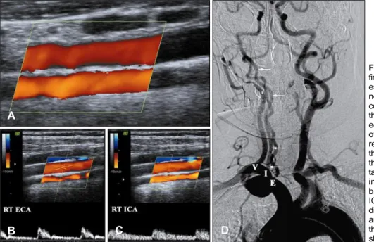
No significant stenotic flow or plaque formation was observ- ed in the left CCA, ECA, or ICA. However, the right CCA could not be found; instead, there were two vessels of ap- proximately equal size in close proximity to each other. The more medially oriented vessel exhibited a low-resistance flow pattern consistent with the ICA, and the more laterally orie- nted vessel exhibited a high-resistance flow pattern consistent with the ECA (Fig. 1A-C). There was no significant stenotic flow or plaque formation in either of the two vessels. The ce- rebral angiographic findings were consistent with the ultra- sonographic findings. The ECA and ICA originated directly from the brachiocephalic trunk, and the ECA arose prox- imal to the ICA (Fig. 1D).
Discussion
The embryologic development of this anomaly has been dis-
Common Carotid Artery Agenesis
150 J Clin Neurol 2009;5:149-150
cussed previously by Lie.5 The CCA may be absent if the ductus caroticus (an embryologic vascular remnant) persists and there is involution of the distal portion of the third bran- chial arch. An alternative mechanism involves a failure of the ECA to migrate laterally and join the ICA (which arises from the third aortic arch) during development. As shown in this case, the resistive indices and spectral waveforms of the ICA and ECA are determined mainly by the regions and their supplied organs and not by the CCA.1,6 CCA agenesis is usu- ally encountered during the diagnostic process. Color-coded duplex ultrasonography appears to be an effective and sensi- tive method for detecting absence of the CCA. In cases of carotid stenting, the technical problem of a lack of distal wire support in the ECA may lead to a higher risk of dislocation of the plaque and debris embolization in the ICA during the unprotected phase of the procedure.7 Endovascular treatment of these cases may be reserved for patients with stable pla- ques. We believe that the findings in our patient are instruc- tive and will help to further our understanding of the embryo- logic development of the carotid arteries.
Acknowledgements
This work was supported by the 2006 Inje University research grant.
REFERENCES
1. Woodruff WW 3rd, Strunsky VP, Brown NJ. Separate origins of the left internal and external carotid arteries directly from the aortic arch:
duplex sonographic findings. J Ultrasound Med 1995;14:867-869.
2. Maybody M, Uszynski M, Morton E, Vitek JJ. Absence of common carotid artery: a rare vascular anomaly. AJNR Am J Neuroradiol 2003;
24:711-713.
3. Simons D, Patetsios P, Moglia R, Dietz P. A case of congenital absence of the right common carotid artery: a rare embryologic anomaly. Journal for Vascular Ultrasound 2003;27:106-109.
4. Purkayastha S, Gupta AK, Varma DR, Bodhey NK, Vattoth S. Abse- nce of the left common carotid artery with cervical origin of the right subclavian artery. AJNR Am J Neuroradiol 2006;27:708-711.
5. Lie TA. Congenital anomalies of the carotid arteries. Amsterdam:
Excepta Medica Foundation, 1968.
6. Beach KW, Strandness DE Jr. Carotid artery velocity waveform an- sVascular Disease. St. Louis: Mosby, 1985:410-413.
7. Faggioli GL, Testi G, Ferri M, Rossi C, Stella A. Common carotid age- nesis and internal carotid stenting. Int Angiol 2007;26:290-291.
Fig. 1. Sonographic and angiographic findings of common carofid artery agen- esis. (A) Color-coded duplex ultraso- nography showing absence of the right common carotid artery (CCA). Instead, there are two vessels of approximately equal size in close proximity to each other. One vessel (B) shows a high- resistance flow pattern consistent with the external carotid artery (ECA), while the other vessel (C) shows a low-resis- tance flow pattern consistent with the internal carotid artery (ICA). (D) Cere- bral angiography showing that the ICA (I, arrows) and ECA (E) originate directly from the brachiocephalic trunk, and that the ECA arises proximal to the ICA. The vertebral artery (V) is also shown.
A
B C D