Introduction
Aortic stenosis (AS) is a prototypical disease of pressure overload to the left ventricle, which affects 2--4% of the elder- ly population.1) It is a gradual but constantly progressive dis- ease with a prolonged asymptomatic period. However the prognosis is poor when symptoms develop2) and the only cura- tive measure is replacement of the diseased valve at the right time. Therefore, understanding the mechanism of left ventric- ular (LV) response to pressure overload in these patients is im- portant not only for the optimal timing of surgery but also,
for predicting the outcome and possibly, excavating new ther- apeutic targets.
With a prolonged period of pressure overload, hypertrophy of the myocardium develops as a compensatory mechanism.3) Microscopically, this is associated with increase of myofiber size and interstitial fibrosis in various human4)5) and animal models of ventricular hypertrophy.6) More importantly, the de- gree of hypertrophy in asymptomatic AS patients has been as- sociated with clinical outcome in numerous previous reports7)8) and although commonly regarded as ‘normal’ systolic func- ORIGINAL ARTICLE J Cardiovasc Ultrasound 2014;22(2):72-79
Association of Myocardial Angiogenesis with Structural and Functional
Ventricular Remodeling in Aortic Stenosis Patients with Normal Ejection Fraction
Seung-Pyo Lee, MD, PhD, Hyung-Kwan Kim, MD, PhD, Yong-Jin Kim, MD, PhD, Seil Oh, MD, PhD, and Dae-Won Sohn, MD, PhD
Department of Internal Medicine and Cardiovascular Center, Seoul National University Hospital, Seoul National University College of Medicine, Seoul, Korea
Background: Although rarefaction of myocardial angiogenesis has been shown to be associated with left ventricular (LV) systolic dysfunction in animal models of ventricular hypertrophy, this relationship has not been investigated in depth nor validated in humans. We aimed to analyze the relationship of myocardial angiogenesis with various functional and structural ventricular remodeling parameters in moderate to severe aortic stenosis (AS) patients with normal LV ejection fraction (LVEF).
Methods: A total of 38 moderate or severe AS patients with LVEF > 50% were enrolled for the current study and all patients underwent LV endomyocardial biopsy at the septum during aortic valve replacement. The biopsy specimens were stained for platelet endothelial cell adhesion molecule-1 (CD31) to analyze the density of blood vessels in the myocardium.
Results: The degree of myocardial angiogenesis tended to increase with worse myocardial systolic function, LV filling pressure and progressed ventricular hypertrophy (Spearman’s ρ = -0.388, p = 0.016 for LVEF; Spearman’s ρ = 0.442, p = 0.007 for E/e’; Spearman’s ρ = 0.424, p = 0.008 for LV mass index). The degree of myocardial angiogenesis was also significantly associated with the degree of aortic valve stenosis (Spearman’s ρ = -0.368, p = 0.023). There was significant difference in the degree of myocardial angiogenesis according to the LV geometry (p = 0.016 for mean difference between different LV geometry groups by analysis of variance).
Significant predictors of myocardial blood vessel density were LV mass index (β = 0.398, p = 0.010) and LVEF (β = -0.313, p = 0.028).
Conclusion: There is a close relationship between myocardial angiogenesis and LV remodeling in moderate to severe AS patients with normal LVEF, with angiogenesis increasing with LV hypertrophy. Further studies to demonstrate the mechanism underlying this phenomenon is warranted.
KEY WORDS: Angiogenesis · Aortic stenosis · Echocardiography · Ventricular remodeling.
• Received: April 28, 2014 • Revised: May 28, 2014 • Accepted: May 28, 2014
• Address for Correspondence: Seung-Pyo Lee, Department of Internal Medicine and Cardiovascular Center, Seoul National University Hospital, Seoul National University College of Medicine, 101 Daehak-ro, Jongno-gu, Seoul 110-744, Korea Tel: +82-2-2072-1980, Fax: +82-2-762-9662, E-mail: splee0624@gmail.com
• This is an Open Access article distributed under the terms of the Creative Commons Attribution Non-Commercial License (http://creativecommons.org/licenses/by-nc/3.0) which permits unrestricted non-commercial use, distribution, and reproduction in any medium, provided the original work is properly cited.
tion, some patients do have subclinical LV systolic dysfunc- tion.3)9) These results suggest that investigating the mecha- nism of ventricular hypertrophy, even in patients with ‘normal’
systolic function, may be important for understanding the pro- cess of ventricular remodeling in AS patients. However the microscopic changes associated with the process of ventricular hypertrophy, especially before the LV systolic dysfunction starts, is largely unknown in humans.
Angiogenesis is a dynamic process that goes side-by-side with the growth and regression of an organ. Specifically, an- giogenesis has been shown to be associated with ventricular hypertrophy in various animal models10) and several investiga- tors have tried to harness angiogenesis for treating cardiac hy- pertrophy.11)12) Although the disruption of coordinated ven- tricular hypertrophy and myocardial angiogenesis has been shown to contribute to overt LV systolic dysfunction in ani- mal models6) and also in humans,5) this relationship has not been investigated in depth nor validated in humans with nor- mal LV systolic function. Also, the phenomenon that has been demonstrated in animals has to be correlated with various pa- rameters of ventricular function in humans, in order to be trans- lated into clinical research in the future.
AS is an excellent human model for studying the change of ventricular function and morphology following chronic pres- sure overload.13) In this report, we analyzed the degree of myo- cardial angiogenesis with various ventricular remodeling pa- rameters, in both function and structure, in moderate to severe AS patients with normal LV ejection fraction (LVEF).
Methods
Patient population
A total of 38 patients with moderate to severe AS as per cur- rent guidelines,14) i.e., aortic valve area (AVA) < 1.5 cm2 and transaortic mean pressure gradient > 30 mmHg or transaortic peak velocity > 3 m/sec, were enrolled to this prospective study from September 2009 to September 2012 at Seoul Na- tional University Hospital. Patients with significant concomi- tant valvular disease of more than mild degree, i.e., moderate aortic regurgitation or moderate mitral valve disease, a previous history of cardiac surgery or myocardial infarction and also, pa- tients with significant LV systolic dysfunction (LVEF < 50%) were excluded. All patients gave informed consent to the study, the protocol of which was approved by the Institutional Review Board of Seoul National University Hospital. Baseline laborato- ry tests, anthropometric measures and medical history were tak- en at the time of echocardiographic examination. Body surface area (BSA) was calculated using the Mosteller formula.
Two-dimensional echocardiographic examination
We performed a comprehensive echocardiographic examina- tion of each patient with an adequate commercialized equip-
ment (Vivid 7, GE Medical System, Horten, Norway) accord- ing to the current recommendations and guidelines.15) In brief, end-diastolic/systolic LV diameter was measured at the stan- dard parasternal view. The aortic root, i.e., aortic annulus, si- notubular junction and ascending thoracic aorta diameter were measured at the standard parasternal long-axis view.
After securing an adequate standard four-chamber view, we measured peak early and late diastolic velocity (E, A velocity, respectively) at the tip of mitral valve using a standard pulsed- wave Doppler and also, mitral annular velocity (e’, a’ velocity, respectively) at the septal annulus using tissue Doppler imag- ing. We also measured transaortic mean pressure gradient (PG) and maximal velocity at all possible views, for example apical 5 or 3 chamber, subcostal, right parasternal and supra- sternal notch view. The AVA was calculated using the conti- nuity equation after acquiring time-velocity integral (TVI) at the aortic valve level and also, LV outflow tract (LVOT) level.
Stroke volume was calculated by multiplying TVI at the LVOT level with the cross-sectional area of LVOT and indexed by di- viding it with BSA. Valvuloarterial impedance, a measure of the global LV afterload, was calculated using the following equation; (systolic blood pressure + mean transaortic PG) / in- dexed stroke volume.16) The LV mass was calculated using the equation of Devereux and Reichek.17)
All patients had baseline heart rate < 100 bpm. For patients in sinus rhythm, all measurements were an average of 3 consec- utive beats. For patients in atrial fibrillation, all measurements were an average of 5 beats according to the current recommen- dations. The pattern of ventricular remodeling was classified according to the previous literatures, using LV mass index and relative wall thickness (RWT), into normal geometry, eccentric hypertrophy or concentric hypertrophy.18) Specifically, the cut- off value of LV mass index were 134 g/m2 for men, 109 g/m2 for women and the cut-off value of RWT was 0.45 for both sex.
Two-dimensional speckle tracking imaging analysis
We obtained standard two-dimensional speckle tracking images at a frame rate of 50--100 frame/second from the three standard apical views after securing a steady breath hold. The LV endocardium was tracked at the end-systolic phase with special caution not to include the pericardium. The region-of- interest was defined semi-automatically by an adequate off- line analysis program (EchoPac 5.0.1 for PC, GE Medical Sys- tems, Milwaukee, WI, USA) between the endocardial and epicardial borders. For an adequate measurement of global longitudinal strain (GLS), we traced at least five segments of each windows. An independent observer blinded to the objec- tive of the study obtained the whole strain curves. Peak GLS was defined as the peak negative value of the strain curve in a single cardiac cycle and calculated for the entire U-shaped LV myocardium as follows; global strain = [L (end-systole) - L (end-diastole)] / L (end-diastole) × 100 (%) (L: whole LV myo-
cardium as one big segment).19) Global strain is the myocardi- al deformity of the myocardium as a whole and not an average of each segmental strain as in previous literatures concerning average strain in severe AS patients.20)21) Peak GLS was aver- aged from GLS values analyzed at apical two, four and three chamber views.9)
Endomyocardial biopsy
All patients gave written consent on the intraoperative bi- opsy. In brief, after a standard aortotomy and removal of the diseased aortic valve, 3 mm sized endomyocardial biopsy spec- imen was collected from the basal septum of the LV cavity us- ing a standard bioptome.
Immunostaining and morphometric analysis All samples were stored overnight in 10% formaldehyde so- lution and embedded in paraffin. Four microgram section was cut for immunohistochemistry and treated for antigen activa- tion. Nonspecific binding sites were pre-blocked using 3% hy- drogen peroxide for 30 minutes. The primary antibody used for detection of blood vessel was rabbit anti-human platelet endo- thelial cell adhesion molecule-1 (PECAM-1, 1:250, Millipore, Billerica, MA, USA). The primary antibody was incubated overnight at 4°C and a secondary biotinylated anti-rabbit IgG (1:100, Promega, Sunnyvale, CA, USA) antibody was incubat- ed following the primary incubation. Finally, the staining re- sults were visualized using a standard DAB kit (Vector lab., Burlingame, CA, USA) according to the manufacturer’s recom- mendation. Morphometric measurements and analysis were done with a semi-automatic dedicated software (ImageJ, http://
rsb.info.nih.gov/ij). The result of the morphometry was ex- pressed as the PECAM-1 positive % area of the whole image.
Statistical analysis
Continuous variables are tested for normality with Kol- mogorov-Smirnov test and presented as mean ± standard de-
viation or median (25--75th percentile) as appropriate. The difference between the groups was compared using Student’s t-test or Mann-Whitney U test and analysis of variance (ANOVA) between three groups. Bivariate correlation analy- sis between the parameters of myocardial structure and func- tion are drawn and the strength of correlation presented as Spearman’s ρ. Dichotomous variables are presented as percent- ages and compared using χ2-test. All analysis was done with SPSS version 21.0 (SPSS Inc., Chicago, IL, USA) and two- tailed p-value of < 0.05 was considered statistically significant.
Results
A total of 38 moderate to severe AS patients were prospec- tively enrolled for the current study. The baseline clinical char- acteristics are summarized in Table 1. We included only those with normal LVEF, i.e., LVEF > 50%, to analyze how angio- genesis was specifically related to the process just before the transition to heart failure. In brief, there was no significant difference between the three groups of LV remodeling.
All patients could be divided into 3 groups according to the LV geometry, normal (n = 9), eccentric hypertrophy (n = 11) or concentric hypertrophy (n = 18). There was no patient with concentric remodeling geometry. We analyzed the echocar- diography data in these patients (Table 2). The dimension, wall thickness and mass index of LV were significantly differ- ent between the three groups as expected. Although there were no significant differences in the E velocity, e’ velocity, transaortic peak velocity nor transaortic mean PG between the three groups, the annulus diameter and the AVA was smaller in the concentric hypertrophy group (p = 0.024 for aortic an- nulus diameter; p = 0.043 for AVA by ANOVA).
Next, we analyzed the blood vessel density in the myocardi- um that was taken at the time of aortic valve replacement.
There was a wide range of vessel density in the given myocardi- al sample that ranged from 1% to nearly 4% of the whole sec- tion. Various echocardiography parameters including parame-
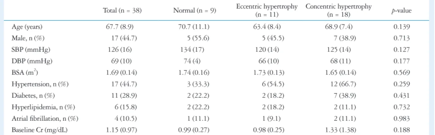
Table 1. Baseline clinical characteristics of the study participants
Total (n = 38) Normal (n = 9) Eccentric hypertrophy
(n = 11) Concentric hypertrophy
(n = 18) p-value
Age (years) 67.7 (8.9) 70.7 (11.1) 63.4 (8.4) 68.9 (7.4) 0.139
Male, n (%) 17 (44.7) 5 (55.6) 5 (45.5) 7 (38.9) 0.713
SBP (mmHg) 126 (16) 134 (17) 120 (14) 125 (14) 0.127
DBP (mmHg) 69 (10) 74 (4) 66 (10) 68 (11) 0.177
BSA (m2) 1.69 (0.14) 1.74 (0.16) 1.73 (0.13) 1.65 (0.14) 0.569
Hypertension, n (%) 17 (44.7) 3 (33.3) 6 (54.5) 12 (66.7) 0.259
Diabetes, n (%) 11 (28.9) 2 (22.2) 2 (18.2) 7 (38.9) 0.431
Hyperlipidemia, n (%) 6 (15.8) 2 (22.2) 2 (18.2) 2 (11.1) 0.732 Atrial fibrillation, n (%) 4 (10.5) 1 (11.1) 1 (9.1) 2 (11.1) 0.983
Baseline Cr (mg/dL) 1.15 (0.97) 0.99 (0.27) 0.98 (0.25) 1.33 (1.38) 0.188
The difference of baseline clinical characteristics between patients with distinct patterns of remodeling, i.e., concentric remodeling, eccentric hypertrophy, concentric hypertrophy was calculated using Student’s t-test, Mann-Whitney U test or χ2-test as appropriate and the results presented as p-value. SBP: systolic blood pressure, DBP: diastolic blood pressure, BSA: body surface area, Cr: creatinine
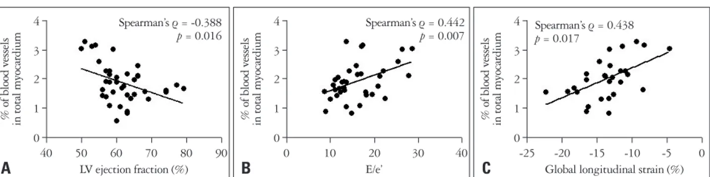
ters of systolic and diastolic function, i.e., LVEF, E/e’, the degree of aortic valve stenosis, i.e., mean transaortic PG, maximal transaortic velocity, AVA and also, the degree of LV hypertrophy was analyzed for correlation with the blood vessel density. Of the several parameters, various parameters of ventricular func- tion, such as, LVEF (Spearman’s ρ = -0.388, p = 0.016) (Fig.
1A) and E/e’ (Spearman’s ρ = 0.442, p = 0.007) (Fig. 1B) showed significant correlation with the blood vessel density.
Strain analysis using two dimensional-speckle tracking image was possible in 30 patients, the results of which demonstrated good correlation between GLS and vessel density (Spearman’s ρ
= 0.438, p = 0.017) (Fig. 1C).
Calculated LV mass index (Spearman’s ρ = 0.424, p = 0.008) (Fig. 2A) significantly correlated with myocardial blood vessel density. Although transaortic mean PG and peak velocity did not show significant correlation with the myocardial vessel
density, AVA and indexed AVA also showed good correlation with the vessel density (Spearman’s ρ = -0.368, p = 0.023 for AVA, Spearman’s ρ = -0.330, p = 0.046 for indexed AVA) (Fig. 2B). These findings demonstrate that the blood vessels may grow according to the hypertrophy of the LV and also, ag- gravation of both LV systolic and diastolic function (Fig. 3).
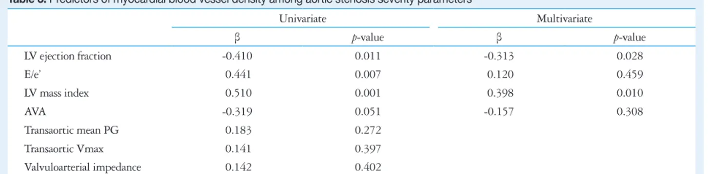
With the above parameters, linear regression analysis was done to determine the factor responsible for myocardial angio- genesis (Table 3). Although E/e’ and AVA was nonsignificant, LVEF and LV mass index remained as significant determi- nants of the degree of angiogenesis (β = -0.317, p = 0.026 for LVEF, β = 0.394, p = 0.009 for LV mass index). This was not changed even after adding the baseline clinical parameters (β = -0.338, p = 0.027 for LVEF, β = 0.460, p = 0.018 for LV mass index).
When the patients were subdivided into the LV geometry
Table 2. Echocardiographic parameters of the study participants
Total (n = 38) Normal (n = 9) Eccentric hypertrophy
(n = 11) Concentric hypertrophy
(n = 18) p-value
LVEDD (mm) 50.5 (5.2) 49.4 (1.8) 54.9 (4.4) 48.2 (5.1) 0.001
LVESD (mm) 31.3 (5.1) 30.7 (1.6) 35.1 (3.1) 29.5 (6.1) 0.015
LVEF (%) 61.7 (6.7) 61.2 (3.9) 60.8 (4.4) 62.6 (8.8) 0.769
IVST (mm) 12.1 (2.4) 10.2 (1.0) 10.7 (1.4) 13.8 (2.2) < 0.001
PWT (mm) 11.6 (1.9) 10.1 (1.4) 10.7 (1.2) 12.9 (1.7) < 0.001
Annulus diameter (mm) 20.8 (1.9) 21.5 (1.4) 21.8 (2.0) 20.0 (1.8) 0.024
E (m/sec) 0.73 (0.19) 0.63 (0.17) 0.76 (0.24) 0.75 (0.15) 0.215
Deceleration time (ms) 254 (66) 262 (62) 253 (80) 250 (63) 0.918
e’ (cm/sec) 4.7 (1.4) 4.9 (1.4) 4.8 (1.5) 4.5 (1.4) 0.710
Vmax (m/sec) 4.7 (0.7) 4.3 (0.3) 4.8 (0.9) 4.9 (0.6) 0.139
AVA (cm2) 0.73 (0.23) 0.86 (0.28) 0.77 (0.20) 0.64 (0.18) 0.043
AVA index (cm2/m2) 0.43 (0.13) 0.49 (0.15) 0.46 (0.16) 0.39 (0.11) 0.131
Transaortic mean PG (mmHg) 55.8 (19.4) 48.5 (12.7) 54.5 (22.7) 60.4 (19.7) 0.320
LV mass index (g/m2) 152.5 (50.8) 108.3 (12.3) 145.1 (28.8) 179.1 (56.9) 0.001
Relative wall thickness 0.46 (0.08) 0.41 (0.05) 0.39 (0.04) 0.53 (0.05) < 0.001
Global longitudinal strain (%) -13.5 (3.6) -15.0 (1.7) -14.8 (4.0) -11.7 (3.5) 0.071
The difference of baseline echocardiographic characteristics between patients with distinct patterns of remodeling, i.e., concentric remodeling, eccentric hypertrophy, concentric hypertrophy was calculated using Mann-Whitney U test. LVEDD: left ventricular end-diastolic diameter, LVESD: left ventricular end- systolic diameter, LVEF: left ventricular ejection fraction, IVST: interventricular septal thickness, PWT: posterior wall thickness, Vmax: maximal transaortic velocity, AVA: aortic valve area, PG: pressure gradient
Fig. 1. Correlation between ventricular function and myocardial blood vessel density. Significant negative correlation between left ventricular (LV) ejection fraction and myocardial blood vessel density (A), in contrast to significant positive correlation between E/e’ and myocardial blood vessel density (B) and also, LV global longitudinal strain (C).
0 0 0
1 1 1
2 2 2
3 3 3
4 4 4
40 50 60 70 80 90 0 10 20 30 40 -25 -20 -15 -10 -5 0
LV ejection fraction (%) E/e’ Global longitudinal strain (%)
% of blood vessels in total myocardium % of blood vessels in total myocardium % of blood vessels in total myocardium
A B C
Spearman’s ρ = -0.388
p = 0.016 Spearman’s ρ = 0.442
p = 0.007 Spearman’s ρ = 0.438 p = 0.017
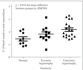
as in the previous literature,18) there was significant difference of the degree of myocardial angiogenesis between the three groups (p = 0.016) (Fig. 4).
Discussion
The main findings of the current study are that 1) within
the similar ‘normal’ systolic function, a wide degree of myo- cardial angiogenesis exists in patients with severe AS, 2) the degree of myocardial angiogenesis correlates with both LV sys- tolic and diastolic function, 3) the degree of myocardial angio- genesis also correlates with the degree of ventricular hypertro- phy and finally, 4) there is a significant difference of the degree
Fig. 3. Examples of myocardial blood vessel density according to left ventricular (LV) geometric remodeling. A: An example of a myocardial sample from a male patient with normal LV geometry [LV mass index 112.9 g/m2 and relative wall thickness (RWT) 0.45]. The LV ejection fraction (LVEF) was 64% and E/e’ 7.5. The myocardial blood vessel density was 1.59% of the total myocardium analyzed. B: An example of a myocardial sample from a male patient with eccentric hypertrophy (LV mass index 134.3 g/m2 and RWT 0.38). The LVEF was 58% and E/e’ 16.1. The myocardial blood vessel density was 2.17% of the total myocardium analyzed. C: An example of a myocardial sample from a female patient with concentric hypertrophy (LV mass index 161.9 g/m2 and RWT 0.49). The LVEF was 55% and E/e’ 25.4. The myocardial blood vessel density was 2.62% of the total myocardium analyzed. All specimens were stained for platelet endothelial cell adhesion molecule-1 immunostaining and visualized under 100 × field.
Fig. 2. Correlation between structural parameters of aortic stenosis and myocardial blood vessel density. Significant positive correlation between left ventricular mass index and myocardial blood vessel density (A), in contrast to significant positive correlation between aortic valve area and myocardial blood vessel density (B).
Table 3. Predictors of myocardial blood vessel density among aortic stenosis severity parameters
Univariate Multivariate
β p-value β p-value
LV ejection fraction -0.410 0.011 -0.313 0.028
E/e’ 0.441 0.007 0.120 0.459
LV mass index 0.510 0.001 0.398 0.010
AVA -0.319 0.051 -0.157 0.308
Transaortic mean PG 0.183 0.272
Transaortic Vmax 0.141 0.397
Valvuloarterial impedance 0.142 0.402
The predictors of myocardial blood vessel density were analyzed with linear regression analysis. LV: left ventricle, AVA: aortic valve area, PG: pressure gradient, Vmax: maximal velocity
A B C
0 0
1 1
2 2
3 3
4 4
0 100 200 300 400 0.0 0.5 1.0 1.5 2.0
LV mass index (g/m2) Aortic valve area (cm2)
% of blood vessels in total myocardium % of blood vessels in total myocardium
A B
Spearman’s ρ = 0.424 p = 0.008
Spearman’s ρ = -0.368 p = 0.023
of myocardial angiogenesis between the different LV geome- try. Although there have been various animal and human studies with AS investigating the correlation of angiogenesis with overt heart failure,22) there has been no studies showing that angiogenesis is associated with subclinical LV dysfunction in ‘normal systolic function’ human hearts.
With chronic pressure overload to the LV in AS, the myofi- brils tend to get thicker, which results in ventricular hypertro- phy.13) This also means that LV hypertrophy is a dynamic pro- cess involving various complex intracellular signals.23)24) Angiogenesis is a dynamic process that is closely related to or- gan growth and metabolism of the cells needs ingrowth of blood vessels to support this process.25) Specifically, the disrup- tion of the coordination between ventricular hypertrophy and angiogenesis has shown to be a cause of overt heart failure in murine models,6) which support the previous hypothesis con- cerning angiogenesis and ventricular hypertrophy. Further- more, the decreased expression of myocardial vascular endo- thelial growth factor has been pointed out as the culprit of systolic dysfunction.26)27)
In this report, we have shown that the degree of myocardial angiogenesis is closely associated with adverse remodeling. Spe- cifically, within patients with normal EF, the systolic/diastolic function, the LV mass and the degree of valve stenosis were all closely associated with the degree of myocardial angiogenesis.
Following the natural concept that angiogenesis follows the growth of an organ25) and that the disruption of angiogenic cy- tokine in the cardiomyocytes leads to systolic dysfunction,26) it can be said that if the systolic function are within normal limits, a certain degree of angiogenesis follows the adverse remodeling of the myocardium with chronic pressure overload. This is sup- ported by some old animal data demonstrating evidence of cap-
illary growth in hypertensive rodent models.28) Furthermore, it is a relatively well-known concept that the coronary vascular re- sistance/reserve is much reduced with the progression of ven- tricular hypertrophy, both in animals,29) and in humans.30) Therefore, it may be logical to say that angiogenesis, which in- evitably involves the sprouting of new vessels, may be a com- pensatory mechanism for the decrease of coronary vascular re- sistance/reserve.
It was interesting to find that the degree of myocardial an- giogenesis is associated with the degree of adverse LV remod- eling, for example LV systolic and diastolic function parame- ters. Specifically, the moderate degree of correlation in nearly all of the echocardiographic data tested shows that the process of LV remodeling, although it may sometimes differ from in- dividual-to-individual, follows a fairly universal process. Fol- lowing a chronic pressure overload to the LV due to the pro- gression of AS, the ventricle starts to get thicker whilst the systolic and diastolic function deteriorate.3)13) Although our data involves only patients with normal LVEF, the results of our analysis demonstrates that the ventricles are undergoing extensive pathological remodeling. Increased angiogenesis may be a compensatory effort of the cardiomyocytes to endure the prolonged stress, which in the long-term may fail.6) There- fore, as previous reports have demonstrated,9) employing more sensitive parameters, such as strain, for assessing the ventricu- lar remodeling may be needed as a guide for effective early treatment,3) which is supported by our data as well. Of course, this should not be mistaken that LVEF is a poor parameter to assess the gross LV systolic dysfunction.
Another interesting finding was that there was a significant difference in the degree of myocardial angiogenesis between the different LV geometry. Previous data have demonstrated that longitudinal strain, a marker of subendocardial fiber con- tractility, is decreased in patients with concentric hypertrophy compared with other types of geometry.31) However, neither the circumferential nor the radial strain were affected by the geometry. This finding, in concert with our previous findings9) and also, the current analysis, demonstrates that the subendo- cardial layer may be the one that undergoes extensive remod- eling in response to chronic pressure overload. It is also nota- ble that all of the biopsy specimens used in our analysis were from endomyocardial biopsy taken at the subendocardium.
Our findings are not without limitations. First, all hemody- namic data, especially LVEF and E/e’, are all load-dependent parameters of LV function. However, all of our patients were in euvolemic status and did not having resting dyspnea on echocardiographic examination. Furthermore, the association of myocardial angiogenesis with systolic function is corrobo- rated by our strain analysis, a relatively load-independent pa- rameter. Second, although we have suggested a possible mech- anism for increased angiogenesis following adverse ventricular remodeling in severe AS, we did not prove a definite data for coronary flow reserve nor resistance. Third, although we have
Fig. 4. Difference of the myocardial blood vessel density according to left ventricular (LV) remodeling pattern. There was significant difference of blood vessel density according to the LV geometry (p = 0.016 for mean difference between groups with ANOVA). ANOVA: analysis of variance.
0 1 2 3 4
Normal Eccentric
hypertrophy Concentric hypertrophy Geometry
% of blood vessels in total myocardium
p = 0.016 for mean difference between groups by ANOVA
provided a relationship between LV remodeling parameters and myocardial angiogenesis, we have not provided a mecha- nistic data on how this happens. We plan to present these data in the near future. Fourth, although the degree of myocardial angiogenesis correlated with AVA, there was no significant cor- relation between the degree of angiogenesis and other AS severi- ty parameters such as transaortic mean PG and peak velocity.
Conversely, these data may demonstrate that the degree of LV hypertrophy rather than the hemodynamic stress parameters are important for myocardial angiogenesis, which is partially dem- onstrated by the results of the multivariate linear regression analysis. It would be interesting to investigate whether this holds true in non-AS left ventricular hypertrophy (LVH) pa- tients, such as patients with LVH by hypertension.
In conclusion, our analysis results demonstrate that there is a close correlation between the degree of myocardial angio- genesis following adverse remodeling of the LV in severe AS patients with normal EF. Specifically, myocardial angiogenesis increases as the degree of adverse LV remodeling increases.
Further study is warranted on the mechanism of myocardial angiogenesis following chronic pressure overload in humans and how this is related to outcome in the future.
• Acknowledgements
We thank Seon-Jin Kim, RN for her assistance in gathering the echocar- diographic data, Tae-Kyung Lee, RN for her help in management of the database. We are also indebted to Jin-Hee Kim and Hye-Jung Kang for their helpful assistance in gathering the myocardial specimens.
This study was supported by a grant of the Korean Health Technology R&D Project (A120753), Ministry for Health, Welfare & Family Affairs, Republic of Korea and an Industry-Academy grant of the Korean Society of Echocardiography.
References
1. Freeman RV, Otto CM. Spectrum of calcific aortic valve disease: pathogen- esis, disease progression, and treatment strategies. Circulation 2005;111:
3316-26.
2. Otto CM. Valvular aortic stenosis: disease severity and timing of interven- tion. J Am Coll Cardiol 2006;47:2141-51.
3. Pibarot P, Dumesnil JG. Improving assessment of aortic stenosis. J Am Coll Cardiol 2012;60:169-80.
4. Krayenbuehl HP, Hess OM, Monrad ES, Schneider J, Mall G, Turi- na M. Left ventricular myocardial structure in aortic valve disease before, intermediate, and late after aortic valve replacement. Circulation 1989;79:
744-55.
5. Hein S, Arnon E, Kostin S, Schönburg M, Elsässer A, Polyakova V, Bauer EP, Klövekorn WP, Schaper J. Progression from compensated hy- pertrophy to failure in the pressure-overloaded human heart: structural dete- rioration and compensatory mechanisms. Circulation 2003;107:984-91.
6. Shiojima I, Sato K, Izumiya Y, Schiekofer S, Ito M, Liao R, Colucci WS, Walsh K. Disruption of coordinated cardiac hypertrophy and angio- genesis contributes to the transition to heart failure. J Clin Invest 2005;115:
2108-18.
7. Orsinelli DA, Aurigemma GP, Battista S, Krendel S, Gaasch WH.
Left ventricular hypertrophy and mortality after aortic valve replacement for aortic stenosis. A high risk subgroup identified by preoperative relative wall thickness. J Am Coll Cardiol 1993;22:1679-83.
8. Cioffi G, Faggiano P, Vizzardi E, Tarantini L, Cramariuc D, Gerdts E, de Simone G. Prognostic effect of inappropriately high left ventricular mass in asymptomatic severe aortic stenosis. Heart 2011;97:301-7.
9. Lee SP, Kim YJ, Kim JH, Park K, Kim KH, Kim HK, Cho GY, Sohn DW, Oh BH, Park YB. Deterioration of myocardial function in paradoxical low-flow severe aortic stenosis: two-dimensional strain analysis.
J Am Soc Echocardiogr 2011;24:976-83.
10. Tomanek RJ. Response of the coronary vasculature to myocardial hypertro- phy. J Am Coll Cardiol 1990;15:528-33.
11. Flanagan MF, Aoyagi T, Arnold LW, Maute C, Fujii AM, Currier J, Bergau D, Warren HB, Rakusan K. Effects of chronic heparin adminis- tration on coronary vascular adaptation to hypertension and ventricular hy- pertrophy in sheep. Circulation 1999;100:981-7.
12. Friehs I, Margossian RE, Moran AM, Cao-Danh H, Moses MA, del Nido PJ. Vascular endothelial growth factor delays onset of failure in pres- sure-overload hypertrophy through matrix metalloproteinase activation and angiogenesis. Basic Res Cardiol 2006;101:204-13.
13. Ozkan A, Kapadia S, Tuzcu M, Marwick TH. Assessment of left ven- tricular function in aortic stenosis. Nat Rev Cardiol 2011;8:494-501.
14. American College of Cardiology; American Heart Association Task Force on Practice Guidelines (Writing Committee to revise the 1998 guidelines for the management of patients with valvular heart disease); Society of Cardiovascular Anesthesiologists, Bonow RO, Carabello BA, Chatterjee K, de Leon AC Jr, Faxon DP, Freed MD, Gaasch WH, Lytle BW, Nishimura RA, O’Gara PT, O’Rourke RA, Otto CM, Shah PM, Shanewise JS, Smith SC Jr, Jacobs AK, Adams CD, Anderson JL, Antman EM, Fuster V, Halperin JL, Hiratzka LF, Hunt SA, Lytle BW, Nishimura R, Page RL, Riegel B. ACC/AHA 2006 guidelines for the management of patients with valvular heart disease:
a report of the American College of Cardiology/American Heart Association Task Force on Practice Guidelines (writing Committee to Revise the 1998 guidelines for the management of patients with valvular heart disease) devel- oped in collaboration with the Society of Cardiovascular Anesthesiologists endorsed by the Society for Cardiovascular Angiography and Interventions and the Society of Thoracic Surgeons. J Am Coll Cardiol 2006;48:e1-148.
15. Lang RM, Bierig M, Devereux RB, Flachskampf FA, Foster E, Pel- likka PA, Picard MH, Roman MJ, Seward J, Shanewise J, Solomon S, Spencer KT, St John Sutton M, Stewart W; American Society of Echocardiography’s Nomenclature and Standards Committee; Task Force on Chamber Quantification; American College of Cardiology Echocardiography Committee; American Heart Association; Euro- pean Association of Echocardiography, European Society of Cardiol- ogy. Recommendations for chamber quantification. Eur J Echocardiogr 2006;7:79-108.
16. Hachicha Z, Dumesnil JG, Bogaty P, Pibarot P. Paradoxical low- flow, low-gradient severe aortic stenosis despite preserved ejection fraction is associated with higher afterload and reduced survival. Circulation 2007;
115:2856-64.
17. Devereux RB, Reichek N. Echocardiographic determination of left ven- tricular mass in man. Anatomic validation of the method. Circulation 1977;55:613-8.
18. Chambers JB. Aortic stenosis. Eur J Echocardiogr 2009;10:i11-9.
19. Reisner SA, Lysyansky P, Agmon Y, Mutlak D, Lessick J, Friedman Z. Global longitudinal strain: a novel index of left ventricular systolic func- tion. J Am Soc Echocardiogr 2004;17:630-3.
20. Delgado V, Tops LF, van Bommel RJ, van der Kley F, Marsan NA, Klautz RJ, Versteegh MI, Holman ER, Schalij MJ, Bax JJ. Strain analysis in patients with severe aortic stenosis and preserved left ventricular ejection fraction undergoing surgical valve replacement. Eur Heart J 2009;
30:3037-47.
21. Lancellotti P, Donal E, Magne J, O’Connor K, Moonen ML, Cosyns B, Pierard LA. Impact of global left ventricular afterload on left ventricu- lar function in asymptomatic severe aortic stenosis: a two-dimensional speck- le-tracking study. Eur J Echocardiogr 2010;11:537-43.
22. De Boer RA, Pinto YM, Van Veldhuisen DJ. The imbalance between
oxygen demand and supply as a potential mechanism in the pathophysiology of heart failure: the role of microvascular growth and abnormalities. Micro- circulation 2003;10:113-26.
23. Frey N, Olson EN. Cardiac hypertrophy: the good, the bad, and the ugly.
Annu Rev Physiol 2003;65:45-79.
24. Selvetella G, Hirsch E, Notte A, Tarone G, Lembo G. Adaptive and maladaptive hypertrophic pathways: points of convergence and divergence.
Cardiovasc Res 2004;63:373-80.
25. Adams RH, Alitalo K. Molecular regulation of angiogenesis and lym- phangiogenesis. Nat Rev Mol Cell Biol 2007;8:464-78.
26. Giordano FJ, Gerber HP, Williams SP, VanBruggen N, Bunting S, Ruiz-Lozano P, Gu Y, Nath AK, Huang Y, Hickey R, Dalton N, Peterson KL, Ross J Jr, Chien KR, Ferrara N. A cardiac myocyte vas- cular endothelial growth factor paracrine pathway is required to maintain cardiac function. Proc Natl Acad Sci U S A 2001;98:5780-5.
27. Yoon YS, Uchida S, Masuo O, Cejna M, Park JS, Gwon HC, Kirch- mair R, Bahlman F, Walter D, Curry C, Hanley A, Isner JM, Losor- do DW. Progressive attenuation of myocardial vascular endothelial growth
factor expression is a seminal event in diabetic cardiomyopathy: restoration of microvascular homeostasis and recovery of cardiac function in diabetic car- diomyopathy after replenishment of local vascular endothelial growth factor.
Circulation 2005;111:2073-85.
28. Tomanek RJ, Searls JC, Lachenbruch PA. Quantitative changes in the capillary bed during developing, peak, and stabilized cardiac hypertrophy in the spontaneously hypertensive rat. Circ Res 1982;51:295-304.
29. Breisch EA, White FC, Nimmo LE, Bloor CM. Cardiac vasculature and flow during pressure-overload hypertrophy. Am J Physiol 1986;251(5 Pt 2):H1031-7.
30. Steadman CD, Jerosch-Herold M, Grundy B, Rafelt S, Ng LL, Squire IB, Samani NJ, McCann GP. Determinants and functional sig- nificance of myocardial perfusion reserve in severe aortic stenosis. JACC Car- diovasc Imaging 2012;5:182-9.
31. Cramariuc D, Gerdts E, Davidsen ES, Segadal L, Matre K. Myocar- dial deformation in aortic valve stenosis: relation to left ventricular geometry.
Heart 2010;96:106-12.