152
Introduction
Takotsubo cardiomyopathy, also called ‘stress-induced cardio- myopathy’, has clinical features that resemble an acute coronary syndrome, such as chest pain, ST-segment changes observed on electrocardiography, mild elevation of serum cardiac enzymes, and transient left ventricular (LV) dysfunction with marked apical dyskinesis and ballooning; however, angiographic features of sig- nificant coronary artery disease are absent.1-5) The general progno- sis is considered to be favorable, although some investigators have reported cases with various complications, including death.4)6)
LV thrombus is a known complication of stress-induced car- diomyopathy.7-9) However, the clinical significance and therapy of LV thrombus in stress-induced cardiomyopathy remain un- clear. Authors experienced a 76-year-old woman who had em- bolic cerebral infarction following LV thrombus with stress-in- duced cardiomyopathy. Therefore, we report this case with review of literature.
Case
A 76-year-old woman, with a past medical history of hyper- tension and diabetes mellitus, visited the emergency depart- ment for worsening nausea and abdominal discomfort. On ad-
mission, her mental status was alert, the blood pressure was 90/60 mmHg, respiratory rate was 22 per minute, pulse rate was 110 per minute, and temperature was 38.1°C. Serum cre- atinin was 2.94 mg/dL. The serum liver enzyme and bilirubin levels were also elevated. Endoscopic retrograde cholangio-pan- creatography revealed suppurative cholangitis, which was treated by biliary stenting.
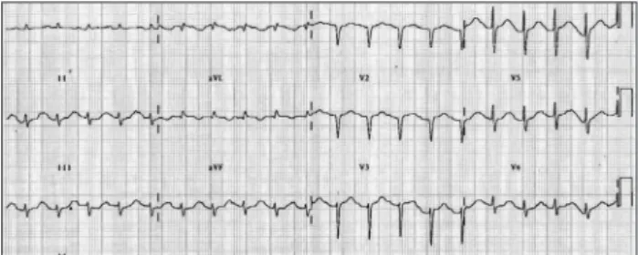
On admission, the electrocardiogram showed an abnormal pathologic Q wave in leads V1-2 and a prolonged QT interval (Fig. 1). She did not complain chest pain or shortness of breath.
The troponin T level was elevated at 0.29 ng/dL (reference lev- el, < 0.01 ng/dL), while creatine kinase (CK) and CK-MB lev-
pISSN 1975-4612/ eISSN 2005-9655 Copyright © 2011 Korean Society of Echocardiography www.kse-jcu.org http://dx.doi.org/10.4250/jcu.2011.19.3.152
CASE REPORT J Cardiovasc Ultrasound 2011;19(3):152-155
Left Ventricular Thrombus Associated with Takotsubo Cardiomyopathy:
A Cardioembolic Cause of Cerebral Infarction
Seoung-Nam Shin, MD, Kyeong Ho Yun, MD, Jum Suk Ko, MD, Sang Jae Rhee, MD, Nam Jin Yoo, MD, Nam-Ho Kim, MD, Seok Kyu Oh, MD and Jin-Won Jeong, MD
Department of Cardiovascular Medicine, Regional Cardiocerebrovascular Center, Wonkwang University Hospital, Iksan, Korea
Takotsubo cardiomyopathy, also called stress-induced cardiomyopathy, usually occurs in patients with severe emotional or physiologic stress. The prognosis is favorable, and the wall motion abnormlities normalize within weeks. However, stress-induced cardiomyopathy is rarely assosicated with left ventricular thrombus and thromboembolic complications. Here, we report a case of stress-induced cardiomyopathy with left ventricular thrombus that embolized to cause cerebral infarction.
KEY WORDS: Takotsubo cardiomyopathy · Thrombus · Cerebral infarction.
• Received: March 13, 2011 • Revised: May 24, 2011 • Accepted: August 17, 2011
• Address for Correspondence: Kyeong Ho Yun, Department of Cardiovascular Medicine, The Heart Center of Wonkwang University Hospital, 895 Muwang-ro, Iksan 570-711, Korea Tel: +82-63-859-2524, Fax: +82-63-852-8480, E-mail: [email protected]
• This is an Open Access article distributed under the terms of the Creative Commons Attribution Non-Commercial License (http://creativecommons.org/licenses/by-nc/3.0) which permits unrestricted non-commercial use, distribution, and reproduction in any medium, provided the original work is properly cited.
Fig. 1. An electrocardiogram showing an abnormal Q wave in the anterior precordial leads and a prolonged QT interval.
Stress Induced Cardiomyopathy with Embolic Stroke | Seoung-Nam Shin, et al.
153 els were normal. Transthoracic echocardiography (TTE) re-
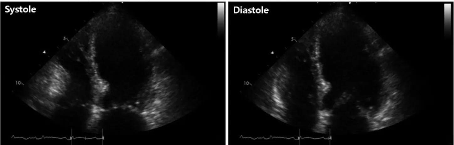
vealed that wall motion was abnormal with mid and apical akinesis, and the ejection fraction (EF) was estimated to be 12% (Fig. 2). Stress-induced cardiomyopathy was diagnosed, and supportive therapy for infection and LV dysfunction was initiated. After biliary stenting and antibiotics therapy, her
general conditions were recovered. However, ten days after ad- mission, her mental state was changed into semicoma state.
Brain magnetic resonance imaging revealed multiple brain em- bolic infarction (Fig. 3). Echocardiography was repeated to de- tect the intracardiac embolic source. TTE revealed mild im- provement of the LV systolic function (EF 44%), with a 24 ×
Fig. 2. Initial transthoracic echocardiographic image in the apical 4-chamber view showing left ventricular apical ballooning and dyskinesis.
Fig. 3. Diffusion image of magnetic resonance imaging showed multiple diffusion restrictive lesions in right cerebellar hemisphere (A), right internal capsule (B), right occipital lobe (C), and left parietal lobe (D).
C A
D B
Journal of Cardiovascular Ultrasound 19 | September 2011
154
25 mm sized thrombus in the LV apex (Fig. 4).
Low molecular weight heparin and warfarin therapy was started. Three days later, her metal state became alert. TTE performed 7 days after the start of anticoagulant therapy re- vealed a near normal LV wall motion (EF 55%) and complete resolution of the apical thrombus (Fig. 5).
Discussion
Takotsubo cardiomyopathy (stress-induced cardiomyopa- thy) is a relatively novel cardiac syndrome characterized by pe- culiar transient LV dysfunction. Approximately 1-3% of the patients with stress-induced cardiomyopathy show symptoms that initially mimic acute coronary syndrome.3)5) In this case, we did not performed coronary angiography, but we could tentatively diagnose as stress-induced cardiomyopathy because of the absent of cardiovascular symptom and no serial changes of cardiac biomarkers in septic patient.
Despite the favorable prognosis, certain serious complica- tions have been reported in patients with stress-induced cardio- myopathy, such as acute decompensated heart failure, ventricu- lar arrhythmia, LV rupture, and LV thrombus.4)7-12) Thrombus formation in such cases was probably related to transient api- cal asynergy combined with increased sympathetic activation,
which alters the coagulation cascade.
To date, the true incidence and clinical significance of LV thrombus and the related embolic outcomes in patients with stress-induced cardiomyopathy have not been fully estab- lished. Haghi et al.11) reported an 8% incidence of LV throm- bus in the study population, but a much lower incidence of accompanying embolic complications. They concluded that LV thrombus can occur at the initial presentation or any time later during the disease. In our patient, the initial echocardio- gram showed only apical ballooning and akinesia without any evidence of LV apical thrombus; however, thrombus formation occurred after a week and led to cerebral infarction. In a sys- tematic review, de Gregorio et al.13) found that among 15 Ta- kotsubo cardiomyopathy patients with ventricular thrombo- sis, 5 patients suffered from thromboembolic events, 3 of whom developed stroke. Therefore, physicians should be aware of this complication.
The current treatment of stress-induced cardiomyopathy consists of supportive care and standard treatments for LV sys- tolic dysfunction. The role of anticoagulation therapy has not yet been defined. To the best of our knowledge, there are no published guidelines for the management of stress-induced cardiomyopathy with LV thrombus. However, some reports
Fig. 4. Transthoracic echocardiographic image obtained after cerebral infarction developed, shows a 24 × 25 mm thrombus (arrow) in the left ventricular apex.
Fig. 5. Transthoracic echocardiographic image obtained after 1 week of anticoagulation therapy shows near normal left ventricular wall motion and complete resolution of the apical thrombus.
Stress Induced Cardiomyopathy with Embolic Stroke | Seoung-Nam Shin, et al.
155 mention that short-term anticoagulation therapy with heparin
and warfarin for several weeks resolved LV thrombus.8)9)11) In this case, LV thrombus was resolved after 1 week of anticoagu- lation therapy.
From our review, we conclude that patients with stress-in- duced cardiomyopathy appear to be at a significant risk for de- velopment of thrombus and subsequent stroke because of the marked apical wall motion abnormality. Thus, all patients with stress-induced cardiomyopathy should be evaluated for the presence of a ventricular thrombus. We cannot observe the LV thrombus even the patients with depressed LV function;
however, we can observe the LV thrombus even the LV func- tion was more improved in follow up echocardiography. The patient did not be followed by echocardiography during 10 days, so we cannot clarify when the LV thrombus was devel- oped. The physicians have to keep in mind that frequent echo- cardiographic follow up should be needed in the cases with stress-induced cardiomyopathy not only a period of markedly reduced LV function but also after clinically improvement.
Although no specific data exist regarding the role of antico- agulation in stress-induced cardiomyopathy, short-term antico- agulation therapy has been indicated as a treatment for patients with LV thrombus. Further research is needed to determine the true incidence of LV thrombus and the role of short-term anti- coagulant therapy in patients with stress-induced cardiomyop- athy with LV thrombus.
References
1. Lee JW, Kim JY. Stress-induced cardiomyopathy: the role of echocardiogra- phy. J Cardiovasc Ultrasound 2011;19:7-12.
2. Wittstein IS, Thiemann DR, Lima JA, Baughman KL, Schulman SP, Gerstenblith G, Wu KC, Rade JJ, Bivalacqua TJ, Champion HC. Neurohumoral features of myocardial stunning due to sudden emotion-
al stress. N Engl J Med 2005;352:539-48.
3. Prasad A. Apical ballooning syndrome: an important differential diagnosis of acute myocardial infarction. Circulation 2007;115:e56-9.
4. Akashi YJ, Tejima T, Sakurada H, Matsuda H, Suzuki K, Kawasaki K, Tsuchiya K, Hashimoto N, Musha H, Sakakibara M, Nakazawa K, Miyake F. Left ventricular rupture associated with Takotsubo cardiomy- opathy. Mayo Clin Proc 2004;79:821-4.
5. Kim DH, Bang DW, Ahn JH, Park SH, Oh HS, Yoon YJ, Hyon MS, Kim SK, Kwon YJ. Three cases of stress induced transient LV dys- function: stress induced cardiomyopathy. J Korean Soc Echocardiogr 2005;
13:83-6.
6. Sharkey SW, Windenburg DC, Lesser JR, Maron MS, Hauser RG, Lesser JN, Haas TS, Hodges JS, Maron BJ. Natural history and ex- pansive clinical profile of stress (tako-tsubo) cardiomyopathy. J Am Coll Cardiol 2010;55:333-41.
7. Iengo R, Marrazzo G, Rumolo S, Accadia M, Di Donato M, As- cione L, Tuccillo B. An unusual presentation of “tako-tsubo cardiomyopa- thy”. Eur J Echocardiogr 2007;8:491-4.
8. Yoshida T, Hibino T, Fujimaki T, Oguri M, Kato K, Yajima K, Ohte N, Yokoi K, Kimura G. Tako-tsubo cardiomyopathy complicated by apical thrombus formation: a case report. Int J Cardiol 2009;132:e120-2.
9. Tibrewala AV, Moss BN, Cooper HA. A rare case of tako-tsubo cardio- myopathy complicated by a left ventricular thrombus. South Med J 2006;
99:70-3.
10. Bybee KA, Kara T, Prasad A, Lerman A, Barsness GW, Wright RS, Rihal CS. Systematic review: transient left ventricular apical ballooning: a syndrome that mimics ST-segment elevation myocardial infarction. Ann In- tern Med 2004;141:858-65.
11. Haghi D, Papavassiliu T, Heggemann F, Kaden JJ, Borggrefe M, Suselbeck T. Incidence and clinical significance of left ventricular thrombus in tako-tsubo cardiomyopathy assessed with echocardiography. QJM 2008;
101:381-6.
12. Tobar R, Rotzak R, Rozenman Y. Apical thrombus associated with Ta- kotsubo cardiomyopathy in a young woman. Echocardiography 2009;26:
575-80.
13. de Gregorio C, Grimaldi P, Lentini C. Left ventricular thrombus forma- tion and cardioembolic complications in patients with Takotsubo-like syn- drome: a systematic review. Int J Cardiol 2008;131:18-24.