Background and Purpose Hemifacial spasm (HFS) is mostly caused by the compression of the facial nerve by cerebral vessels, but the significance of spasm severity remains unclear. We investigated the clinical significance of spasm severity in patients with HFS who underwent mi- crovascular decompression (MVD).
Methods We enrolled 636 patients with HFS who underwent MVD between May 2010 and December 2013 at Samsung Medical Center (SMC), Seoul, Korea. Subjects were divided into two groups based on spasm severity: severe (SMC grade 3 or 4) and mild (SMC grade 1 or 2).
We compared demographic, clinical, and surgical data between these two groups.
Results The severe-spasm group was older and had a longer disease duration at the time of MVD compared to the mild-spasm group. Additionally, hypertension and diabetes mellitus were more common in the severe-spasm group than in the mild-spasm group. Regarding surgi- cal findings, there were more patients with multiple offending vessels and more-severe indenta- tions in the severe-spasm group than in the mild-spasm group. Even though the surgical out- comes did not differ, the incidence of delayed facial palsy after MVD was higher in the severe- spasm group than in the mild-spasm group. Logistic regression analysis showed that severe- spasm was correlated with longer disease duration, hypertension, severe indentation, multiple offending vessels, and delayed facial palsy after MVD.
Conclusions Spasm severity does not predict surgical outcomes, but it can be used as a marker of pathologic compression in MVD for HFS, and be considered as a predictor of delayed facial palsy after MVD.
Key Words hemifacial spasm, microvascular decompression, severity, indentation, facial palsy.
Severe Hemifacial Spasm is a Predictor of Severe Indentation and Facial Palsy after Microdecompression Surgery
INTRODUCTION
Hemifacial spasm (HFS) is a movement disorder characterized by involuntary, irregular, and recurring contractions of the muscles innervated by the facial nerve.1 HFS mainly re- sults from the compression of ipsilateral facial nerves by cerebral vessels,2,3 which rarely re- solves spontaneously.4 Even though HFS is not a life-threatening disease, it can severely impair the quality of life.5,6 HFS is directly related to sleep disturbance and difficulty in performing daily activities such as reading and driving.7,8 Moreover, HFS is associated with various psychologic symptoms including low self-esteem, social embarrassment, social iso- lation, and depression.6,7
Despite the wide-ranging impact of HFS, the significance of its clinical features has not been clearly demonstrated. Only a few clinical characteristics of HFS have been reported previously and no consensus has been reached,9-11 while various surgical findings including intraoperative severity of indentation or resolution of the lateral spread response were sug- gested to be related to surgical outcome.10-13 It is particularly notable that the severity of HFS Boo Suk Naa,b
Jin Whan Choa,b Kwan Parkb,c Soonwook Kwona,b Ye Sel Kima,b Ji Sun Kima,b Jinyoung Youna,b
a Departments of Neurology and
c Neurosurgery, Samsung Medical Center, Sungkyunkwan University
School of Medicine, Seoul, Korea
b Neuroscience Center, Samsung Medical Center, Seoul, Korea
pISSN 1738-6586 / eISSN 2005-5013 / J Clin Neurol 2018;14(3):303-309 / https://doi.org/10.3988/jcn.2018.14.3.303
Received July 20, 2017 Revised January 14, 2018 Accepted January 16, 2018 Correspondence Jinyoung Youn, MD, PhD Department of Neurology, Samsung Medical Center, Sungkyunkwan University School of Medicine, 81 Irwon-ro, Gangnam-gu, Seoul 06351, Korea Tel +82-2-3410-0245 Fax +82-2-3410-0055 E-mail [email protected]
cc This is an Open Access article distributed under the terms of the Creative Commons Attribution Non-Com- mercial License (http://creativecommons.org/licenses/by-nc/4.0) which permits unrestricted non-commercial use, distribution, and reproduction in any medium, provided the original work is properly cited.
JCN
Open Access ORIGINAL ARTICLEThe Meaning of Hemifacial Spasm Severity
JCN
is a clinical barometer that can be easily assessed in a clinic, but there is no consensus on how to evaluate spasm severity.
We previously proposed the Samsung Medical Center (SMC, Seoul, Korea) grading system as a tool for evaluating HFS se- verity, and found that spasm severity was strongly correlated with quality of life.14
With the aim of elucidating the clinical meaning of HFS se- verity, we used the SMC grading system to evaluate HFS se- verity and compared clinical features, surgical findings, and outcomes between patients with severe- and mild-spasm.
METHODS
Subjects
This study was approved by the Institutional Review Board of SMC (2016-08-184). We retrospectively reviewed the med- ical records of patients with primary HFS, who 1) underwent microvascular decompression (MVD) between May 2010 and December 2013 at SMC and 2) were followed up for at least 6 months after MVD (Supplementary Fig. 1 in the online- only Data Supplement). Primary HFS is presumably caused by vascular compression of the ipsilateral facial nerve, and is the most common type among the patients who visited the clinic at SMC for HFS evaluations.15,16 Brain magnetic reso- nance imaging (MRI) was applied to all subjects to evaluate neurovascular compression and to exclude any organic causes of HFS. MVD was considered in all primary HFS patients with neurovascular compression on brain MRI, unless their physi- cal status was worse than American Society of Anesthesiolo- gists class II (normal healthy patients or subjects with mild systemic disease).17 The application of these criteria resulted in 636 patients being recruited for this study.
We divided the enrolled subjects into two groups based on HFS severity as reported previously14: mild-spasm (SMC grade 1 or 2) and severe-spasm (SMC grade 3 or 4). The 636 enrolled subjects were divided into 327 in the mild-spasm group and 309 in the severe-spasm group.
Clinical and surgical data assessment
Demographic and clinical data including age at the time of MVD, disease duration, involved side, and comorbidities were collected. The clinical course of HFS was classified into three types: periocular onset (spasm presenting first in the periocular area), perioral onset (perioral involvement at symptom onset), and hemi onset (hemifacial area at onset).18 We also assessed the presence of HFS-related tinnitus result- ing from spasm of the stapedius muscle, which is innervat- ed by facial nerves.
The surgical findings of the patients were reviewed. The offending vessels and the presence/extent of indentation of
the facial nerve root exit zone by compressing vessels were as- sessed in the surgical field and categorized into three grades:
grade 1, no or mild indentation; grade 2, moderate indenta- tion; and grade 3, severe indentation with discoloration.
The degree of indentation reflects the severity of intraoper- ative neurovascular compression.12
Surgical outcomes were assessed at the outpatient clinic at 6 months after surgery. The subjects were divided into two groups based on their surgical outcome: successful (subjects with HFS resolved by at least 90%) and unsuccessful (patients with spasm relief of up to 90%).19
Patients were divided into the following six categories of complications: delayed facial palsy, hearing loss, ear effusion, tinnitus, surgery-related (infection, wound problems, leakage of cerebral spinal fluid, or hemorrhage), and other. Facial palsy with a grade of III or more on the House-Brackmann grading system was regarded as significant.20 Pure-tone audi- ometry (PTA) and the Speech Discrimination Score (SDS) were evaluated before and after MVD (usually at 3–10 days after surgery).21 A decrease of more than 15 dB from the postoperative bone-conduction average PTA threshold (at 500, 1,000, 2,000, and 3,000 Hz) or a decrease of more than 20% in the SDS relative to the baseline for each patient was defined as hearing loss.22,23
Statistical analyses
All data are presented as mean±standard-deviation values.
The significance of differences between the mild- and se- vere-spasm groups was evaluated using the independent Student’s t-test or the Mann-Whitney U test for continuous and ordinal variables, while Pearson’s chi-square test or Fish- er’s exact test was used for categorical variables. To identify variables related to HFS severity, multivariate analysis was carried out with a logistic regression model. The mild- and severe-spasm groups were included as the dependent vari- ables, whereas age, involved side, disease duration, clinical course, hypertension (HTN), diabetes mellitus (DM), heart/
lung disease, stroke, complications (facial palsy, hearing loss, ear effusion, tinnitus, surgery-related, and other), and out- come were included as controlled variables. The logistic re- gression results are presented using odds ratios with 95%
confidence intervals. Probability values of p<0.05 were con- sidered statistically significant. All statistical analyses were performed using commercially available software (PASW for Windows, version 18.0, SPSS Inc., Chicago, IL, USA).
Na BS et al.
JCN
RESULTS
Comparison of clinical features between the mild- and severe-spasm groups
The demographic and clinical data of the mild- and severe- spasm groups are presented in Table 1. The severe-spasm group was older at the time of MVD and had a longer dis- ease duration than the mild-spasm group. The age at symp- tom onset did not differ significantly between the mild-spasm group (46.2±11.1 years) and the severe-spasm group (44.7±
9.9 years, p=0.073). There was also no significant intergroup difference in any of the other demographic data or HFS clini- cal features. Of the examined comorbidities, HTN, and DM were more common in the severe-spasm group than in the mild-spasm group.
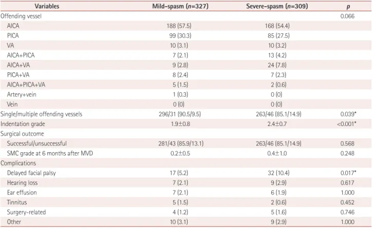
Comparison of surgical findings and outcomes between the mild- and severe-spasm groups
While there was no significant difference in the number of offending vessels, multiple offenders were more common in the severe-spasm group than in the mild-spasm group (Ta- ble 2). In addition, indentation was more severe in the se- vere-spasm group than in the mild-spasm group.
A successful outcome after MVD was reported in 86.9% of the 636 enrolled subjects, with no difference in rate of suc- cessful outcome between the mild- and severe-spasm groups.
There were more complications in the severe-spasm group
(18.1%) than in the mild-spasm group (14.4%), but the dif- ference was not statistically significant. The complication of delayed facial palsy was more common in the severe-spasm group than in the mild-spasm group, while there were no in- tergroup differences in any of the other complications.
Multivariate analysis of the relationship between HFS severity and other variables
We performed logistic regression analysis to identify variables associated with HFS severity. The mild- and severe-spasm groups were included as the dependent variables, while age, involved side, disease duration, clinical course, HTN, DM, heart/lung disease, stroke, complications (facial palsy, hearing loss, ear effusion, tinnitus, surgery-related, and other), and outcome were included as controlled variables (Table 3). Se- vere-spasm was significantly associated with longer disease duration, HTN, multiple offending vessels, severe indenta- tion, and more delayed facial palsy after MVD.
DISCUSSION
This is the first study to investigate the clinical implications of HFS severity in patients with HFS. Although many previ- ous studies have examined predictors of the prognosis of HFS after MVD, they predominantly investigated intraoperative findings rather than clinical features.10-13 Unlike previous studies, we focused on the clinical characteristics of HFS. Our Table 1. Comparison of demographic data between the mild- and severe-spasm groups
Variables Mild-spasm (n=327) Severe-spasm (n=309) p
Age at MVD (years) 49.2±11.0 52.1±9.7 <0.001*
Sex (male/female) 78/249 (23.9/76.1) 87/222 (28.2/71.8) 0.240
Age at onset (years) 46.2±11.1 44.7±9.9 0.073
Disease duration (years) 3.1±2.6 7.5±5.6 <0.001*
Side (right/left) 142/185 (43.4/56.6) 157/152 (50.8/49.2) 0.068
Clinical course (onset) 0.324
Periocular 319 (97.6) 299 (96.8)
Perioral 2 (0.6) 0 (0)
Hemifacial 6 (1.8) 10 (3.2)
SMC grade 1.8±0.4 3.31±0.4 <0.001*
Tinnitus related to HFS 83 (25.4) 87 (28.2) 0.473
ASA class 1.3±0.5 1.5±0.5 0.001*
Comorbidities
Hypertension 72 (22.0) 98 (31.7) 0.007*
Diabetes mellitus 8 (2.4) 19 (6.1) 0.029*
Stroke 1 (0.3) 2 (0.6) 0.614
Heart disease 18 (5.5) 24 (7.8) 0.267
Lung disease 26 (8.0) 33 (10.7) 0.274
Data are n (%) or mean±standard-deviation values.
*p<0.05.
ASA: American Society of Anesthesiologists, HFS: hemifacial spasm, MVD: microvascular decompression, SMC: Samsung Medical Center.
The Meaning of Hemifacial Spasm Severity
JCN
reasoning was that the treatment modality should be decided based on both clinical features and intraoperative findings.
Our patients with severe HFS had a longer disease duration and more-severe indentation, and were also more likely to have multiple offending vessels and HTN. A particularly in- teresting finding was that among the surgical complications examined, delayed facial palsy was related to spasm severity whereas the rate of successful outcomes was not.
HFS severity has previously been shown to be indepen- dently related to quality of life,24 but the role of spasm severi- ty in patients with HFS has not been investigated. Only one study found that severe HFS was related to headaches in pa- tients with HFS.25 Since HFS can present with various clini-
cal characteristics, such as clonic or tonic spasms, differences in the involved area or fluctuations therein could make it dif- ficult to assess all spasms on a single grading scale. Moreover, there is currently no consensus on how to evaluate HFS se- verity. A previous study suggested using the SMC grade as a measure of HFS severity, and found this to be strongly corre- lated with the quality of life in patients with HFS.14 Addition- ally, higher SMC grade was associated with longer disease duration, which is in accordance with our results. However, none of the previous studies directly evaluated the relationships between spasm severity and surgical findings, outcomes, and complications.
We found that HTN and DM were more common in the se- Table 2. Comparison of surgical findings and outcomes between the mild- and severe-spasm groups
Variables Mild-spasm (n=327) Severe-spasm (n=309) p
Offending vessel 0.066
AICA 188 (57.5) 168 (54.4)
PICA 99 (30.3) 85 (27.5)
VA 10 (3.1) 10 (3.2)
AICA+PICA 7 (2.1) 13 (4.2)
AICA+VA 9 (2.8) 24 (7.8)
PICA+VA 8 (2.4) 7 (2.3)
AICA+PICA+VA 5 (1.5) 2 (0.6)
Artery+vein 1 (0.3) 0 (0)
Vein 0 (0) 0 (0)
Single/multiple offending vessels 296/31 (90.5/9.5) 263/46 (85.1/14.9) 0.039*
Indentation grade 1.9±0.8 2.4±0.7 <0.001*
Surgical outcome
Successful/unsuccessful 281/43 (85.9/13.1) 263/46 (85.1/14.9) 0.568
SMC grade at 6 months after MVD 0.2±0.5 0.4±1.0 0.248
Complications
Delayed facial palsy 17 (5.2) 32 (10.4) 0.017*
Hearing loss 7 (2.1) 9 (2.9) 0.617
Ear effusion 7 (2.1) 6 (1.9) 1.000
Tinnitus 5 (1.5) 2 (0.6) 0.452
Surgery-related 4 (1.2) 5 (1.6) 0.746
Other 10 (3.1) 9 (2.9) 1.000
Data are n (%) or mean±standard-deviation values.
*p<0.05.
AICA: anterior inferior cerebellar artery, MVD: microvascular decompression, PICA: posterior inferior cerebellar artery, VA: vertebral artery.
Table 3. Results of a multivariate analysis to identify variables associated with the severe-spasm group
Variables OR (95% CI) p
Disease duration, annual increase 1.380 (1.286–1.482) <0.001
Indentation, per severity 1.807 (1.389–2.351) <0.001
Multiple offending vessels (compared with a single offending vessel) 1.845 (1.013–3.361) 0.045
Delayed facial palsy after MVD 2.455 (1.187–5.075) 0.015
Hypertension 1.623 (1.011–2.607) 0.045
Age, involved side, disease duration, clinical course, comorbidities (hypertension, diabetes mellitus, heart/lung disease, and stroke), complications (de- layed facial palsy, hearing loss, ear effusion, tinnitus, surgery-related, and other), and outcome were included as controlled variables.
CI: confidence interval, MVD: microvascular decompression, OR: odds ratio.
Na BS et al.
JCN
vere-spasm group than in the mild-spasm group. Moreover, HTN was significantly associated with severe-spasm in the regression analysis. Similarly, when we compared clinical fea- tures between elderly patients with HFS and younger patients in a previous study, the elderly patients with HFS showed more severe-spasms and were more likely to have HTN and DM.18 Considering that the age at MVD was not associated with spasm severity in the multivariate analysis performed in the present study, the severity of HFS might not be directly related to age, but rather to other conditions such as HTN or disease duration. Previous studies have also focused on the as- sociation between vascular dolichoectasia and HFS, conclud- ing that dolichoectasia is usually associated with HTN.26,27 However, conflicting results have been obtained regarding the relationship between HFS and HTN,28-30 and no study has focused on HTN and spasm severity in patients with HFS. Future studies investigating the association between HTN and HFS pathophysiology will help to elucidate this re- lationship. Regarding DM, there have been only a few report- ed cases supporting the possibility of a relationship between hyperglycemia and HFS,31,32 and DM was not associated with spasm severity in the multivariate analysis in the present study; caution is therefore required when interpreting these results.
When we compared surgical findings between the mild- and severe-spasm groups, we found that severe indentation and multiple offending vessels were associated with severe spasms. Although the surgical outcomes did not differ be- tween the severe- and mild-spasm groups in our study, the degree of indentation and multiple offenders could be im- portant because many previous studies have found these sur- gical findings to be associated with surgical outcome.11-13,33 Moreover, indentation has been reported to be associated with delayed facial palsy and disease duration,34 and we also found that HFS severity was independently related to dis- ease duration, indentation degree, multiple offending vessels, and delayed facial palsy. Based on our results, spasm severity could be regarded as a biomarker of the degree of pathologic compression in patients with HFS.
Another particularly interesting finding of the present study was that the severe-spasm group was independently associat- ed with delayed facial palsy. Although the etiology of delayed facial palsy has not been clearly elucidated, it has been shown to be associated with a higher degree of indentation and lon- ger disease duration.34 Considering that severe indentation with longer disease duration could aggravate demyelination of the facial nerve, more time would be needed to repair the damage after the offending vessel is removed.34 Additionally, more delayed facial palsy has been reported in HFS patients with multiple offending vessels, although this difference was
not significant.34 That study suggested that the presence of multiple offenders hinders surgery, resulting in a greater risk of facial nerve damage. However, we found that severe-spasm was independently associated with multiple offenders and delayed facial palsy, indicating that there might be direct re- lationships between severe-spasm, multiple offending vessels, and delayed facial palsy. Various other risk factors for delayed facial palsy after MVD have also been suggested,35-37 and so further studies are required to elucidate the pathophysiology of delayed facial palsy after MVD.
Besides delayed facial palsy, spasm severity was not associ- ated with any other complication or surgical outcome in our study, in contrast to previous studies finding indentation and multiple offending vessels to be associated with surgical out- come.11-13,33 This discrepancy can be attributed to the effects of various factors including age, disease duration, comorbid- ities, and psychologic status on surgical outcomes and com- plications.9,18 Although we observed that facial palsy was de- layed in the severe-spasm group, the complication rate was still low and so MVD could be an effective treatment option for patients with HFS. Since treatment modalities are selected and complications are predicted based on clinical or laborato- ry findings, rather than on intraoperative findings, more stud- ies focusing on the clinical features of HFS—like the present study—are important for providing doctors with the best information possible for making appropriate decisions.
MVD was performed in 626 patients with primary HFS in this study, of whom 554 (85.5%) reported complete relief or minimal symptoms. The rate of successful outcomes in the present study was slightly lower than those in our previ- ous studies.12,13,38 However, considering the delayed cure after MVD and the relatively short follow-up period (6 months af- ter MVD) in this study, our results are comparable with those of previous studies. Furthermore, we used different tools to evaluate the surgical outcomes compared to previous studies due to the lack of a consensus on how to assess surgical out- comes. The complication rates in the current study were similar to those in a previous study.38
This study was subject to some limitations. First, we only enrolled patients with HFS who underwent MVD and were followed up for at least 6 months. Considering that the cure was delayed after MVD, the improvements might have been greater and the complication rate lower if we had used a longer follow-up period.13 However, since we wanted to fo- cus on the clinical characteristics of HFS in as many subjects as possible, we assessed surgical outcomes and complica- tions for only 6 months after MVD. Second, the severe-spasm group was older than the mild-spasm group. As we reported previously, elderly patients may exhibit specific characteristics for HFS.18 However, a previous study found no difference in
The Meaning of Hemifacial Spasm Severity
JCN
the prevalence of delayed facial palsy between elderly and younger patients with HFS. Moreover, to minimize possible confounding effects of age or comorbidities, we used multi- variate logistic regression analysis in this study. Third, recall bias might have been present in the clinical history because most of the collected data were self-reported and the overall disease duration was 5.2±4.9 years.
In conclusion, although MVD is an effective treatment op- tion for patients with severe HFS as well as for patients with mild-spasm, surgeons should carefully monitor patients with severe HFS due to the greater risk of delayed facial palsy. In addition, spasm severity could be used as a marker of patho- logic compression in patients with HFS who need MVD.
Supplementary Materials
The online-only Data Supplement is available with this arti- cle at https://doi.org/10.3988/jcn.2018.14.3.303.
Conflicts of Interest
The authors have no financial conflicts of interest.
REFERENCES
1. Wang A, Jankovic J. Hemifacial spasm: clinical findings and treatment.
Muscle Nerve 1998;21:1740-1747.
2. Gardner WJ. Concerning the mechanism of trigeminal neuralgia and hemifacial spasm. J Neurosurg 1962;19:947-958.
3. Jannetta PJ. Observations on the etiology of trigeminal neuralgia, hemifacial spasm, acoustic nerve dysfunction and glossopharyngeal neuralgia. definitive microsurgical treatment and results in 117 pa- tients. Neurochirurgia 1977;20:145-154.
4. Mauriello JA Jr, Leone T, Dhillon S, Pakeman B, Mostafavi R, Yepez MC. Treatment choices of 119 patients with hemifacial spasm over 11 years. Clin Neurol Neurosurg 1996;98:213-216.
5. Reimer J, Gilg K, Karow A, Esser J, Franke GH. Health-related quality of life in blepharospasm or hemifacial spasm. Acta Neurol Scand 2005;
111:64-70.
6. Tan EK, Fook-Chong S, Lum SY, Thumboo J. Validation of a short disease specific quality of life scale for hemifacial spasm: correlation with SF-36. J Neurol Neurosurg Psychiatry 2005;76:1707-1710.
7. Heuser K, Kerty E, Eide PK, Cvancarova M, Dietrichs E. Microvascu- lar decompression for hemifacial spasm: postoperative neurologic fol- low-up and evaluation of life quality. Eur J Neurol 2007;14:335-340.
8. Tan EK, Fook-Chong S, Lum SY, Lim E. Botulinum toxin improves quality of life in hemifacial spasm: validation of a questionnaire (HFS- 30). J Neurol Sci 2004;219:151-155.
9. Jin Y, Zhao C, Su S, Zhang X, Qiu Y, Jiang J. Residual hemifacial spasm after microvascular decompression: prognostic factors with emphasis on preoperative psychological state. Neurosurg Rev 2015;38:567-572.
10. Thirumala PD, Shah AC, Nikonow TN, Habeych ME, Balzer JR, Crammond DJ, et al. Microvascular decompression for hemifacial spasm: evaluating outcome prognosticators including the value of in- traoperative lateral spread response monitoring and clinical charac- teristics in 293 patients. J Clin Neurophysiol 2011;28:56-66.
11. Lv MY, Deng SL, Long XF, Liu ZL. Long-term outcome of microvas- cular decompression for hemifacial spasm. Br J Neurosurg 2017;31:322- 12. Kim HR, Rhee DJ, Kong DS, Park K. Prognostic factors of hemifacial 326.
spasm after microvascular decompression. J Korean Neurosurg Soc
2009;45:336-340.
13. Jo KW, Kong DS, Park K. Microvascular decompression for hemifa- cial spasm: long-term outcome and prognostic factors, with empha- sis on delayed cure. Neurosurg Rev 2013;36:297-301.
14. Lee JA, Jo KW, Kong DS, Park K. Using the new clinical grading scale for quantification of the severity of hemifacial spasm: correlations with a quality of life scale. Stereotact Funct Neurosurg 2012;90:16-19.
15. Yaltho TC, Jankovic J. The many faces of hemifacial spasm: differen- tial diagnosis of unilateral facial spasms. Mov Disord 2011;26:1582- 1592.
16. Lu AY, Yeung JT, Gerrard JL, Michaelides EM, Sekula RF Jr, Bulsara KR. Hemifacial spasm and neurovascular compression. Scientific- WorldJournal 2014;2014:349319.
17. Doyle DJ, Garmon EH. American Society of Anesthesiologists Classifi- cation (ASA Class). Treasure Island (FL): StatPearls Publishing LLC, 2017.
18. Youn J, Kwon S, Kim JS, Jeong H, Park K, Cho JW. Safety and effec- tiveness of microvascular decompression for the treatment of hemifa- cial spasm in the elderly. Eur Neurol 2013;70:165-171.
19. Huh R, Han IB, Moon JY, Chang JW, Chung SS. Microvascular de- compression for hemifacial spasm: analyses of operative complica- tions in 1582 consecutive patients. Surg Neurol 2008;69:153-157.
20. House JW, Brackmann DE. Facial nerve grading system. Otolaryngol Head Neck Surg 1985;93:146-147.
21. Jo KW, Kim JW, Kong DS, Hong SH, Park K. The patterns and risk factors of hearing loss following microvascular decompression for hemifacial spasm. Acta Neurochir 2011;153:1023-1030.
22. Chen YC, Thaler D, Nixon PD, Stern CE, Passingham RE. The func- tions of the medial premotor cortex. II. the timing and selection of learned movements. Exp Brain Res 1995;102:461-473.
23. Park K, Hong SH, Hong SD, Cho YS, Chung WH, Ryu NG. Patterns of hearing loss after microvascular decompression for hemifacial spasm. J Neurol Neurosurg Psychiatry 2009;80:1165-1167.
24. Rudzińska M, Wójcik M, Malec M, Grabska N, Szubiga M, Hartel M, et al. Factors affecting the quality of life in hemifacial spasm patients.
Neurol Neurochir Pol 2012;46:121-129.
25. Peeraully T, Tan SF, Fook-Chong SM, Prakash KM, Tan EK. Headache in hemifacial spasm patients. Acta Neurol Scand 2013;127:e24-e27.
26. Kim KJ, Kim JM, Bae YJ, Bae HJ, Jeon B, Kim JH, et al. The associa- tion between vertebrobasilar dolichoectasia and hemifacial spasm.
Parkinsonism Relat Disord 2016;32:54-59.
27. Crabtree GS, Gish D, Goldberg D. Hemifacial spasm in a patient with basilar artery dolichoectasia caused by uncontrolled hyperten- sion. J Community Hosp Intern Med Perspect 2016;6:32686.
28. Leong JL, Li HH, Chan LL, Tan EK. Revisiting the link between hy- pertension and hemifacial spasm. Sci Rep 2016;6:21082.
29. Oliveira LD, Cardoso F, Vargas AP. Hemifacial spasm and arterial hy- pertension. Mov Disord 1999;14:832-835.
30. Rudzińska M, Wójcik-Pędziwiatr M, Malec-Litwinowicz M, Grabska N, Hartel M, Flak M, et al. Is hypertension a risk factor of hemifacial spasm? Neurol Neurochir Pol 2016;50:69-74.
31. Bandyopadhyay SK, Dutta A. Hemifacial spasm complicating dia- betic ketoacidosis. J Assoc Physicians India 2005;53:649-650.
32. Chakrabarti S. Hemifacial spasm due to non-ketotic hyperglycemia.
Int J Adv Med Health Res 2014;1:90-92.
33. Li S, Feng B, Xie C, You C, Wei X, Zheng X. Good surgical outcomes of hemifacial spasm patients with obvious facial nerve indentation and color change. World Neurosurg 2016;92:218-222.
34. Hua Z, Da TY, Hui WX, Tingting Y, Jin Z, Yan Y, et al. Delayed facial palsy after microvascular decompression for hemifacial spasm. J Cra- niofac Surg 2016;27:781-783.
35. Liu LX, Zhang CW, Ren PW, Xiang SW, Xu D, Xie XD, et al. Progno- sis research of delayed facial palsy after microvascular decompression for hemifacial spasm. Acta Neurochir 2016;158:379-385.
36. Prasad GL, Kumar V, Menon G. Delayed facial palsy after microvascu-
Na BS et al.
JCN
lar decompression: report of two cases. J Neurosci Rural Pract 2017;8:
461-465.
37. Furukawa K, Sakoh M, Kumon Y, Teraoka M, Ohta S, Ohue S, et al.
[Delayed facial palsy after microvascular decompression for hemifacial spasm due to reactivation of varicella-zoster virus]. No Shinkei Geka
2003;31:899-902.
38. Lee MH, Jee TK, Lee JA, Park K. Postoperative complications of mi- crovascular decompression for hemifacial spasm: lessons from expe- rience of 2040 cases. Neurosurg Rev 2016;39:151-158.