Review Article
Current status in the management of uterine corpus cancer in Korea
Nan-Hee Jeong, Jong-Min Lee, Seon-Kyung Lee
Department of Obstetrics and Gynecology, Kyung Hee University School of Medicine, Seoul, Korea
Uterine corpus cancer has increased in prevalence in Korean women over the last decade. Recently, elegant studies have been reported from many institutes. To improve treatment strategies, a review of our own data is warranted.
This work will discuss the risks and prognostic factors for uterine corpus cancer, and the radiologic evaluation, prediction of lymph node metastasis, systematic lymphadenectomy, minimally invasive surgery, ovarian-saving surgery, fertility-sparing treatment, and adjuvant treatment in women with uterine cancer.
Key Words: Uterine neoplasms, Review
Received June 3, 2010, Accepted June 27, 2010 Correspondence to Jong-Min Lee
Department of Obstetrics and Gynecology, East-West Neo Medical Center, Kyung Hee University, 149 Sangil-dong, Gangdong-gu, Seoul 134-890, Korea
Tel: 82-2-440-6140, Fax: 82-2-440-7894 E-mail: [email protected]
This research received the Grand Prize from the Korean Research Foundation of Gynecologic Cancer at the 25th Annual Meeting of the Korean Society of Gynecologic Oncology and Colposcopy.
INTRODUCTION
Uterine corpus cancer is one of the most common female genital tract malignancies in many developed countries and has become more prevalent in Korean women over the last decade.1,2 Uterine corpus cancer accounts for approximately 4% of all newly diagnosed cancers and approximately 1.7% of cancer deaths in women worldwide. In 2002, an estimated 199,000 new cases of uterine corpus cancer and 50,000 cases of cancer-related deaths were expected worldwide.3 The asso- ciation between obesity and increased risk of uterine corpus cancer is more significant than any other obesity-related can- cer in reviews based on Western populations.4-8 Considering that the obesity is on the rise and uterine corpus cancer has be- come more prevalent, accounting for 3.7% of all newly diag- nosed cancers in Korean women in 2005,9 it is anticipated that uterine corpus cancer will become a more significant public health problem in the future in Korea.
Based on the American College of Obstetrics and Gynecolo- gists (ACOG) recommendations and the newly revised surgi- cal staging system by the International Federation of Gyneco-
logy and Obstetrics (FIGO), the cornerstone of treatment for uterine corpus cancer patients is surgery, including peritoneal cytology, hysterectomy, bilateral salpingo-oophorectomy, lymphadenectomy, and complete resection of all extrauterine disease.10,11 Nevertheless, debate continues regarding surgi- cal staging procedures, especially in terms of the role of, can- didates for, and extent of lymphadenectomy.
The data in support of treatment strategies of uterine corpus cancer are conflicting. For example, several retrospective stu- dies have suggested a therapeutic benefit associated with lym- phadenectomy in early stage uterine corpus cancer,12,13 but re- cent randomized trials have failed to prove a survival advan- tage.14,15 Moreover, several randomized trials have shown no evidence of benefit for adjuvant radiation for early stage ute- rine corpus cancer in terms of overall survival.16,17 Thus, we are still at chaos with the limited randomized controlled trials and sometimes, contradictory treatment guidelines for ute- rine corpus cancer.
Considering the ethnic and epidemiologic difference in the susceptibility to uterine corpus cancer, and the relatively low incidence of uterine corpus cancer compared to cervical can- cer, we need to analyze and understand the results of Korean studies. Recently, the interest of Korean researchers in ute- rine corpus cancer has increased and there have been many clinically meaningful results. Thus, this article reviews the current status of the management of uterine corpus cancer in Korea based on the recently published results.
RISK FACTORS
Nulliparity, obesity, unopposed estrogen use, late meno- pause, and diabetes are the well-established clinical risk fac-
Table 1. Association of genetic polymorphisms with uterine corpus cancer risk in Korean women
Author, year Cases (n) Controls (n) Gene, position Genotype OR (95% CI)
Roh (2004)26 95 285 p53, P72R Arg/Pro+Pro/Pro vs. Arg/Arg 3.56 (2.10-6.04)
p21, S31R Ser/Ser vs. Arg/Arg+Arg/Ser 2.68 (1.59-4.51)
Kang (2005)27 77 154 cyclin D1, G870A AA vs. GG+AG 2.63 (1.04-2.66)
Jo (2007)28 102 302 ERCC1, C19007T CT+TT vs. CC 0.91 (0.57-1.44)
Tong (2009)29 125 302 HER-2, rs1801200 GA vs. AA 1.13 (0.59-2.16)
HER-2, rs1810132 TT vs. CC 0.97 (0.44-2.14)
HER-2, rs2517951 TT vs. CC 1.40 (0.67-2.00)
HER-2, rs1058808 GG vs. CC 1.39 (0.68-2.87)
OR: odds ratio, CI: confidence interval, ERCC1: excision repair cross-complementing group 1.
tors for the development of uterine corpus cancer.18 Most of these risk factors are primarily associated with prolonged ex- posure to unopposed estrogen. Obesity is an important risk factor for uterine corpus cancer. In Western populations, obe- sity has been associated with a 2- to 5-fold increase in endo- metrial cancer risk in both pre- and post-menopausal women, because extra-ovarian estrogen derived from androgens ar- omatized in adipose tissue plays an important role in the de- velopment of uterine corpus cancer.4 In Korea, Jeong et al.19 studied the effect of obesity on uterine corpus cancer risk in an age-matched case-control study, and found that both obese (BMI≥25 kg/m2) and overweight (23 kg/m2≤BMI<25 kg/m2) women had a significantly increased risk for endome- trioid uterine cancer compared to non-obese women (BMI
<23 kg/m2).
Recently, antioxidant micronutrients have received atten- tion as an environmental and lifestyle risk factor for uterine corpus cancer.20,21 Two small Korean case-control studies have reported that plasma levels of β-carotene, lycopene, fo- late, and vitamin B12 are inversely associated with uterine corpus cancer risk;22,23 however, they failed to prove an associ- ation between the intake of folic acid and vitamin B12 and dis- ease risk.23
Studies involving genetic single nucleotide polymorphisms (SNPs) for uterine corpus cancer have helped to explain the modest risk and differences in individual cancer susceptibility.
Candidate genes may be involved in DNA damage repair, es- trogen metabolism, carcinogen metabolism, cell-cycle control, apoptosis, and steroid receptor activation pathways.24 In this way, several results have indicated common genetic poly- morphisms in Korean women that augment the effects of risk- factor exposure, such as p53, p21, CCND1, ERCC1, and HER- 2 genes; the results are summarized in Table 1.25-29 p53, which is one of the representative tumor suppressor genes, and p21, which is a downstream mediator of p53, were examined by Roh et al.26 They found that the p53 genotypes containing the Pro allele at codon 72 and homozygous carriers of the p21 Ser allele at codon 31 were significantly associated with an in- creased risk of uterine corpus cancer. CCND1 is known as the gene involved in the normal cell cycle (the product of CCND1 is cyclin D1). An increased risk of uterine corpus cancer for
the AA genotype of the G870A SNP in CCND1 was described by Kang et al.27 One study involving the C19007T SNP of ex- cision repair cross-complementing group 1 (ERCC1) showed no evidence of an association with uterine corpus cancer risk.28 Two interesting studies reported that HER-2 SNPs did not significantly affect the risk for uterine corpus cancer and these results were repeated with an additional analysis in rela- tion to body mass index and patient age.25,29
These studies were characterized by a single ethnicity, hos- pital-based case-control design, relatively small sample size, selection of hospital controls, and adjustment of clinical varia- bles (eg, age, body mass index, reproductive history, and exog- enous hormone use). Therefore, these findings remain to be confirmed by an additional population-based study.
PREDICTION OF LYMPH NODE METASTASIS Lymph node (LN) metastasis, one of the most significant prognostic factors, was observed in approximately 13% of pa- tients with uterine corpus cancer in a multicenter Korean study.30 Prediction of LN metastasis represents critical steps for planning the extent of surgery. To avoid unnecessary LN dissection and the associated surgical morbidity, several mo- dalities, such as CT, MRI, PET/CT, and serum CA-125 levels, have been suggested for the prediction of LN metastasis (Table 2).31-37
Selman et al.38 conducted a meta-analysis to evaluate the ac- curacy of radiologic imaging to predict LN metastases, and found that MRI and sentinel node biopsy were more accurate than CT scanning. Similarly, there have been several efforts involving the performance of MRI for prediction of LN meta- stasis and deep myometrial invasion. Considering the clinical significance of LN metastasis, the tests to predict LN meta- stasis should have a high sensitivity and negative predictive value. The sensitivity and negative predictive value of MRI for LN metastasis were reported as 45-78% and 91-95%, respec- tively (Table 2). This uncertainty might be caused by difficulty in differentiating metastatic nodes from hyperplastic nodes.39 Thus, we suggest that MRI shows unsatisfactory results in the prediction of LN metastasis and cannot replace surgical staging. This error with respect to LN metastasis was also ob-
Table 2. Performance of magnetic resonance imaging (MRI), positron emission tomography/computed tomography (PET/CT), and CA-125 in prediction of lymph node metastasis
Author, year Modality Cases (n) Se (%) Sp (%) PPV (%) NPV (%) Accuracy (%)
Chung (2007)31 MRI 120 68.8 97.1 78.6 95.3 93.3
Yoo (2009)33 MRI* 99 77.7 85.6 35.0 94.7 -
Han (2010)34 MRI or CT 297 53.3 92.9 45.7 94.7 -
Cho (2010)35 MRI 166 45.0 80.8 24.3 91.4 -
Park (2008)32 MRI 53 46.2 87.9 28.6 94.0 83.9
PET/CT 53 69.2 90.3 42.9 96.6 88.3
Choi (2005)36 CA-125† 42 100 87.2 37.5 100 -
Chung (2006)37 CA-125‡ 92 61.5 94.9 - - -
Han (2010)34 CA-125§ 219 78.6 78.0 34.4 96.1 -
Se: sensitivity, Sp: specificity, PPV: positive predictive value, NPV: negative predictive value.
*Only pelvic nodes were included. †The cut-off value was determined to be 50 U/mL. ‡The cut-off value was determined to be 28.5 U/mL.
§The cut-off value was determined to be 28 U/mL (if ≥50 years of age) and 20 U/mL (if ≤49 years of age).
Table 3. Performance of magnetic resonance imaging (MRI) in prediction of myometrial invasion
Author, year Modality Cases (n) Se (%) Sp (%) PPV (%) NPV (%) Accuracy (%)
Kim (1995)41 MRI 26 90 88 - - 89
Ahn (2006)42 MRI 43 87.5 85.7 58.3 96.8 -
Chung (2007)31 MRI 120* 50.6 89.2 91.3 44.6 62.5
Yoo (2009)33 MRI 99 46.6 84.5 35 89.8 -
Hwang (2009)43 MRI 53 50.0 89.7 63.6 83.3 79.2
Suh (2009)44 MRI 301* 68.8 74.4 86.9 49.2 59.2
MRI 301 55.9 92.4 50.0 93.9 88.0
Han (2010)34 MRI 219 36.4 88.3 25.8 93.6 -
Cho (2010)35 MRI 182 66.7 95.2 77.4 92.1 -
Se: sensitivity, Sp: specificity, PPV: positive predictive value, NPV: negative predictive value.
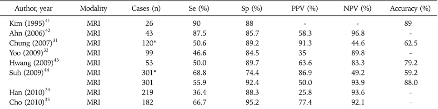
*Both deep and superficial myometrial invasion was considered; others only included deep myometrial invasion.
served in clinical stage I disease (45.0% sensitivity, n=166), thus some investigators warned that an incorrect decision might be made for a number of patients based on a pre-oper- ative MRI.35 MRI was not effective in the detection of para- aortic node metastasis as well. Park et al.32 described that 1 of 7 positive para-aortic LN areas were detected by MRI (sensi- tivity, 14.3%), compared to 5 of 6 pelvic node areas (sensitiv- ity, 83%).32 Yoo et al.33 reported that MRI failed to detect any of 3 positive para-aortic nodes among 99 patients. On the oth- er hand, Park et al.32 compared PET/CT with MRI for accuracy in detecting retroperitoneal metastasis in 53 women, and found that PET/CT had a higher sensitivity than MRI in de- tecting para-aortic or pelvic node metastasis, although the dif- ference was insignificant (69.2% vs. 46.2%, p=0.250).
The sensitivity of CA-125 had a wide range of variation (Table 2). Nevertheless, CA-125 was suggested as an additive tool to predict LN metastasis. Han et al.34 examined the fol- lowing 6 pre-operative factors to predict LN metastasis: age ≥ 55 years; serum CA-125 level (≥20 U/mL if <50 years of age;
≥28 U/mL if ≥50 years of age); non-endometrioid histology;
grade 3; metastatic LN assessed by pelvic MRI or CT; and deep myometrial invasion assessed by pelvic MRI only. In an analy- sis of 300 women, they reported 100% sensitivity and neg-
ative predictive value combining the 6 pre-operative tests.
Additionally, LN metastasis is highly correlated with the depth of myometrial invasion,30,40 and commonly used for the assessment of the risk of LN metastasis (Table 3).31,33-35,41-44
Prediction of myometrial invasion is also crucial in decisions as to whether conservative treatment should be administered to uterine corpus cancer patients. In this case, MRI is the pre- ferred imaging modality in the prediction of myometrial in- vasion (Table 3). However, the sensitivity (36-90%) and neg- ative predictive values (83-94%) of MRI in the detection of deep myometrial invasion was not satisfactory. Underestima- tion of myometrial invasion was associated with an isotense junctional zone-to-myometrium (p<0.001), and the pres- ence of polypoid tumors (p=0.037) on MRI.31 In particular, false-positive cases were frequently found in the detection of any degree of myometrial invasion, and these results gives us attention in performing fertility-sparing treatment as the pri- mary therapy of uterine corpus cancer.44
As a result, previous results for the prediction of LN meta- stasis and myometrial invasion are not sufficiently reliable, thus leaving room for further study. On the other hand, the risk of LN metastasis based on pathological findings, such as myometrial invasion and tumor grade in 834 Korean patients
with endometrioid uterine cancers, were analyzed by Lee et al.30 Of the 215 patients with no myometrial invasion and tu- mor grade I/II, only 1 patient (0.5%) had LN metastasis and the other patients had significant risk, that is, at least more than 3.5%. Therefore, all uterine corpus cancers, except endo- metrioid uterine cancers with no myometrial invasion and tu- mor grade 1-2, would require complete pelvic and para-aortic lymphadenectomy for surgical staging.
ROLE OF SYSTEMATIC LYMPHADENECTOMY Systematic lymphadenectomy has been reported to have an important role in the surgical staging of uterine corpus cancer.45 It is used as a prognostic indicator and suggests guid- ance for adjuvant treatment. Retrospective studies have re- ported that more extensive lymphadenectomy is associated with improved survival in patients with uterine corpus cancer, including high-risk, early-stage disease.13,46 However, the therapeutic value of systematic lymphadenectomy for stage I uterine cancer is not evident in all studies. Data from the Surveillance, Epidemiology, and End Results (SEER) pro- gram also showed that lymphadenectomy is associated with an improved disease-specific survival in stage I disease with tumor grade 3.47 On the other hand, systematic lymphadenec- tomy was not recommended as a routine procedure for ther- apeutic purposes in patients with stage I disease in the A Study in the Treatment of Endometrial Cancer (ASTEC) trial.15 The negative results from this trial may be caused by the fact that all histologic subtypes were included, para-aortic LN dis- section was optional and inconsistently harvested, an in- adequate number of LNs was retrieved in 1/3 of patients, and most patients were in the low-risk group.48,49
A Korean multi-center retrospective study involving 758 pa- tients surgically treated for early-stage endometrioid uterine cancer, with a median follow-up of 35 months, reported that systematic lymphadenectomy did not provide a therapeutic benefit in terms of overall survival in all of the patients, while the systematic lymphadenectomy group showed improved overall survival in high-risk patients.50 In a retrospective anal- ysis of 303 women with stage I disease, Seo et al.51 concluded that complete surgical staging, including lymphadenectomy, did not improve overall survival significantly. For non-endo- metrioid uterine cancer, 112 patients who underwent surgical staging were retrospectively reviewed. The systematic lym- phadenectomy group did not have improved overall survival and distant failure was the prominent pattern of disease spread irrespective of systematic lymphadenectomy.50
To evaluate a therapeutic role for para-aortic lymphadenec- tomy, Chang et al.52 compared 85 women who underwent sur- gical staging with pelvic and para-aortic lymphadenectomy with 75 women who received surgical staging with pelvic lym- phadenectomy alone in stage I-III uterine cancer. They pro- posed that patients who underwent para-aortic lymphadenec- tomy had improved 5-year disease-free survival and overall
survival in patients with intermediate- and high-risk uterine cancer. However, a consensus has not been reached regarding the issue of whether to perform para-aortic lymphadenectomy on intermediate- and high-risk patients.53
Therefore, we suggest that systematic lymphadenectomy is effective in detecting micro or occult LN metastasis, and thus improve surgical staging, and make it possible to accurately predict the prognosis in patients with uterine corpus cancer.
ROLE OF MINIMALLY INVASIVE SURGERY Since laparoscopy was first proposed as an option for appa- rently early-stage uterine corpus cancer in 1993,54 several Korean researchers have published their experience on the feasibility of this approach (Table 4).55-58 These results collec- tively suggest that laparoscopic staging is as feasible and effec- tive as laparotomy. Recently, the laparoscopic approach has been more commonly used in Korea. According to results of the Korean Gynecologic Oncology Group (KGOG) survey in 2009,59 laparoscopic staging was routinely performed by about one-half of Korean gynecologic oncologists. This trend was more common in young surgeons with 10 years of experi- ence or less.
Cho et al.58 retrospectively analyzed 388 women with clin- ical stage I-II treated by laparoscopy or laparotomy between 1997 and 2006. Compared with laparotomy, the laparoscopy group had shorter hospital stays and fewer complications, and had no difference in operative time and number of harvested LNs. Nevertheless, most intra-operative complications, in- cluding injuries to the great vessels, bladder, ureters, and bowel serosa were more frequent in the laparoscopy group;
most intra-operative complications were repaired by laparo- scopic methods. Post-operative complications, such as wound problems, intra-abdominal abscesses, ileus, and thromboem- bolic events were more common in the laparotomy group.
They found that the survival outcome and recurrence rate af- ter laparoscopy was also similar to the laparotomy group (Table 5). In another retrospective study involving stage I-III disease, there was better survival in the laparoscopy group, but the results were limited because of favorable tumor char- acteristics in the laparoscopy group.56
Although laparoscopic staging has become an accepted alter- native to traditional laparotomy, it has not been determined whether laparoscopic staging using a uterine manipulator is associated with positive peritoneal cytology or not. Regarding this issue, it has been suggested that laparoscopic staging us- ing a uterine manipulator with a balloon might be associated with positive cytologic conversion.60 They showed that 2 of 46 patients had positive cytology conversion after the insertion of a uterine manipulator, but neither of the 2 women had recurrences.
On the other hand, robotic surgery using the da Vinci system (Intuitive Surgical Inc., Sunnyvale, CA, USA) has received at- tention as a new surgical option for the treatment of uterine
Table 5. Survival outcomes of Korean studies comparing laparoscopy with laparotomy for the management of uterine corpus cancer Author, year Study period Stage No. subjects Follow-up periods (mo)
Survival outcome Laparoscopy Laparotomy Laparoscopy Laparotomy
Cho (2007)58 Hahn (2010)56
1997-2006 1996-2007
I-II I-III
165 140
144 325
28 (1-98) 40 (1-145)
51 (1-113) 67 (1-149)
PFS, 95.5% vs. 96.5%
DFS, 97.7% vs. 90.8%
PFS: progression-free survival, DFS: disease-free survival.
Table 4. Feasibility of laparoscopy for the management of uterine corpus cancer
Author, year Subjects (n) Stage No. harvested LNs Freq. of complications Operating time (min)
Hospital stay (days)
Pelvic PA IOC (%) POC (%)
Laparoscopy
Cho (2007)58* 165 I-II 26 5 6 (3.6) 10 (6.1) 155 10
Lee (2008)57 35 I-III 22 7 1 (2.9) 3 (8.6) 150 8
Hahn (2010)56 140 I-III 28 13 NA 23 (16.4) 160 10
Jung (2010)55 25 I 18 4 0 2 (8.0) 165 8
Robotic surgery
Jung (2010)55 28 I 21 8 0 2 (7.1) 193 8
LNs: lymph nodes, PA: para-aortic, IOC: intra-operative complication, POC: post-operative complication, EBL: estimated blood loss, NA: not applicable.
*They used mean values and the others used median values.
corpus cancer. Robotic surgery has been proposed as an in- novative technology that uses a three-dimensional visual sys- tem and laparoscopic instruments with wrist-like mechanisms.
The feasibility of robotic surgery was investigated by one Korean comparative study.55 They described that robotic sur- gery using three robotic arms is a feasible approach for the uterine corpus cancer because both the robotic surgery and laparoscopy groups had a similar number of harvested LNs, operative times, complications, and hospital stays.
ROLE OF OVARIAN-SAVING SURGERY
The incidence of ovarian metastasis in patients with uterine corpus cancer has been reported to be approximately 5%, and more infrequent in early-stage disease.40,61 Early-stage, well- differentiated uterine corpus cancer has been reported to be most commonly encountered in younger patients. Even though bilateral salpingo-oophorectomy is recommended as a part of surgical staging for the treatment of uterine corpus cancer, the issue of ovarian-saving surgery for early-stage ute- rine corpus cancer needs further consideration.59,62 In two case-control Korean studies regarding this issue, it was con- cluded that ovarian-saving surgery in early-stage disease did not affect disease-free survival and overall survival with a mean of 39 months follow-up63 and recurrence.64 They sug- gested young patients with a low pre-operative CA-125 and low tumor grade are considered as candidates for ovarian-sav- ing surgery.64
In a nationwide survey under the influence of the Korean Gynecologic Oncology Group, Lee et al.48 analyzed outcomes
of 175 women undergoing ovarian-saving surgery. During a median follow-up of 55 months, recurrence-free survival and overall survival were 94.3% and 93.3%, respectively. All 7 re- current cases had risk factors, including non-endometrioid histology (4/7), deep myometrial invasion (5/7), cervical stromal invasion (4/7), and inadequate adjuvant treatment (4/7). They proposed the indications of ovarian-saving sur- gery for uterine corpus cancer to be as follows: women who desire to retain ovarian function; no gross intra-operative ex- tra-uterine spread; no gross abnormalities in bilateral ovaries, negative results of frozen biopsy for lymph nodes suspicious for metastasis; endometrioid-type histology on pre-operative biopsy; and women without a genetic predisposition to breast or ovarian cancer.
ROLE OF ADJUVANT THERAPY
Whereas adjuvant treatment in advanced-stage uterine cor- pus cancer is commonly accepted, the role of adjuvant radiation in early-stage uterine cancer is a matter of debate. Most trials have been able to show the same trend of adjuvant radiation to reduce locoregional recurrence, but have not been able to con- vert these results into improvement in overall survival.16,65-67 In an aforementioned Korean study involving 758 patients for early-stage endometrioid uterine cancer, the effect of adjuvant radiation on overall survival in 547 endometrioid uterine cancer patients who underwent systematic lymphadenectomy was analyzed.50 The 5-year survival rate of the adjuvant radiation versus the no-adjuvant radiation group was similar and this re- sult was repeated, even in the high-risk group. Therefore, it is
Table 6. Performance of positron emission tomography/computed tomography (PET/CT) and PET for the evaluation of uterine corpus cancer
Author, year Modality No. of
patients Validity Se Sp PPV NPV Accuracy
(%)
Park (2008)32 PET/CT 53 Primary lesion 90 51 93 38 85
PALNM 57 88 57 88 81
PLNM 83 91 36 99 88
Total LNM 69 90 43 97 88
Chung (2008)80 PET/CT 31 Recurrent lesion 100 95 92 100 97
Park (2008)81 PET/CT or PET 24* Recurrent lesion 100 83 96 95 100
64† Recurrent lesion 100 100 100 100 100
Se: sensitivity, Sp: specificity, PPV: positive predictive value, NPV: negative predictive value, PALNM: para-aortic lymph node metastasis, PLNM: pelvic lymph node metastasis, LNM: lymph node metastasis.
*Patients were suspected to have recurrence based on tumor markers or CT. †Patients were asymptomatic with no evidence of recurrence.
thought that adjuvant radiation is of limited value in controlling distant recurrences of uterine corpus cancer,68 especially in sys- tematic lymphadenectomized patients. In view of the limited value of adjuvant radiation in systematic lymphadenectomized patients, the role of adjuvant radiation should be re-evaluated based on the recurrence risk and disease spread pattern for ear- ly-stage endometrioid uterine cancer.
On the other hand, adjuvant chemotherapy or chemoradiation may be proposed as a reasonable option in these high-risk patients.69,70 There have been two Korean studies involving this issue.71,72 In an analysis of 46 patients with high- risk early dis- ease, the adjuvant chemoradiation or chemotherapy group showed better disease-free survival than the adjuvant radiation group.71 Another study for stage I-IV disease suggested that ad- juvant paclitaxel/platinum chemotherapy had a similar activity as adjuvant radiation and acceptable toxicity for the treatment of uterine corpus cancer.72
ROLE OF FERTILITY-SPARING TREATMENT Since it has been proposed that young women (under 40 years of age) with endometrioid uterine cancer may be treated conservatively with progestin therapy,73,74 a few Korean stud- ies have supported this hypothesis.75-78 Recently, Hahn et al.77 demonstrated the efficacy of progestin therapy in early-stage, grade 1 endometrioid uterine cancer. They found that the effi- cacy of 250-1500 mg of medroxyprogesterone acetate (MPA) or 160 mg of megestrol for grade 1 endometrioid uterine can- cer (35 cases) presumed to be stage IA was complete remission (CR) in 22 women (63%). However, 9 of the 22 patients (41%) with CR developed recurrent disease and the median time-to- recurrence was 12 months (range, 8 to 48 months). This result was comparable with one phase II trial by Ushijima et al.74 showing 55% CR and 47% recurrence after CR. Thus, in spite of the proven efficacy of fertility-sparing treatment with high-dose of MPA, close follow-up is needed because of the substantial rate of recurrence, even in responders. For preg- nancy outcomes, fertility-preserving therapy is considered a good option in women with early-stage uterine corpus cancer.
According to two retrospective Korean studies, approximately 80% of women who tried to conceive were successful with nor- mal pregnancies, and 8 of 14 pregnancies resulted in live births.77,78
POSITRON EMISSION TOMOGRAPHY/COMPUTED TOMOGRAPHY (PET/CT)
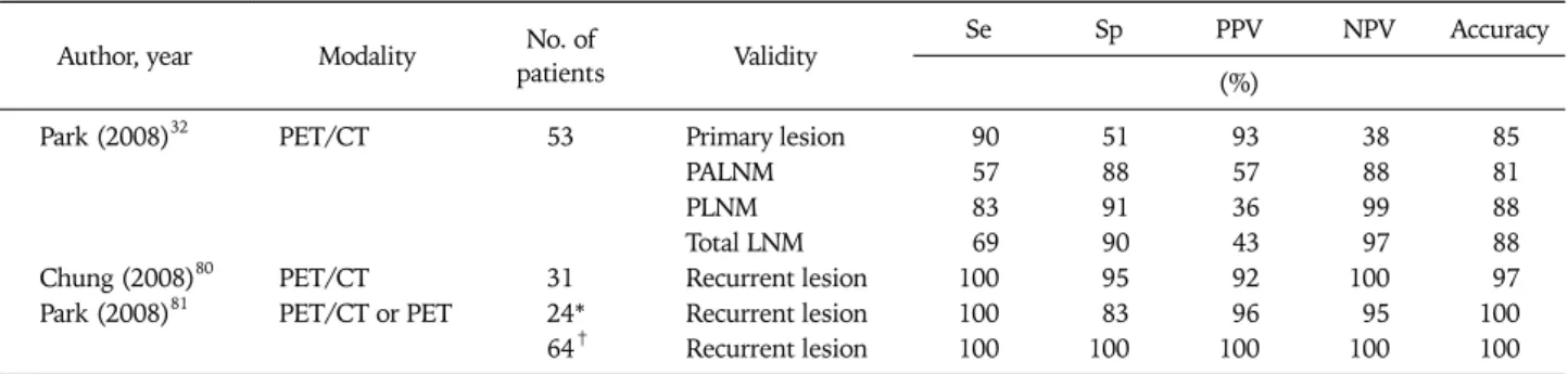
Recently, investigative work involving molecular imaging technologies has received much attention. PET uses the glucose analogue, 18-fluorodeoxyglucose (FDG), as a radioisotope and relies on increased metabolism of tumor cells, and thus in- creased glucose uptake.79 PET/CT provides the synergistic ben- efits of morphologic and molecular imaging because of local- ization of increased FDG uptake with anatomical specificity.80 Substantial evidence has now emerged documenting the role of PET/CT in the management of uterine corpus cancer pa- tients, at the pre-operative evaluation as well as at the post- treatment surveillance (Table 6).32,80,81 Park et al.32 performed a retrospective comparison of surgical staging with PET/CT in 53 uterine corpus cancer patients. They showed that pre-oper- atively, PET/CT detected lymph node metastasis with 69% sen- sitivity and 90% specificity. According to other studies for the detection of recurrent disease,80,81 PET/CT had excellent per- formance with 100% sensitivity and 83-100% specificity. The use of PET/CT has been shown to detect recurrences even in asymptomatic women with 100% sensitivity and specificity,81 and can provide important prognostic information for women after primary treatment.
PROGNOSTIC FACTORS
The prognostic impact of age, histologic subtype, grade, and FIGO stage has been established in Western populations.82 However, according to a recent retrospective survey, uterine corpus cancer of Korean women is characterized by a younger age in peak incidence and less obesity compared to Western countries.19 Thus, the prognostic significance of the afore- mentioned factors in Korean patients with endometrioid ute-
Table 7. Hazard ratios of endometrioid uterine cancer based on clinico-pathological variables in Korea
Variables Univariate Multivariate
HR (95% CI) p-value HR (95% CI) p-value
BMI <23.0 297 1.00 1.00
23.0-24.9 207 0.98 (0.42-2.27) 0.96 0.93 (0.37- 2.30) 0.87
≥25.0 433 0.82 (0.40-1.66) 0.58 0.87 (0.41-1.83) 0.70
Age ≤50 376 1.00 1.00
>50 561 5.14 (2.01-13.11) <0.01 2.66 (0.84-8.50) 0.10
Menopause No 396 1.00 1.00
Yes 505 3.08 (1.41-6.73) <0.01 1.51 (0.57-3.99) 0.41
Stage I+II 777 1.00 1.00
III+IV 142 6.50 (3.50-12.09) <0.01 3.37 (1.55-7.35) <0.01
ADJTX No 590 1.00 1.00
Yes 340 4.30 (2.22-8.34) <0.01 1.30 (0.53-3.22) 0.57
Grade I 644 1.00 1.00
II 199 2.20 (1.06-4.55) 0.03 1.46 (0.65-3.24) 0.36
III 81 4.50 (1.92-10.57) <0.01 1.96 (0.79-4.88) 0.15
LNM No 629 1.00
Yes 80 6.30 (2.93-13.53) <0.01
TD ≤2 cm 299 1.00
>2 cm 485 2.02 (0.94-4.36) 0.07
MI No 273 1.00
Yes 639 3.81 (1.49-9.73) 0.01
CE No 816 1.00
Yes 107 3.05 (1.49-6.24) <0.01
LVSI No 734 1.00 1.00
Yes 184 5.86 (3.11-11.0) <0.01 2.75 (1.27-5.95) 0.01
AE No 840 1.00
Yes 53 5.80 (2.74-12.26) <0.01
PPC No 836 1.00
Yes 39 4.54 (1.89-10.91) <0.01
RS ER−/PR− 91 1.00
ER+/PR− 30 0.60 (0.07-5.13) 0.64
ER−/PR+ 63 0.38 (0.07-1.99) 0.25
ER+/PR+ 213 0.68 (0.23-2.00) 0.49
HR: hazard ratio, CI: confidence interval, BMI: body mass index, ADJTX: adjuvant treatment, LNM: lymph node metastasis, TD: tumor diameter, MI: myometrial invasion, CE: cervical extension, LVSI: lympho-vascular space invasion, AE: adnexal involvement, PPC: positive peritoneal cytology, RS: receptor status, ER: estrogen receptor, PR: progesterone receptor.
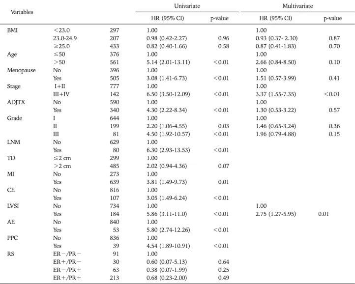
rine cancer is different. For example, FIGO stage (hazard ra- tios, 3.37; p<0.01) and lymphovascular invasion (hazard ra- tios, 2.75; p=0.01) were significant prognostic factors for overall survival, whereas age provided no significant impact in prognostic ability (Table 7). Even though age is well-known as one of the prognostic factors for uterine corpus cancer, this result did not show independent prognostic ability. This re- sult may reflect the original characteristics of Korean women, i.e., Korean patients were younger compared to Western patients. Therefore, the prognostic effect of age could be lessened.
To further improve treatment and follow-up for uterine cor- pus cancers, a number of molecular markers have been ex- tensively studied. DNA ploidy, hormone receptors, p53, bcl-2, and proliferation markers have already been shown with consistent results to be prognostic factors through retro-
spective studies.82 In Korea, there have been several efforts to define the molecular factors for uterine corpus cancer (Table 8).37,83-88 Whereas cyclooxygenase-2 (COX-2), mammalian target of rapamycin (mTOR), and microsatellite instability (MSI) have shown no prognostic ability, several other mark- ers, including p53, pre-operative CA-125, and osteopontin, have been shown to have significance as prognostic factors for uterine corpus cancers.
Immunohistochemical overexpression of p53, one of the most frequently detected tumor suppressor genes in human cancer, has a strong association with tumor aggressiveness.83 Similarly, based on the results of 131 patients, 29.8% of pa- tients had p53 alteration determined by p53 overexpression or exonal mutation, and p53 alteration was associated not on- ly with aggressive tumor behavior, but with poor disease-free survival.86
Table 8. Molecular prognosticators for uterine corpus cancer in Korea Author,
year Target Cases Materials Tool Results Outcome Prognostic
ability Jeon
(2004)83
COX-2 152 Paraffin-embedded tissue
IHC 17.8% of patients were COX- 2 positive
Possible association with carcinogenesis during the postmenopausal period
Null
p53 152 Paraffin-embedded tissue
IHC 20.4% of patients showed overexpression
Strong association with can- cer aggressiveness
Null
Chung (2006)37
CA-125 92 Serum RIA DFS of patients with ≤28.5
IU/mL was worse prog- nosis than patients with > 28.5 IU/mL
Independent prognostic mar- ker for DFS
DFS, log rank;
p=0.004
An (2007)87
MSI 100 Paraffin-embedded tissue
TMA MSI-high phenotype was re- lated with LVSI, DMI, and higher clinical stages
Possible role as a prognostic factor
Null
Kim
(2008)88 PTTG 43 Paraffin-embedded
tissue IHC 7.5% of patients had nuclear PTTG overexpression with a lower OS
Possible role as a prognostic
factor OS, log-rank;
p=0.04
Cho (2009)85
Osteopontin 56 Frozen tissue, paraffin-embedded tissue, plasma
Real-time PCR, IHC, ELISA
62.1% of stage I patients were seropositive that were not detected by CA125;
AUC 0.758 for plasma os- teopontin levels
Useful diagnostic marker for uterine cancer, independent prognostic marker for DFS
DFS, HR=3.18;
p=0.0035
No (2009)84
mTOR 141 Paraffin-embedded tissue
IHC 7.1% of patients showed overexpression
Association with COX-2 overexpression
Null
Lee
(2010)86 p53 131 Paraffin-embedded
tissue PCR, IHC 29.8% of patients had p53 al-
teration with a lower DFS Independent prognostic mar-
ker for DFS DFS, HR=17.7;
p=0.027 COX-2: cyclooxygenase-2, IHC: immunohistochemistry, RIA: radioimmunoassay, DFS: disease-free survival, MSI: microsatellite instability, TMA:
tissue microarray, LVSI: lympho-vascular space invasion, DMI: deep myometrial invasion, PTTG: pituitary tumor-transforming gene, OS: overall survival, PCR: polymerase chain reaction, ELISA: enzyme-linked immunosorbent assay, HR: hazard ratio, mTOR: mammalian target of rapamycin.
Preoperative serum CA-125 has been shown to be a sig- nificant prognostic factor.89 According to Chung et al.,37 pa- tients with a serum CA-125 ≤ 28.5 U/mL have a significantly better 5-year disease-free survival than patients with a CA-125>28.5 U/mL (85.6% vs. 60.0%, p=0.004). Therefore, they suggested a preoperative CA-125 as a useful tool for opti- mal individualized cancer treatment.
Osteopontin, a kind of phosphoglycoprotein, is known as a mediator of tumor transformation and malignant progression in human cancers, and its overexpression in tissues or blood of cancer patients may indicate the presence of a tumor.90 Cho et al.85 investigated the diagnostic and prognostic role of os- teopontin in uterine corpus cancer. They proved that osteo- pontin was seropositive in 62.1% of patients with stage I dis- ease who were not detected by CA-125, and that osteopontin positivity was an independent prognosticator for disease-free survival in patients with uterine corpus cancer.
MANAGEMENT OF RECURRENT DISEASE Most uterine corpus cancers are diagnosed at an early stage and have a favorable prognosis. However, a substantial num- ber of patients undergo disease recurrence after primary treat- ment. According to one Korean retrospective study of 301 pa-
tients,91 approximately 26 (9%) of patients develop recurrent disease and the mean time-to-recurrence is 29 months. Among women with recurrences, 22 (85%) had distant failure and most women with recurrences presented at an advanced stage at the time of diagnosis. After diagnosing a recurrence, 18 women received systemic chemotherapy or chemoradiation, 2 women had radiation, and only one woman underwent surgi- cal treatment. Among the 18 women with chemotherapy, the taxane-based group had a better median survival than the non-taxane group (22 vs. 15 months, p=0.047).92 In the plan- ning for patients with recurrent disease, it is important to con- sider the site of recurrence, performance status of the patient, and prior therapy the patient has received. After specific con- siderations for each patient, combined modalities, as well as surgical cytoreduction, radiation therapy, hormonal treat- ment, systemic chemotherapy, and palliative therapy may be used. In this era, research should focus on developing new chemotherapeutic and molecular targets active against ute- rine corpus cancer.
HORMONE REPLACEMENT THERAPY
Because there has been no conclusive data to prove the bene- fit or safety of hormone replacement therapy in patients with
uterine corpus cancer, this question remains unanswered. To date, an overall lower recurrence rate or a non-significant dif- ference in recurrence between hormone-users and -non-users has been noted in several retrospective studies93-96 and in one randomized controlled trial.97 A retrospective Korean study which evaluated tibolone in patients with uterine corpus can- cer also showed no significant difference in disease-free sur- vival and overall survival between the two groups.98 This find- ing should be used along with other information for counsel- ing patients regarding the risks of hormone replacement ther- apy in uterine corpus cancer.
CONCLUSION
Like other malignancies, uterine corpus cancer must be af- fected by genetic differences. Thus, if possible, treatment guidelines should be individualized based on ethnic-specific treatment results. Even though there have been a number of advances in treatment strategies and research products for uterine corpus cancers in Korea, most of our data are charac- terized by retrospective design, small sample size, single in- stitute experience, and insufficient follow-up. Thus, pro- spective multi-center trials should be performed to make more progress in the treatment of gynecologic cancer patients, including uterine corpus cancer. In addition, we also should be interested in primary and secondary prevention to improve cancer control, and in treatment-related symptom manage- ment, palliation, and end-of-life care to improve quality of life.
CONFLICT OF INTEREST
No potential conflict of interest relevant to this article was reported.
ACKNOWLEDGEMENTS
This research was supported by the Kyung Hee University Research Fund in 2010 (KHU-20100651).
REFERENCES
1. Parkin DM, Pisani P, Ferlay J. Estimates of the worldwide in- cidence of 25 major cancers in 1990. Int J Cancer 1999; 80:
827-41.
2. SOG Gynecologic Oncology Committee. Annual report of gyne- cologic cancer registry program in Korea for 2004 (Jan. 1st, 2004-Dec. 31st, 2004). Korean J Obstet Gynecol 2007; 50:
28-78.
3. Sankaranarayanan R, Ferlay J. Worldwide burden of gynaeco- logical cancer: the size of the problem. Best Pract Res Clin Obstet Gynaecol 2006; 20: 207-25.
4. Kaaks R, Lukanova A, Kurzer MS. Obesity, endogenous hor- mones, and endometrial cancer risk: a synthetic review. Cancer Epidemiol Biomarkers Prev 2002; 11: 1531-43.
5. Lindemann K, Vatten LJ, Ellstrom-Engh M, Eskild A. Body mass, diabetes and smoking, and endometrial cancer risk: a fol- low-up study. Br J Cancer 2008; 98: 1582-5.
6. Bjorge T, Engeland A, Tretli S, Weiderpass E. Body size in rela- tion to cancer of the uterine corpus in 1 million Norwegian women. Int J Cancer 2007; 120: 378-83.
7. Furberg AS, Thune I. Metabolic abnormalities (hypertension, hyperglycemia and overweight), lifestyle (high energy intake and physical inactivity) and endometrial cancer risk in a Norwegian cohort. Int J Cancer 2003; 104: 669-76.
8. Goodman MT, Hankin JH, Wilkens LR, Lyu LC, McDuffie K, Liu LQ, et al. Diet, body size, physical activity, and the risk of endometrial cancer. Cancer Res 1997; 57: 5077-85.
9. Jung KW, Won YJ, Park S, Kong HJ, Sung J, Shin HR, et al.
Cancer statistics in Korea: incidence, mortality and survival in 2005. J Korean Med Sci 2009; 24: 995-1003.
10. ACOG practice bulletin, clinical management guidelines for ob- stetrician-gynecologists, number 65, August 2005: manage- ment of endometrial cancer. Obstet Gynecol 2005; 106: 413-25.
11. Pecorelli S. Revised FIGO staging for carcinoma of the vulva, cervix, and endometrium. Int J Gynaecol Obstet 2009; 105:
103-4.
12. Mohan DS, Samuels MA, Selim MA, Shalodi AD, Ellis RJ, Samuels JR, et al. Long-term outcomes of therapeutic pelvic lymphadenectomy for stage I endometrial adenocarcinoma.
Gynecol Oncol 1998; 70: 165-71.
13. Cragun JM, Havrilesky LJ, Calingaert B, Synan I, Secord AA, Soper JT, et al. Retrospective analysis of selective lymphadenec- tomy in apparent early-stage endometrial cancer. J Clin Oncol 2005; 23: 3668-75.
14. Benedetti Panici P, Basile S, Maneschi F, Alberto Lissoni A, Signorelli M, Scambia G, et al. Systematic pelvic lymphadenec- tomy vs. no lymphadenectomy in early-stage endometrial carci- noma: randomized clinical trial. J Natl Cancer Inst 2008; 100:
1707-16.
15. Kitchener H, Swart AM, Qian Q, Amos C, Parmar MK. Efficacy of systematic pelvic lymphadenectomy in endometrial cancer (MRC ASTEC trial): a randomised study. Lancet 2009; 373:
125-36.
16. Creutzberg CL, van Putten WL, Koper PC, Lybeert ML, Jobsen JJ, Warlam-Rodenhuis CC, et al. Surgery and postoperative ra- diotherapy versus surgery alone for patients with stage-1 endo- metrial carcinoma: multicentre randomised trial. PORTEC Study Group. Post Operative Radiation Therapy in Endometrial Carcinoma. Lancet 2000; 355: 1404-11.
17. Blake P, Swart AM, Orton J, Kitchener H, Whelan T, Lukka H, et al. Adjuvant external beam radiotherapy in the treatment of endometrial cancer (MRC ASTEC and NCIC CTG EN.5 rando- mised trials): pooled trial results, systematic review, and meta-analysis. Lancet 2009; 373: 137-46.
18. Henderson BE, Feigelson HS. Hormonal carcinogenesis.
Carcinogenesis 2000; 21: 427-33.
19. Jeong NH, Lee JM, Lee JK, Kim JW, Cho CH, Kim SM, et al.
Role of body mass index as a risk and prognostic factor of en- dometrioid uterine cancer in Korean women. Gynecol Oncol 2010; 118: 24-8.
20. Xu WH, Dai Q, Xiang YB, Zhao GM, Ruan ZX, Cheng JR, et al.
Nutritional factors in relation to endometrial cancer: a report from a population-based case-control study in Shanghai, China.
Int J Cancer 2007; 120: 1776-81.
21. McCann SE, Freudenheim JL, Marshall JR, Brasure JR, Swanson MK, Graham S. Diet in the epidemiology of endo- metrial cancer in western New York (United States). Cancer Causes Control 2000; 11: 965-74.
22. Jeong NH, Song ES, Lee JM, Lee KB, Kim MK, Yun YM, et al.
Preoperative levels of plasma micronutrients are related to en- dometrial cancer risk. Acta Obstet Gynecol Scand 2009; 88:
434-9.
23. Lee KH, Lee JK, Kim JH, Lee JM, Song ES, Kim MK. Folic acid and vitamin B12 intake in relation to risk of endometrial can- cer: case-control study. Korean J Obstet Gynecol 2008; 51:
1103-11.
24. Meyer LA, Westin SN, Lu KH, Milam MR. Genetic poly- morphisms and endometrial cancer risk. Expert Rev Anticancer Ther 2008; 8: 1159-67.
25. Lee JM, Lee JH, Tong SY, Ki KD, Lee SK, Huh CY. The effect of HER-2 polymorphism according to age on the risk and patho- logic feature of endometrial cancer. Korean J Obstet Gynecol 2009; 52: 529-37.
26. Roh JW, Kim JW, Park NH, Song YS, Park IA, Park SY, et al.
p53 and p21 genetic polymorphisms and susceptibility to endo- metrial cancer. Gynecol Oncol 2004; 93: 499-505.
27. Kang S, Kim JW, Park NH, Song YS, Kang SB, Lee HP. Cyclin D1 polymorphism and the risk of endometrial cancer. Gynecol Oncol 2005; 97: 431-5.
28. Jo H, Kang S, Kim SI, Kim JW, Park NH, Song YS, et al. The C19007T polymorphism of ERCC1 and its correlation with the risk of epithelial ovarian and endometrial cancer in Korean womenL a case control study. Gynecol Obstet Invest 2007; 64:
84-8.
29. Tong SY, Ha SY, Ki KD, Lee JM, Lee SK, Lee KB, et al. The ef- fects of obesity and HER-2 polymorphisms as risk factors for endometrial cancer in Korean women. BJOG 2009; 116:
1046-52.
30. Lee KB, Ki KD, Lee JM, Lee JK, Kim JW, Cho CH, et al. The risk of lymph node metastasis based on myometrial invasion and tumor grade in endometrioid uterine cancers: a multi- center, retrospective Korean study. Ann Surg Oncol 2009; 16:
2882-7.
31. Chung HH, Kang SB, Cho JY, Kim JW, Park NH, Song YS, et al.
Accuracy of MR imaging for the prediction of myometrial in- vasion of endometrial carcinoma. Gynecol Oncol 2007; 104:
654-9.
32. Park JY, Kim EN, Kim DY, Suh DS, Kim JH, Kim YM, et al.
Comparison of the validity of magnetic resonance imaging and positron emission tomography/computed tomography in the preoperative evaluation of patients with uterine corpus cancer.
Gynecol Oncol 2008; 108: 486-92.
33. Yoo SC, Kim WY, Yoon JH, Kim HY, Lee EJ, Chang SJ, et al.
Accuracy of preoperative magnetic resonance imaging in as- sessing lymph node metastasis and myometrial invasion in pa- tients with uterine cancer. Eur J Gynaecol Oncol 2009; 30:
167-70.
34. Han SS, Lee SH, Kim DH, Kim JW, Park NH, Kang SB, et al.
Evaluation of preoperative criteria used to predict lymph node metastasis in endometrial cancer. Acta Obstet Gynecol Scand 2010; 89: 168-74.
35. Cho H, Kim YT, Kim JH. Accuracy of preoperative tests in clin- ical stage I endometrial cancer: the importance of lymphade- nectomy. Acta Obstet Gynecol Scand 2010; 89: 175-81.
36. Choi YS, Koh SB, Ahn JY, Yi CM, Shin IH, Lee TS. Usefulness of preoperative CA 125 level in decision making of lymphade- nectomy in endometrial cancer patients. Korean J Obstet Gynecol 2005; 48: 2877-87.
37. Chung HH, Kim JW, Park NH, Song YS, Kang SB, Lee HP. Use of preoperative serum CA-125 levels for prediction of lymph node metastasis and prognosis in endometrial cancer. Acta Obstet Gynecol Scand 2006; 85: 1501-5.
38. Selman TJ, Mann CH, Zamora J, Khan KS. A systematic review of tests for lymph node status in primary endometrial cancer.
BMC Womens Health 2008; 8: 8.
39. Girardi F, Petru E, Heydarfadai M, Haas J, Winter R. Pelvic lym- phadenectomy in the surgical treatment of endometrial cancer.
Gynecol Oncol 1993; 49: 177-80.
40. Creasman WT, Morrow CP, Bundy BN, Homesley HD, Graham JE, Heller PB. Surgical pathologic spread patterns of endo- metrial cancer: a Gynecologic Oncology Group Study. Cancer 1987; 60: 2035-41.
41. Kim SH, Kim HD, Song YS, Kang SB, Lee HP. Detection of deep myometrial invasion in endometrial carcinoma: comparison of transvaginal ultrasound, CT, and MRI. J Comput Assist Tomogr 1995; 19: 766-72.
42. Ahn JY, Yi CM, Lee TS, Koh SB, Park YC, Jung KJ, et al.
Analysis of factors to affect on MR assessment of myometrial invasion and cervical involvement in endometrial cancer.
Korean J Obstet Gynecol 2006; 49: 113-21.
43. Hwang JH, Lee NW, Lee KW, Lee JK. Magnetic resonance imag- ing for assessment of deep endometrial invasion for patients with endometrial carcinoma. Aust N Z J Obstet Gynaecol 2009;
49: 537-41.
44. Suh DS, Kim JK, Kim KR, Kim DY, Kim JH, Kim YM, et al.
Reliability of magnetic resonance imaging in assessing my- ometrial invasion absence in endometrial carcinoma. Acta Obstet Gynecol Scand 2009; 88: 990-3.
45. Hacker NF. Uterine cancer. In: Berek JS, Hacker NF, editors.
Berek & Hacker's gynecologic oncology. 5th ed. Philadelphia:
Lippincott Williams & Wilkins; 2010. p.400-17.
46. Kilgore LC, Partridge EE, Alvarez RD, Austin JM, Shingleton HM, Noojin F 3rd, et al. Adenocarcinoma of the endometrium:
survival comparisons of patients with and without pelvic node sampling. Gynecol Oncol 1995; 56: 29-33.
47. Chan JK, Wu H, Cheung MK, Shin JY, Osann K, Kapp DS. The outcomes of 27,063 women with unstaged endometrioid ute- rine cancer. Gynecol Oncol 2007; 106: 282-8.
48. Lee TS, Kim JW, Kim TJ, Cho CH, Ryu SY, Ryu HS, et al.
Ovarian preservation during the surgical treatment of early stage endometrial cancer: a nation-wide study conducted by the Korean Gynecologic Oncology Group. Gynecol Oncol 2009;
115: 26-31.
49. Tangjitgamol S, Anderson BO, See HT, Lertbutsayanukul C, Sirisabya N, Manchana T, et al. Management of endometrial cancer in Asia: consensus statement from the Asian Oncology Summit 2009. Lancet Oncol 2009; 10: 1119-27.
50. Lee JM, Lee JK, Kim JW, Cho CH, Kim SM, Park SY, et al.
Current status in the management of uterine cancer in Korea: a multi-center retrospective Korean study. The 25th Annual Meeting of Korean Society of Gynecologic Oncology and Colposcopy 2010, Abstract 10S-006.
51. Seo MW, Lee HY, Kim DY, Suh DS, Kim JH, Kim YM, et al. The effect of lymphadenectomy on the survival rate of clinical stage I endometrial cancer. Korean J Gynecol Oncol 2006; 17: 227-33.
52. Chang SJ, Kim WY, Yoon JH, Yoo SC, Chang KH, Ryu HS.
Para-aortic lymphadenectomy improves survival in patients with intermediate to high-risk endometrial carcinoma. Acta Obstet Gynecol Scand 2008; 87: 1361-9.
53. Fujimoto T, Nanjyo H, Nakamura A, Yokoyama Y, Takano T, Shoji T, et al. Para-aortic lymphadenectomy may improve dis- ease-related survival in patients with multipositive pelvic lymph node stage IIIc endometrial cancer. Gynecol Oncol 2007;
107: 253-9.
54. Childers JM, Brzechffa PR, Hatch KD, Surwit EA. Laparoscopi- cally assisted surgical staging (LASS) of endometrial cancer.
Gynecol Oncol 1993; 51: 33-8.
55. Jung YW, Lee DW, Kim SW, Nam EJ, Kim JH, Kim JW, et al.
Robot-assisted staging using three robotic arms for endo- metrial cancer: comparison to laparoscopy and laparotomy at a single institution. J Surg Oncol 2010; 101: 116-21.
56. Hahn HS, Kim HJ, Yoon SG, Kim WC, Choi HJ, Kim HS, et al.