Effect of span length on the fit of zirconia
framework fabricated using CAD/CAM system
Jeong-Yol Lee, DDS, MSc, PhD, Sang-Jin Choi, DDS, Min-Soo Kim, DDS, Ha-Young Kim, DDS, Young-Soo Kim, DDS, PhD, Sang-Wan Shin*, DDS, MPH, PhD, MSc
Postgraduate School of Clinical Dentistry, Institute for Clinical Dental Research, Korea University, Seoul, Republic of Korea
PURPOSE. The purpose of this study was to evaluate the effect of the span length on the fit of zirconia framework fabricated using CAD/CAM system. MATERIALS AND METHODS. Abutments for single, 4-unit and 6-unit fixed partial prostheses were fabricated. Ten zirconia frameworks were fabricated for each group. The marginal and internal gap were presented by means of replica technique and measured by measuring microscope (AXIO®, Carl Zeiss, Rochester, NY) and software (I-solution®, IMT i-solution Inc., Vancouver, BC, Canada). The results were statistically analyzed by multivariate analysis test and Dunnett T3 test for post hoc test (α=.05). RESULTS. There were statistically significant differences at 2, 4, 7, 8 points (mesio-distal section) and b, d, e, f, g (labio-lingual section). In some marginal reference points of 6-unit group (P<.05), the marginal gap were larger than 120 µm.
CONCLUSION. Span length of zirconia core may have an influence on marginal and internal fit. Within the limitation of this study, the increase of span length of zirconia framework of 6 or more-unit fixed partial denture may decrease the marginal and internal fit. [J Adv Prosthodont 2013;5:118-25]
KEY WORDS: Zirconia; CAD/CAM; Span length; Fit
INTRODUCTION
Recently, esthetic requirements in dental restorative treat- ment are on the rise. For this reason, recent attention-draw- ing material is zirconia which is a biocompatible material with stable structure. Flexural strength and fracture tough- ness of zirconia reaches up to 900-1200 MPa and 9-10 MPa·m1,2 respectively, and zirconia has been proven to be much more excellent in its strength than that of glass infil-
trated full porcelain which is widely used in esthetic restora- tion.1,2 Material properties of zirconia like these show that zirconia can be substituted for metal and can be surely used even in long span fixed prosthesis.3,4
CAD/CAM system which was introduced to dentistry in 1980’s has brought cost and time reduction in fabricating restoration by using computer for inputting, designing and cutting the form of restoration. In order to overcome dis- advantage of previous method of framework fabrication in which post-sintered zirconia block was cut, pre-sintered zir- conia block is being used recently. By doing so, fabrication time and consumption of bur have been reduced and thus cost and time was also reduced. This method, however, has disadvantage in that approximately 15-30% shrinkage occur in the procedure of sintering and hardening the cut block using CAM unit for the increased density of zirconia block.5 When using partially sintered block, therefore, it is more important to calculate shrinkage rate accurately than any other things in order to compensate shrinkage which inevitably occurs in sintering process, and also to gain pre- cise product when using partially sintered block.
It is considered that internal and marginal fit plays a key role in longevity of prosthesis. In previous studies, many authors said that 120 µm or less marginal gap of prosthesis was clinically acceptable.6,7 According to a study on fit of
Corresponding author:
Sang-Wan Shin
Institute for Clinical Dental Research, Korea University Hospital, 97 Gurodong-gil, Guro-gu, Seoul, 152-703, Republic of Korea Tel. 82226261922: e-mail, [email protected]
Received October 19, 2012 / Last Revision May 7, 2013 / Accepted May 13, 2013
© 2013 The Korean Academy of Prosthodontics
This is an Open Access article distributed under the terms of the Creative Commons Attribution Non-Commercial License (http://creativecommons.
org/licenses/by-nc/3.0) which permits unrestricted non-commercial use, distribution, and reproduction in any medium, provided the original work is properly cited.
This study was funded by development project of original industrial technology by the Ministry of Knowledge Economy (assignment number:10032032).
zirconia framework which was used in all ceramic crown, marginal gap was reported to be 64-83 µm,8,9 and in another study on clinical fit of 3-unit fixed prosthesis, zirconia prosthesis (LAVA, 3M ESPE, Seefeld, Germany) for which CAD/CAM was used reportedly showed 80µm of mean marginal gap.10 The reason why marginal fit is important is because increased marginal leakage may cause secondary caries, periodontitis, pulpitis etc. and also cause esthetic problem and eventually cause failure of prosthesis. Internal fit also is very important for longevity of prosthesis. It influences retention and support of prosthesis. There was a report from one previous study saying the thicker the cement became due to a large internal gap, the weaker the porcelain became.11 This effect is shown also in zirconia.
Excessive thickness of cement increases radial crack12,13 and causes fracture of veneering porcelain.14 Fracture of veneer- ing porcelain is considered to be one of the greatest rea- sons for removing zirconia restoration.15-18 As opposed to this, too small internal gap may cause unstable seating of prosthesis.19 Abduo et al.20 said that factors which influ- enced fit of prosthesis were fabrication system of zirconia, veneering, configuration, span length of zirconia etc.
Although the use of zirconia has increased and are being used more in multiple areas where the tooth are miss- ing, studies on fit of zirconia fixed partial denture until the now have been limited to 3-unit fixed partial denture with nearly linear form; there has been few study on internal or marginal fit of 4 or more unit fixed partial denture with curved form.20 Particularly, there was no study in which both internal and marginal fit depending on the span length were simultaneously evaluated.
The purpose of this study, therefore, was to measure marginal and internal fit of single, 4-unit, 6-unit zirconia fixed partial denture core which had been fabricated using CAD/CAM system by using replica technique and to evalu- ate the effect that span length on fit while evaluating whether measured marginal gap was in clinically acceptable range or not.
MATERIALS AND METHODS
Experimental groups were divided into single, 4-unit and 6-unit groups, and preparation of each abutment teeth were done in order to make single crown of upper right central incisor, 4-unit fixed partial denture with abutment teeth of upper right and left lateral incisor where upper right and left central incisors were missing, and 6-unit fixed partial denture with abutment teeth of upper right and left canines where upper right, left central and lateral incisors were missing (Table 1). For tooth preparation, surveyor and dia- mond bur were used, and total taper of each abutment teeth was intended to be 6˚. Preparation amount was 2 mm on incisal part and 1 mm on axial wall, and 1 mm width deep chamfer margin was formed.
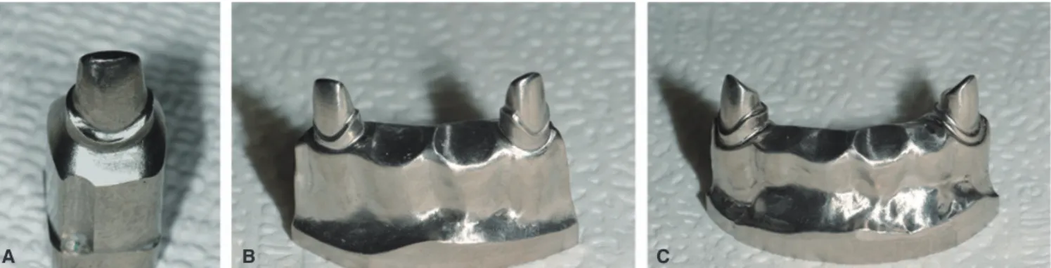
In order to prevent possible wear or fracture of master models from repetitive impression taking procedure, denti- formmodels (Nissin Dental Prod. Inc., Japan) where abut- ment preparation were done were duplicated and titanium master model weremade (Addtech Co., Seoul, Korea) (Fig. 1).
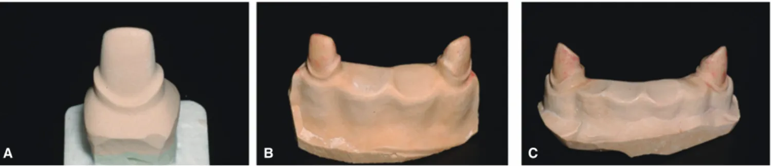
Impression of titanium model were taken with conven- tional method by using addition silicone impression materi- al (Imprint II, 3M ESPE, St. Paul, MN, USA) and ready- made tray, and total 30 improved stone (Fuji rock® EP, GC Corp, Tokyo, Japan) models including 10 models per each group were made (Fig. 2).
Table 1. Test group, missing teeth, abutment and numbers of sample zirconia cores
Group Missing teeth Abutment N
Single #11 10
4-unit fixed partial denture #11, 21 #12, 22 10 6-unit fixed partial denture #12, 11, 21, 22 #13, 23 10
Fig. 1. Titanium master model fabricated using CAD/CAM (Addtech Co., Seoul, Korea). A: single, B: 4-unit, C: 6-unit.
A B C
After scanning of the manufactured plaster model using CAD/CAM system, partially sintered zirconia blocks were cut and sintered, and finally zirconia cores were fabricated (Fig. 3). When fabricating cores, CAD/CAM system of Orapix (Seoul, Korea) and zirconia blocks were used.
Thickness of core and cement space were set as 0.6 mm and 40 µm respectively. Internal adjustment was not done so that fit of core that was fabricated by only CAD/CAM could be merely evaluated. After core fabrication was done, whether there were remnants inside, defects or distortions were checked and then cleaned with high pressure steaming.
After positioning fabricated zirconia cores on each models, jigs for positioning the zirconia coreon the model were made using Pattern resin® (GC Dental, Japan) so that zirconia could always be positioned at a same spot on the models. After filling fit checking material (Fit checker, GC Dental, Japan) in inner surface of core, it was placed on abutment tooth. After that, pre-made resin jig was posi- tioned above the core. While maintaining adapted position by hand pressure, it was place in Universal testing machine (Shimadzu corporation, Kyoto, Japan) immediately.
Universal testing machine was set to measurement mode, and its compressive force was limited to 40 N, and regular force (40 N) was maintained for 5 minutes until Fit checker was completely hardened.
After 5 minutes, the core was removed from the model
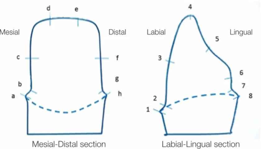
carefully. When doing so, the silicon film of Fit checker has to be fully attached to silicone core. Filling inner surface of silicone film of core with regular bodied addition silicone impression material (AquasilLV, Densply Caulk, USA) and hardening the filled material increase strength andalso enables gaining of stable film layer. Each 8 measuring points were established for labial and mesio-distal side respectively (Fig. 4), and cutting was done right in the cen- ter of the model labialy and mesio-distally (Fig. 5). By using measuring microscope (AXIO®, Carl Zeiss, Rochester, NY, USA) and I-SolutionTM (IMT I-Solution Inc., Vancouver, BC, Canada), thickness of fit checker was measured at 16 measuring points of each abutment teeth (Fig. 6). Mean value was documented after 3 times measured by 2 experi- menter for each measuring points.
Mean value and standard deviation value were calculated by adding measurement values of right and left abutment teeth of each group which was divided into bucco-lingual and mesio-distal.
Whether there was statistically significant difference between groups which were divided into bucco-lingual and mesio-distal or not was analyzed by multivariate analysis.
After that, if there had been statistically significant differ- ence, post-hoc test was done using Dunnett T3 test per each measuring points. In all analyses, statistical significance was accepted at 5% probability level.
Fig. 2. Duplicated master models with dental stone. A: single, B: 4-unit, C: 6-unit.
A B C
Fig. 3. Zirconia cores fabricated using CAD/CAM (Orapix, Seoul, Korea). A: single, B: 4-unit, C: 6-unit.
A B C
RESULTS
Twenty seven specimens including nine single, nine 4-unit and nine 6-unit were used in fit measurement. There existed 1 specimen per each group of which measurement could not be done because fracture had occurred in the process of applying force after putting Fit checker inside the core and placing it on abutment.
Statistical analysis was performed by adding each mea- surement values of right and left abutment teeth of each group. By doing so, number of specimen of each group became nine single, and eighteen 4-unit and eighteen 6-unit.
Statistical analysis was performed separately for bucco-lin- gual and mesio-distal.
Mean value and standard deviation value of gap at buc- co-lingual measuring point were calculated (Table 2). As a result of multivariate analysis there was significant differ- ence in at least one or more point. Which measuring point showed statistically significant difference was checked using ANOVA. As a result, statistically significant difference was
shown at point 2, 4, 7 and 8. In order to investigate between which groups of point 2, 4, 7 and 8 showed the difference, post-hoc test was performed using Dunnett T3 test (Table 3).
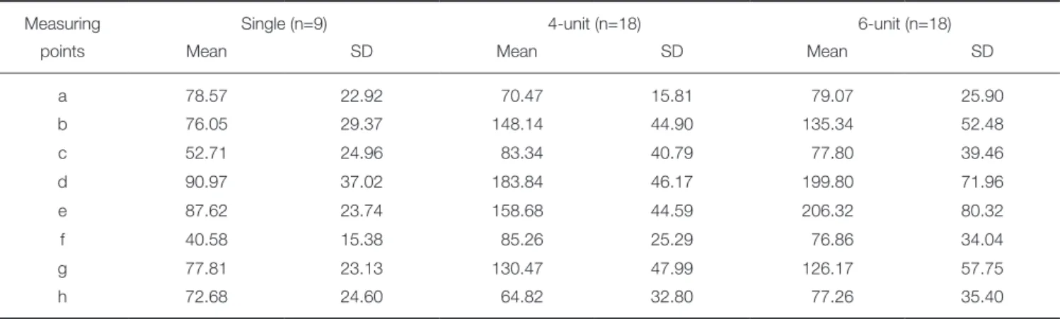
Mean value and standard deviation value of gap at mesio-distal measuring point were calculated (Table 4).
There was significant difference in at least one or more point in multivariate analysis. The result of ANOVA showed statistically significant differences at point 2, 4, 7 and 8 by span length factor. In order to investigate between which groups of point 2, 4, 7 and 8 showed the difference, post-hoc test was performed using Dunnett T3 test (Table 5).
Fig. 4. Reference points to measure the thickness of fit checker (Left: mesio-distal, Right: labio-lingual).
Mesial Distal Labial Lingual
Mesial-Distal section Labial-Lingual section
Fig. 5. Directions and positions of section of replica materials (Left: directions of labio-lingual and mesio- distal section, Right: view of labio-lingual section).
Fig. 6. Measurement of thickness of Fit checker® at each reference point (A: labial reference point, B:
incisalreference point, C: marginal reference point, D:
lingual reference point).
A B
C D
As a result of measuring point analysis which showed statistically significant difference, measurement value increased from single, 4-unit and 6-unit in order in point 2, 4, 7 when bucco-lingual analysis was done. But in point 8,
the gap of 4-unit group was measured to be the smallest. In the result of mesio-distal analysis, measurement value increased from single, 6-unit and 4-unit in order in point b, f and g (4-unit group showed the largest value).
Table 2. Mean and standard deviation (SD) of gaps (µm) at bucco-lingual reference point
Measuring Single (n=9) 4-unit (n=18) 6-unit (n=18)
points Mean SD Mean SD Mean SD
1 87.64 23.01 68.99 25.37 69.53 32.64
2 109.26 29.41 132.69 45.63 166.13 74.40
3 86.62 27.34 76.00 43.69 80.60 46.24
4 95.12 27.65 165.35 51.70 224.20 83.52
5 104.66 20.94 116.13 37.09 140.21 55.02
6 85.38 19.82 79.61 27.04 99.31 71.26
7 108.32 35.53 128.07 39.11 203.06 79.43
8 104.67 56.66 57.91 23.16 133.28 82.03
Table 3. The results of multiple comparison test (Dunnett T3)
Position Group Single 4-unit 6-unit
2 Single *
4-unit
6-unit *
4 Single *** ***
4-unit *** *
6-unit *** *
7 Single **
4-unit **
6-unit ** **
8 Single
4-unit **
6-unit **
*** means P<.001, and ** means P<.01, and * means P<.05.
Table 4. Mean and standard deviation (SD) of gaps (µm)at mesio-distal reference point
Measuring Single (n=9) 4-unit (n=18) 6-unit (n=18)
points Mean SD Mean SD Mean SD
a 78.57 22.92 70.47 15.81 79.07 25.90
b 76.05 29.37 148.14 44.90 135.34 52.48
c 52.71 24.96 83.34 40.79 77.80 39.46
d 90.97 37.02 183.84 46.17 199.80 71.96
e 87.62 23.74 158.68 44.59 206.32 80.32
f 40.58 15.38 85.26 25.29 76.86 34.04
g 77.81 23.13 130.47 47.99 126.17 57.75
h 72.68 24.60 64.82 32.80 77.26 35.40
DISCUSSION
Zirconia is an esthetical and biocompatible material, and its physical property is so strong that it can be used for fixed prosthesis of posterior region. Because of its property like this, it has become one of the most noteworthy materials in esthetic prosthetics. In order for zirconia to be used as prosthetic restoration material that can last long and func- tion stably, not only its physical property but also the fit of prosthesis made by it should meet clinical requirements.
In fabrication of zirconia prosthesis using CAD/CAM, completely sintered block was used initially. However, this method has disadvantages in time and cost although fit could be more excellent than when partially sintered block which is used in most fabrication system is used. On the other hand, if partially sintered block is used, it will be bet- ter off when it comes to time and cost factor. However, if cutting is not done by accurate calculation due to shrinkage in sintering process, level of fit will decrease significantly.
According to previous studies, 120 µm or smaller mar- ginal gap is clinically accepted.6,7 In the studies on fit of sin- gle or 3-unit fixed partial denture zirconia prosthesis fabri- cated by CAD/CAM system, marginal gap showed approxi- mately 64-83 µm as a result.8,9 These show that zirconia can be used for fixed partial denture restoration adequately.
However, these were the studies which had been mostly proceeded on linear type fixed prosthesis which was shorter than 3-unit. Study on prosthesis of which span is longer than this has not been done.10 In this study, therefore, whether fit of zirconia prosthesis is influenced by the span length was investigated using anterior teeth model.
As methods for measuring marginal or inner fit, a meth- od of direct inspection, a method of inspection after cut- Table 5. The results of multiple comparison test (Dunnett T3)
Position Group Single 4-unit 6-unit
b Single *** **
4-unit ***
6-unit **
d Single *** ***
4-unit ***
6-unit ***
e Single *** ***
4-unit 6-unit
f Single *** **
4-unit ***
6-unit **
g Single ** *
4-unit **
6-unit *
*** means P<.001, and ** means P<.01, and * means P<.05.
ting, a method of evaluation by impression taking, a meth- od of evaluation using explorer and so on were suggested.
Although a method of inspection after cutting may be the most accurate way, there is a shortcoming in that more specimens have to be made for the increase of the number of measurement. Therefore, replica technique where mea- surement of fit of various parts could be done easily with only small number of specimens was used in this study.
This method had been considered to have low accuracy in the past,21 but it became known to be reliable compared to other methods for measuring fit by Rahme et al.22 and Laurent et al.23 study. Specimens for measurement were made using Replica technique, and thickness of fit checker was measured at 16 measuring points which were designat- ed per each abutment teeth, and 2 experimenters recorded mean value after 3 times of measurements so that error could be minimized.
As a result of measurement, overall fit was acceptable (small gap) in marginal and axial wall area, but connection part between margin and axial wall and incisal part showed relatively large gap. Because it was considered that whether it was either right abutment tooth or left abutment tooth could not be a variable for analysis, mean value and stan- dard deviation were calculated by adding measurement val- ues of right and left abutment teeth together for statistical analysis in 4-unit and 6-unit group. In the case of this study, multivariate analysis was used because it was consid- ered that gap of inner surface and margin could not be independent to each other although independent variable was a span length.
As a result of statistical analysis of bucco-lingual (1-8) and mesio-distal (a-h) measuring points, there were statisti- cally significant differences at point 2, 4, 7, 8 and b, d, e, f, g
among the groups. From this result, the fact that span length influenced fit of zirconia core was confirmed. As a result of analysis of each measuring points which showed statistically significant difference, measurement value increased from single, 4-unit to 6-unit in order at 2, 4, 7 point, but measurement value of 4-unit group came out smallest at 8 point. As opposed to this, 4-unit group showed the largest value at b, f, g point as a result of mesio-distal analysis. The value increased from single, 6-unit and 4-unit in order. This is considered to be because of small number of specimens that were used in the experi- ment and measurement error as well. Also, there were more points which showed significant difference inmesio-distal part than inbucco-lingual part. It is guessed that the reason for this is because more error occurred in the process of shrinkage at mesio-distal part since the form of zirconia core was not bucco-lingually but mesio-distally long.
Although there was not enough number of specimen for more accurate multivariate analysis in statistical analysis process, Kruskal-Wallis test was performed to compensate it, and same result as that of multivariate analysis was gained.
It can be said that the meaning of this study lays in the fact that evaluation of effect of span length on fit in fabri- cation of prosthesis. However, the number of specimen was not enough for securing statistical significance which is the weak point of this study. Also, if fit of prosthesis had been measured after veneering, the study would have been closer to clinical setting. In some specimens, measurement was difficult because boundary of silicone film and impres- sion material was blurred. For more accurate measurement, this method needs improvement. Also, in case of fixed par- tial denture, fit is not something that is independent to one another. Therefore, it is considered that comparative evalu- ation on volume will be more necessary than to just observe some part after cutting it. Also, if analysis such as micro CT24 and so on is additionally used, appearance of transformation or distortion in shrinkage process will be able to be evaluated. The further studies to investigate the difference of fit among the different manufacturers with increased number of specimens will be necessary. And improvement of fit through continuous analysis and evalu- ation will advance fit of zirconia.
CONCLUSION
Change of the span length influenced marginal fit and internal fit to some degree. In single or 4-unit fixed partial denture group, mean value of marginal fit was within clini- cally acceptable range. In 6-unit group, however, some mar- gins showed values that were out of clinically acceptable range. Therefore, it was analyzed that the increase of the span length could possibly decrease fit between zirconia core and abutment tooth when fixed partial denture was 6-unit or longer.
REFERENCES
1. Seghi RR, Denry IL, Rosenstiel SF. Relative fracture tough- ness and hardness of new dental ceramics. J Prosthet Dent 1995;74:145-50.
2. Reich S, Kappe K, Teschner H, Schmitt J. Clinical fit of four- unit zirconia posterior fixed dental prostheses. Eur J Oral Sci 2008;116:579-84.
3. Luthardt RG, Holzhüter M, Sandkuhl O, Herold V, Schnapp JD, Kuhlisch E, Walter M. Reliability and properties of ground Y-TZP-zirconia ceramics. J Dent Res 2002;81:487-91.
4. Tinschert J, Natt G, Mautsch W, Spiekermann H, Anusavice KJ. Marginal fit of alumina-and zirconia-based fixed partial dentures produced by a CAD/CAM system. Oper Dent 2001;26:367-74.
5. Suttor D, Bunke K, Hoescheler S, Hauptmann H, Hertlein G.
LAVA-the system for all-ceramic ZrO2 crown and bridge frameworks. Int J Comput Dent 2001;4:195-206.
6. Belser UC, MacEntee MI, Richter WA. Fit of three porce- lain-fused-to-metal marginal designs in vivo: a scanning elec- tron microscope study. J Prosthet Dent 1985;53:24-9.
7. Karlsson S. The fit of Procera titanium crowns. An in vitro and clinical study. Acta Odontol Scand 1993;51:129-34.
8. Reich S, Wichmann M, Nkenke E, Proeschel P. Clinical fit of all-ceramic three-unit fixed partial dentures, generated with three different CAD/CAM systems. Eur J Oral Sci 2005;113:
174-9.
9. Bindl A, Mörmann WH. Marginal and internal fit of all-ce- ramic CAD/CAM crown-copings on chamfer preparations. J Oral Rehabil 2005;32:441-7.
10. Sulaiman F, Chai J, Jameson LM, Wozniak WT. A compari- son of the marginal fit of In-Ceram, IPS Empress, and Procera crowns. Int J Prosthodont 1997;10:478-84.
11. Tuntiprawon M, Wilson PR. The effect of cement thickness on the fracture strength of all-ceramic crowns. Aust Dent J 1995;40:17-21.
12. Lawn BR, Pajares A, Zhang Y, Deng Y, Polack MA, Lloyd IK, Rekow ED, Thompson VP. Materials design in the per- formance of all-ceramic crowns. Biomaterials 2004;25:2885- 92.
13. Thompson VP, Rekow DE. Dental ceramics and the molar crown testing ground. J Appl Oral Sci 2004;12:26-36.
14. Rekow D, Thompson VP. Near-surface damage-a persistent problem in crowns obtained by computer-aided design and manufacturing. Proc Inst Mech Eng H 2005;219:233-43.
15. Sailer I, Fehér A, Filser F, Gauckler LJ, Lüthy H, Hämmerle CH. Five-year clinical results of zirconia frameworks for pos- terior fixed partial dentures. Int J Prosthodont 2007;20:383-8.
16. Tinschert J, Schulze KA, Natt G, Latzke P, Heussen N, Spiekermann H. Clinical behavior of zirconia-based fixed partial dentures made of DC-Zirkon: 3-year results. Int J Prosthodont 2008;21:217-22.
17. Molin MK, Karlsson SL. Five-year clinical prospective evalu- ation of zirconia-based Denzir 3-unit FPDs. Int J Prosthodont 2008;21:223-7.
18. Beuer F, Naumann M, Gernet W, Sorensen JA. Precision of fit: zirconia three-unit fixed dental prostheses. Clin Oral
Investig 2009;13:343-9.
19. Hung SH, Hung KS, Eick JD, Chappell RP. Marginal fit of porcelain-fused-to-metal and two types of ceramic crown. J Prosthet Dent 1990;63:26-31.
20. Abduo J, Lyons K, Swain M. Fit of zirconia fixed partial den- ture: a systematic review. J Oral Rehabil 2010;37:866-76.
21. May KB, Russell MM, Razzoog ME, Lang BR. Precision of fit: the Procera AllCeram crown. J Prosthet Dent 1998;80:
394-404.
22. Rahme HY, Tehini GE, Adib SM, Ardo AS, Rifai KT. In vi- tro evaluation of the “replica technique” in the measurement of the fit of Procera crowns. J Contemp Dent Pract 2008;9:
25-32.
23. Laurent M, Scheer P, Dejou J, Laborde G. Clinical evaluation of the marginal fit of cast crowns-validation of the silicone replica method. J Oral Rehabil 2008;35:116-22.
24. Moldovan O, Luthardt RG, Corcodel N, Rudolph H. Three- dimensional fit of CAD/CAM-made zirconia copings. Dent Mater 2011;27:1273-8.
Three-dimensional finite element analysis of stress distribution for different implant thread slope and implant angulation
Young-Hun Seo1, DDS, PhD, Hyun-Pil Lim1, DDS, PhD, Kwi-Dug Yun1, DDS, PhD, Suk-Ja Yoon2, DDS, PhD, Mong-Sook Vang1*, DDS, MSD, PhD
1Department of Prosthodontics, 2Department of Oral and Maxillofacial Radiology, School of Dentistry, Chonnam National University, Gwangju, Korea
Purpose: The purpose of this study was to find an inclination slope of the screw thread that is favorable in distributing the stresses to alveolar bone by using three dimensional finite element analysis. Materials and methods: Three types modelling changed implant thread with fixed pitch of 0.8 mm is the single thread implant with 3.8° inclination, double thread implant with 7.7° inclination and the triple thread implant with 11.5° inclination. And three types implant angulation is the 0°, 10° and 15°
on alveolar bone. The 9 modelling fabricated for three dimensional finite element analysis that restored prosthesis crown. The crown center applied on 200 N vertical load and 15° tilting load. Results: 1. The more tilting of implant angulation, the more Von-Mises stress and Max principal stress is increasing. 2. Von-Mises stress and Max principal stress is increasing when applied 15° tilting load than vertical load on the bone. 3. When the number of thread increased, the amount of Von-Mises stress, Max principal stress was reduced since the generated stress was effectively distributed. 4. Since the maximum principal stress affects on the alveolar bone can influence deeply on the longevity of the implants. When comparing the magnitude of the maximum principal stress, the triple thread implant had a least amount of stress. This shows that the triple thread implant gave a best result.
Conclusion: A triple thread implant to increase in the thread slope inclination and number of thread is more effective on the distribution of stress than the single and double thread implants especially, implant angulation is more tilting than 10° on alveolar bone. Thus, effective combination of thread number and thread slope inclination can help prolonging the longevity of implant. (J Korean Acad Prosthodont 2013;51:1-10)
Key words: Implant thread angulation; Implant installation angulation; Three-dimensional finite element analysis; Stress distribution
Noteworthy Abstracts of the Current Literature