Case Report
J Gastric Cancer 2015;15(2):139-142 http://dx.doi.org/10.5230/jgc.2015.15.2.139
Correspondence to: Byoung Jo Suh
Department of Surgery, Haeundae Paik Hospital, Inje University College of Medicine, 875 Haeun-daero, Haeundae-gu, Busan 612-862, Korea Tel: +82-51-797-0656, Fax: +82-51-797-0260
E-mail: oltx62@hanmail.net Received February 18, 2015 Revised March 18, 2015 Accepted March 19, 2015
Copyrights © 2015 by The Korean Gastric Cancer Association www.jgc-online.org
This is an open-access article distributed under the terms of the Creative Commons Attribution Non-Commercial License (http://creativecommons.org/
licenses/by-nc/4.0) which permits unrestricted noncommercial use, distribution, and reproduction in any medium, provided the original work is properly cited.
Introduction
Early gastric cancer (EGC) is defined as a lesion that is confined to the mucosa or submucosa regardless of the presence of lymph node metastasis. Endoscopic resection (ER) with endoscopic mu- cosal resection (EMR) or endoscopic submucosal dissection (ESD) is a beneficial EGC treatment in that it is minimally invasive and preserves the stomach, allowing for improved quality of life.1
The recurrence rate of EGC following curative resection is re- ported to be between 1.3% and 13.8%.2 Hematogenous metastasis is the most common cause of relapse, and the liver is the most
common site of recurrence with a reported incidence of 0.4% to 0.7%, followed by the lymph nodes and bones. Submucosal cancers are particularly prone to metastasis because of the abundance of vascular and lymphatic vessels in this space. Therefore, such lesions should be treated with gastrectomy and regional lymph node dis- section regardless of margin involvement following ER.3
We report a case of metachronous liver metastasis in a patient with EGC initially treated by ESD. Since there was submucosal invasion of the primary lesion, the patient underwent a subsequent laparoscopic gastrectomy with regional lymph node dissection.
There were no metastases to the regional lymph nodes at the time of resection. The lesion exhibited human epidermal growth factor receptor 2 (HER2) overexpression.
Case Report
A 61-year-old man was admitted to our hospital for the evalu- ation and treatment of gastric cancer that was initially diagnosed
pISSN : 2093-582X, eISSN : 2093-5641
Metachronous Liver Metastasis Resulting from Early Gastric Carcinoma after Subtotal Gastrectomy
Following Endoscopic Resection: A Case Report
Sung Jin Oh and Byoung Jo Suh
Department of Surgery, Haeundae Paik Hospital, Inje University College of Medicine, Busan, Korea
Hepatic metastasis of early gastric cancer (EGC) following subtotal gastrectomy with lymphadenectomy is rare. We report the case of a 61-year-old male patient who was diagnosed with EGC that was initially treated using endoscopic submucosal dissection (ESD) and subsequently underwent laparoscopic subtotal gastrectomy. Histopathological examination of the patient’s ESD specimen showed a moderately differentiated tubular adenocarcinoma invading the submucosa without lymphatic invasion. The deep margin of the speci- men was positive for adenocarcinoma, and he subsequently underwent laparoscopic distal gastrectomy. The patient developed liver me- tastasis 15 months after the operation and then underwent liver resection. Histology of the resected specimen confirmed the diagnosis of two foci of metastatic adenocarcinoma originating from stomach cancer. Immunohistochemical analysis of the specimen demonstrated overexpression of human epidermal growth factor receptor 2. The patient was treated with trastuzumab in combination with chemo- therapy consisting of capecitabine and cisplatin. Twenty-four months after the operation, the patient remained free of recurrence.
Key Words: Gastric cancer; Liver meastasis; Endoscopic submucosal dissection
Oh SJ and Suh BJ
140
by medical check-up. The patient’s past medical history was un- remarkable except for antihypertensive medication use. Laboratory findings were within their respective normal ranges, including lev- els of the tumor markers serum carcinoembryonic antigen, cancer antigen 19-9, and alpha fetoprotein.
Esophagogastroduodenoscopy revealed a 1.7-cm superficially elevated (IIa)-type of EGC in the gastric antrum on the lesser cur- vature (Fig. 1). This lesion was revealed as a moderately differenti- ated adenocarcinoma on pathological examination. Abdominopelvic computed tomography (CT) showed no evidence of distant metas-
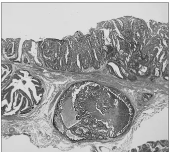
tasis. Given the clinical diagnosis of EGC, ESD was performed by a gastroenterologist. Histopathological examination of the specimen showed a moderately to poorly differentiated adenocarcinoma that invaded the submucosal layer to a depth of 1.5 mm (Fig. 2). The deep margin of the resected specimen had tumor involvement.
There was no evidence of lymphatic, venous, or perineural inva- sion. We therefore performed a laparoscopic distal gastrectomy with lymph node dissection. There was no evidence of residual carcinoma in the resected specimen and no evidence of metastasis in the 38 identified lymph nodes. The postoperative course was un- eventful, and the patient was discharged 14 days after the resection.
Fig. 1. Esophagogastroduodenoscopy showing a small superficial el- evated lesion in the antrum of the stomach.
Fig. 2. Histopathological examination of the specimens resected by endoscopic submucosal dissection revealing tubular adenocarcinoma with submucosal invasion (H&E, × 40).
Fig. 3. An abdominopelvic computed tomography scan showing a 3.5- cm-mass in the liver that demonstrated strong enhancement following the intravenous administration of contrast material.
Fig. 4. 18F-2-deoxy-2-fluoro-D-glucose (FDG) positron emission to- mography image showing intense FDG uptake in the liver.
Liver Metastasis from Early Gastric Cancer
141
The patient was followed-up regularly, and a hepatic metastasis was diagnosed 15 months postoperatively. Abdominopelvic CT re- vealed a 3.5-cm low attenuation lesion in the dome of the liver and a second 1-cm lesion in segment 8 of the liver (Fig. 3). Positron emission tomography-computed tomography (PET-CT) failed to demonstrate any other metastatic disease, except in the liver (Fig. 4).
Ultrasound-guided needle biopsy (TSK ACECUT biopsy needle;
TSK Laboratory, Tochigi, Japan) of the liver lesions confirmed the diagnosis of a metastatic poorly differentiated adenocarcinoma.
The patient underwent right anterior sectionectomy of the liver, and the specimen contained both lesions identified by imaging.
Histopathological examination confirmed the diagnosis of two foci of metastatic adenocarcinoma consistent with a gastric primary (Fig. 5). Immunohistochemical analysis of the lesion demonstrated the presence of HER2. The patient was treated with trastuzumab in combination with chemotherapy consisting of capecitabine and cisplatin. Twenty-four months after the operation, the patient re- mained free of recurrence.
Discussion
Lymph node metastasis is the most significant risk factor for EGC recurrence with a reported incidence of 1.4% to 2.8%.4 EGC patients with submucosal invasion have been reported to have a higher incidence of lymph node metastasis (23.8%). Unfortunately, preoperative diagnostic modalities for the detection of lymph node metastasis remain limited and inaccurate. Thus, when submucosal
invasion is identified in patients who underwent ESD, it is prudent to consider potential curative resection since the possibility of re- sidual tumor and/or lymph node metastasis after EMR/ESD can- not be ignored.1
Vascular invasion within the submucosal layer is a more signifi- cant risk factor for subsequent hepatic metastasis than lymph node or lymphatic invasion. Although venous invasion was not detected in this case, the venous invasion of primary tumor cells entering the portal circulation is presumed to be the cause of liver metasta- sis.3 EGC of the macroscopic elevated type is also associated with liver metastasis.3 Among EGCs, the dominant-elevated type has a relatively high incidence of vascular invasion thought to be due to tumor penetration and expansive growth.5
Hepatic resection for metastatic gastric cancer is not common due to the extremely poor postoperative prognosis. Therefore, liver resection would only benefit highly selected cases of hepatic metastasis from gastric cancer. Sakamoto et al.6 described criteria for considering resection of liver metastases in patients with gastric cancer. These include the absence of any distant metastatic disease including peritoneal dissemination and/or pulmonary involvement and the feasibility of macroscopic, complete gross resection.
In the present case, imaging (CT, magnetic resonance imaging, and PET-CT) revealed two lesions that were consistent with he- patic metastases. While only two metastatic lesions were identified, they were ultimately amenable to resection with a 5-mm safety margin. In patients with liver metastasis from colorectal cancer, liver resection with a tumor-free margin of less than 5 to 10 mm is sufficient because of the rare occurrence of satellite nodules around the main metastatic lesion.7 Meanwhile, in patients with hepatic metastases from gastric cancer the prognostic value of a particular surgical margin is controversial due to the aggressive biological be- havior of the disease.
Overexpression of the HER2/neu proto-oncogene product is identified in 10% to 20% of gastric cancers and is correlated with poor outcome.8 An increased incidence of liver metastasis has been observed in patients with gastric cancer exhibiting HER2 overex- pression. Dang et al.9 reported that 25.7% of gastric cancer patients with liver metastasis had tumors with HER2 overexpression.
Trastuzumab, a recombinant anti-HER2 monoclonal antibody, acts synergistically with appropriate chemotherapy in the treat- ment of HER2-positive gastric cancer. In a recent international phase III clinical trial, the addition of trastuzumab to chemotherapy significantly improved overall patient survival compared with that of patients treated with chemotherapy alone.10 In this report, im- Fig. 5. Histology of metastatic adenocarcinoma showing a glandular
pattern with luminal necrosis (H&E, × 40).
Oh SJ and Suh BJ
142
munohistochemical analysis of ultrasound-guided needle biopsy specimens of the patient’s metastatic liver mass demonstrated the presence of HER2. Therefore, our patient was treated with trastuzumab in combination with capecitabine and cisplatin. In a similar previously published case report, a small EGC removed by ESD showed submucosal invasion with lymphatic invasion and the patient subsequently underwent laparoscopic-assisted distal gas- trectomy with lymph node dissection. One year later, liver resection was performed due to recurrence and the patient was treated with trastuzumab plus capecitabine/cisplatin because the resected speci- men exhibited HER2 overexpression.11 In conclusion, liver me- tastasis rarely occurs in patients with EGC. Although there was no evidence of lymphatic, vascular, or perineural invasion in our case of EGC with submucosal invasion, the involvement of the deep margin and overexpression of HER2 potentially correlated with tumor recurrence.
References
1. Hirasawa T, Gotoda T, Miyata S, Kato Y, Shimoda T, Taniguchi H, et al. Incidence of lymph node metastasis and the feasibility of endoscopic resection for undifferentiated-type early gastric cancer. Gastric Cancer 2009;12:148-152.
2. Youn HG, An JY, Choi MG, Noh JH, Sohn TS, Kim S. Recur- rence after curative resection of early gastric cancer. Ann Surg Oncol 2010;17:448-454.
3. Ishida M, Morita S, Saka M, Fukagawa T, Taniguchi H, Katai H. Metachronous liver metastasis from early gastric cancer. J Gastrointest Surg 2012;16:837-841.
4. Saka M, Katai H, Fukagawa T, Nijjar R, Sano T. Recurrence in early gastric cancer with lymph node metastasis. Gastric Can-
cer 2008;11:214-218.
5. Kodama Y, Inokuchi K, Soejima K, Matsusaka T, Okamura T. Growth patterns and prognosis in early gastric carcinoma.
Superficially spreading and penetrating growth types. Cancer 1983;51:320-326.
6. Sakamoto Y, Ohyama S, Yamamoto J, Yamada K, Seki M, Ohta K, et al. Surgical resection of liver metastases of gastric cancer:
an analysis of a 17-year experience with 22 patients. Surgery 2003;133:507-511.
7. Yamamoto J, Sugihara K, Kosuge T, Takayama T, Shimada K, Yamasaki S, et al. Pathologic support for limited hepatectomy in the treatment of liver metastases from colorectal cancer.
Ann Surg 1995;221:74-78.
8. Kim SY, Kim HP, Kim YJ, Oh do Y, Im SA, Lee D, et al. Trastu- zumab inhibits the growth of human gastric cancer cell lines with HER2 amplification synergistically with cisplatin. Int J Oncol 2008;32:89-95.
9. Dang HZ, Yu Y, Jiao SC. Prognosis of HER2 over-expressing gastric cancer patients with liver metastasis. World J Gastroen- terol 2012;18:2402-2407.
10. Bang YJ, Van Cutsem E, Feyereislova A, Chung HC, Shen L, Sawaki A, et al; ToGA Trial Investigators. Trastuzumab in combination with chemotherapy versus chemotherapy alone for treatment of HER2-positive advanced gastric or gastro- oesophageal junction cancer (ToGA): a phase 3, open-label, randomised controlled trial. Lancet 2010;376:687-697.
11. Namikawa T, Shiga M, Ichikawa K, Kitagawa H, Kobayashi M, Hanazaki K. Metachronous liver and bone metastasis from small early gastric carcinoma without lymph node involve- ment: a case report. Mol Clin Oncol 2013;1:249-252.