Operative Treatment of the Tarsal Tunnel Syndrome Caused by Tarsal Coalition
Duck Joo Kwon, M.D., Sang Wook Park, M.D.
Department of Orthopedic Surgery, Hallym University Sacred Heart Hospital, College of Medicine, Anyang, Korea
=Abstract=
Purpose: Study was to evaluate the operative results for tarsal coalition with tarsal tunnel syndrome.
Materials and Methods: From Jan. 2005 to Mar. 2006, among a number of patients who were diagnosed with tarsal tunnel syndrome caused by tarsal coalition and treated surgically, 5 patients were closely observed for more than 12 months. All cases were talocalcaneal coalition and there were two male and three female patients with a mean age of 36 years (22-50 years). We used the Takakura rating scale as clinical evaluation.
Results: All five patients had a burning pain in the sole or extended to toes and showed positive Tinel’s sign.
Sensory disturbances were observed in the distribution of the medial plantar nerves in four patients and in the area of the medial and lateral plantar nerves in one. Atrophy and weakness of the plantar muscles were seen in two patients. The mean Takakura scale in preoperative and postoperative was 3.4 points (1 to 5 points), 8.6 point (6 to 10 points). The mean follow up was 14.4 months (12 to 16 months). The postoperative results were excellent in two patients, good in two and fair in one. As postoperative complications, there were persistent swelling in one patient and a flexion disturbance of Hallux in one.
Conclusion: The coalition resection performed on tarsal tunnel syndrome caused by tarsal coalition could improve a level of pains and neurological symptoms significantly. However, since there were some undesirable complications, a detailed explanation to patients is required prior to surgical treatment and study of such complications may be required.
Key Words: Talocalcaneal coalition, Tarsal tunnel syndrome, Coalition resection
∙Address for correspondence Sang Wook Park, M.D.
Department of Orthopedic Surgery, Hallym University Sacred Heart Hospital, 896 Pyeongan-dong, Dongan-gu, Anyang, Gyeonggi-do, 431-796, Korea
Tel: +82-31-380-1814 Fax: +82-31-382-1814 E-mail: [email protected]
* 본 논문의 요지는 2007년도 한일정형외과학회에서 발표되었음.
서 론
족근골 결합이란 족근골의 정상적인 분절 부전으로 인하 여, 둘 이상의 족근골이 섬유성, 연골성 또는 골성 결합을 보이는 선천성 족부 이상을 말한다1). 대부분 어릴 때는 증상 이 없다가 성장하면서 체중과 활동이 증가함에 따라 증상이 유발되는 것으로 알려져 있다2). 증상 있는 환자의 대부분은
Table 1. Details of Five Patients
Case Sex/
Age Diagnosis
Duration of symptom (months)
Pain Burning
pain Tinel sign Sensory disturbance
Muscle weakness
Takakura scale (preoperation)
1 F/42 Ta-Ca* 8 ++ + + Medial - 5
2 F/35 Ta-Ca 12 + + + Medial - 4
3 F/22 Ta-Ca 120 ++ ++ + Medial + 1
4 M/50 Ta-Ca 1 + ++ + Whole - 4
5 M/33 Ta-Ca 6 ++ + + Medial +- 3
*Ta-Ca, Talo-calcaneal coalition.
(A) (B)
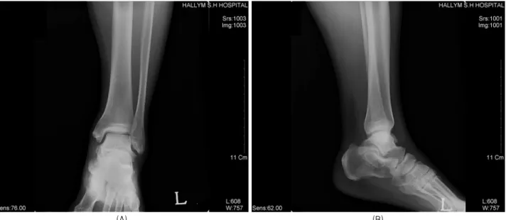
Figure 1. Preoperative plain posteroanterior (A) and lateral (B) roentgenograms of the left ankle revealed a typical characteristic of talocalcaneal coalition.(‘C-sign’)
활동시 악화되는 족부 피로감, 후족부 동통 등의 비전형적 인 족부 동통과 내반 및 외반의 제한을 호소한다1). 하지만 후 경골 신경의 압박으로 인한 이상감각 및 감각저하 등 족 근관 증후군을 보이는 경우도 있다3,4). 족근골 결합에 의한 족근관 증후군의 치료는 Takakura 등이 1991년 최초로 보고하였으며3), 아직까지 치료 결과에 대한 공식적인 국내 보고는 없어 이에 대하여 알아보고자 하였다.
대상 및 방법
1. 연구 대상
2005년 1월부터 2006년 3월까지 본원에서 족근골 결합에 의한 족근 관 증후군으로 진단받고 골 결합 제거술을 시행 받은 환자 중 12개월 이상 추시 관찰 가능하였던 5예를 대
상으로 하였다. 수술시 연령은 평균 36세(22~50세)였고, 남자가 2예, 여자가 3예를 차지하였다. 증상을 나타내고 수 술을 받기까지의 기간은 평균 29.4개월(1개월~10년)이었다 (Table 1).
진단은 일반 방사선 전후면, 측면, 외전 20도 사진을 이용하였다(Fig. 1). 모든 환자에서 컴퓨터 단층 촬영을 시 행하였다(Fig. 2). 컴퓨터 단층 촬영에서 골 결합의 유무 가 명백하지 않았던 1예의 경우, 다른 원인을 감별하기 위 하여 자기 공명 영상 촬영을 하였다(Fig. 3).
연구대상의 모든 환자는 중간 소관절 면의 결합이었고, 관절면의 50%를 침범하지는 않았다. 또한 거골하 관절에 관절염 소견은 관찰되지 않았다.
2. 연구 방법
Figure 2. Nonosseous coalition of middle facet in CT coronal plane was revealed. Joint space narrowing with sclerosis and cortical irregularity was shown.
Figure 3. T2-weighted coronal MR image of a fibrous coalition showed the horizontal orientation of the inferior surface of the sustentaculum tali.
Figure 4. The photo showed the external appearance of medial aspect of the right ankle. The linear line outlined the planned skin incision. The swelling was posteroinferior to the medial malleolus.
The swelling corresponded to the bony prominence of talocalcaneal coalition.
환자들의 평가는 자발통 및 운동통, 작열통, Tinel 징후, 감각이상, 족저 근 위축의 5항목을 기초로 하여 점수를 합산하는 Takakura의 rating scale을 이용하였다. 결과는 우수 10점, 양호 8~9점, 보통 6~7점, 불량 5점 이하로 등급을 나누어서 술 전 및 술 후를 비교하였다.
3. 수술 방법 및 수술 후 처치
환자들 모두 족근 관절 내과 후하방에 족근골 결합에 의 한 무동성 종물이 촉지 되었다(Fig. 4). 골성 족근골 결합 부위 위로 후 경골 신경의 방향에 평행하게 피부절개를 가 하고, 굴곡 지대(flexor retinaculum)를 종적으로 절개하 였다(Fig. 5-A). 장 족지 굴건과 후 경골 신경 사이로 조심 스럽게 박리하여 거종 결합의 내측으로 접근하였다. 후 경 골 신경 및 그 분지들은 거종 결합에 의해 압박되어 후방 전 위되어 있었다(Fig. 5-B). 절골기 및 골겸자 등을 이용하여 골결합을 제거한 후, 작은 골막 박리기를 넣어 중간 소면의 움직임을 확인하였다. 족근 관 감압을 위하여 굴곡 지대를 근위부에서 윈위부까지 신경 확인 후 노출시켜, 후 경골 신 경을 덮고 있는 장무지 외전근 원위부까지 신경을 가동성 있게 만들었다. 골결합을 제거한 후, 감압된 후 경골 신경을 확인할 수 있었다(Fig. 5-C). 혈종에 의한 신경 재 압박을 예방하기 위하여 bone wax로 골 제거 부위에 도포한 후, 흡인 배액관을 유치하였다. 수술 후 단순 방사선 촬영을 통 하여 골결합 제거를 확인하였다.
수술 후 첫 3주간은 단하지 부목 고정 및 목발보행을 실 시하였다. 수술 후 3주째 부목 고정을 제거하였으며, 부분 체중부하 및 족근 관절 운동을 시작하였고, 6주째부터 전 체중부하를 허용하였다.
(A) (B) (C)
Figure 5. Intraoperative photo showed posterior tibial nerve lying directly over talocalcaneal coalition. The nerve was stretched over the bony prominence. Fibrotic thickening of the nerve was observed(A). After excising the coalition, a good articulation of middle facet (B) and a decompressed posterior tibial nerve (C) were observed.
Table 2. Results of Operation
Case Follow-up (months)
Residual findings
Takakura scale
(postoperation) Overall result Pain Burning pain Tinel sign Sensory
disturbance Muscle weakness
1 13 - - - - - 10 Excellent
2 12 - - - + - 9 Good
3 14 + - + + + 6 Fair
4 15 - - - - - 10 Excellent
5 16 - - - + + 8 Good
결 과
1. 임상증상
다섯 환자 모두 내과 후하방에 고정된 단단한 종물이 촉 지 되었으며, 주변부로 동통을 호소하였다. 정도의 차이는 있었으나, 모든 환자가 발바닥 혹은 발가락 쪽으로 뻗치는 작열통을 호소하였고, Tinel sign 양성을 보였다. 내측 족 저 신경 지배영역에 감각이상을 호소한 경우가 4예였으며, 1예에서는 족저 전반(내, 외측 족저 신경 지배영역)에 걸쳐 감각이상을 호소하였다. 2예에서는 족저 근 위축이 관찰 되었다(Table 1).
2. 평가
수술 전 Takakura scale은 평균 3.4점(1~5점)이었고, 수술 후 평균 추시 기간은 14.4개월(12~16개월)이었다(Table 1). 수술 후에 자발통 및 운동통, 작열통은 대부분의 환자 에서 조기에 회복되었으나, 감각이상 및 Tinel 징후, 족저 근 위축은 서서히 완화되는 양상을 보였다. 2예에 있어서는 족저 근 위축 및 감각이상이 15개월 추시 기간 동안 회복 정
도가 완전하지 않았다. 술 후 Takakura scale은 평균 8.6점 (6~10점)이었고, 성적은 우수 2예, 양호 2예, 보통 1예였다 (Table 2).
3. 합병증
수술 부위의 지속적 동통이 발생한 경우가 1예, 수술 후 무지관절의 굴곡장애를 호소한 경우가 1예 있었다. 이중 후 자의 경우 수술 후 약 4주째부터 굴곡기능이 회복되기 시작 했고, 12개월 추시 결과 굴곡력 Grade IV로 거의 정상이며, 일상생활에 전혀 불편함을 호소하지 않았다.
고 찰
족근골 결합의 유병률은 0.03%~1%로 매우 드물고, 대부 분은 종주상 결합과 거종 결합이 차지한다5). 이중 종주상 결합은, 거종 결합에 비해 상대적으로 어린 나이에 증상을 나타내며, 사춘기 이후의 증상을 보이는 환자는 주로 거종 결합으로 보고되고 있다.
거종 결합은 족근골 결합의 흔한 형태 중 하나이다. 대부 분의 환자가 연령이 증가함에 따라 체중 증가 및 족근골
있다3,4).
족근관 증후군은 후 경골 신경 또는 그 분지들이 족근관 내에서 압박되어 발생하는 압박성 신경병증의 한 형태이 다. 족근관은 족근 관절의 내측부로부터 중족부까지 이어 지며, 굴곡 지대를 표층 경계, 거골 및 종골의 내측면, 재거 돌기(sustentaculum tali)를 외측 경계, 무지 외전 근을 하부 경계로 한다. 그 내부에는 족근 관절 내측 건들과 후 경골 신경 및 동정맥이 통과한다6). 후 경골 신경은 이 부 근에서 내, 외측 족저부 및 종부(heel)로 가는 감각신경 분 지를 형성한다7). 족근관 협착이 가장 잘 일어나는 부위는 내, 외측 족저 신경이 방향을 전환하여 족저부로 내려가는 이행부위로 굴곡 지대의 원위 경계부가 이에 해당한다8). 따 라서 이러한 해부학적인 위치관계에 따라 거종결합에 의한 족근관 증후군이 발생할 수 있다. 거종 골결합은 내측 혹은 후방에 발생하게 되며, 이중 전자의 경우가 내측 족저 신경 을 압박하여 족근관 증후군을 더 잘 유발하는 것으로 알려 져 있다4).
족근관 증후군의 원인으로는 종양, 대사이상, 외상, 족부 및 족관절 이상 등이 보고되고 있다. Mann은 전체 환자의 약 60% 정도에서 원인을 찾을 수 있다고 하였다9). 여러 가지 공간점유병소가 족근관 증후군을 일으킬 수 있으며, 색소 융모 결절성 활액막염(pigmented villonodular sy- novitis), 지방종, 활액 낭종, 신경내 퇴행성 낭종(intra- neural degenerative cyst), 결절성 낭종, 신경초종(neu- rilemoma), 골절된 재거 돌기, 그리고 족근골 결합 등이 원 인이 된다9). 따라서, 공간 점유 병소에 의한 족근관 증후군 을 의심할 경우에는 초음파 혹은 핵자기 공명 영상 검사가 치료 방침 및 수술 계획 설정에 도움이 될 수 있다4).
족근관 증후군의 진단은 문진과 이학적 검사, 그리고 방 사선 검사 특히 전산화 단층 촬영 및 자기 공명 영상 결과 를 종합하여 이루어진다. 전형적인 증상은 후 경골 신경 및 그 분지의 지배영역 부위로 활동 시나 혹은 야간에 악화되 는 통증이며10), 같은 부위로 이상감각이나 감각저하 등이 동반되어 있는 경우가 많다. 환자의 약 80%에서 신경전도 검사에서 이상소견을 보이는 것으로 알려져 있으므로11),
어 보존적 요법보다는 수술적 요법이 선호 된다3). 하지만 수술시 연령이 높을수록, 증상 보유기간이 길수록 수술결과 가 불량한 것으로 알려져 있다13). 저자들의 연구에서 보통 의 결과를 나타난 1예는 수술시 연령은 22세로 높지 않았으 나, 증상 보유기간이 약 10년으로 매우 길었고, 수술 전 Takakura scale이 1점으로 최하점을 받았던 환자였다.
족근관 증후군의 수술은 굴근지대를 근위부에서 윈위부 까지 신경 확인 후 노출시켜 후 경골 신경을 덮고 있는 장무 지 외전 근 원위부까지 신경을 가동성 있게 만드는 것이 매우 중요하다고 하였다8). 또한, 골결합을 제거한 후, 작은 골막 박리기를 넣어 중간 소면의 움직임을 확인하고, 골 제거 후 지방, 장 무지굴건과 같은 연부 조직의 개재를 권유 하였다14-16).
족근골 결합의 치료에 대한 합병증으로는 감염, 창상치 유지연 및 괴사, 반사성 교감 신경 이영양증(reflex sym- pathetic dystrophy), 가관절 형성, 재발, 지속적 동통 등 이 보고되고 있다17-19). 저자들의 경우 수술 후 지속적 동통 이 발생한 경우가 1예, 무지 관절의 굴곡 장애를 호소한 경우가 1예 있었다. 이 중 전자의 경우는 지속적인 부종과 함께, 교감 신경 이영양증이 관찰되었으며, 보존적 치료 후 호전되었다. 후자의 경우는 거종 골결합에 의하여 후방 전 위되고 신연되어있던 장 무지 굴건이 골결합의 제거에 따라 다시 전방 전위되면서, 건의 활주와 무지의 운동에 장애가 발생했을 것으로 사료된다. 하지만 아직까지 이런 합병증에 대한 공식적인 보고가 없고 증례가 적어 이에 대한 추가적 인 연구가 필요하겠다.
결 론
족근골 결합에 의한 족근관 증후군의 경우에 골 결합의 제거술은 거골하 관절의 운동을 호전시키며 압박된 후 경골 신경 및 그 분지들의 감압효과도 함께 얻을 수 있다. 하지만 수술 후에 발생 가능한 합병증에 대해 수술 전 환자에게 충분한 설명을 요할 것으로 사료된다.
REFERENCES