ISSN: 2233-601X (Print) ISSN: 2093-6516 (Online)
− 489 −
Received: May 19, 2016, Revised: July 5, 2016, Accepted: July 6, 2016, Published online: December 5, 2016
Corresponding author: Sang-Ho Cho, Department of Thoracic and Cardiovascular Surgery, Kyung Hee University Hospital at Gangdong, 892 Dongnam-ro, Gangdong-gu, Seoul 05278, Korea
(Tel) 82-2-440-6158 (Fax) 82-2-440-8004 (E-mail) [email protected]
© The Korean Society for Thoracic and Cardiovascular Surgery. 2016. All right reserved.
This is an open access article distributed under the terms of the Creative Commons Attribution Non-Commercial License (http://creativecommons.org/
licenses/by-nc/4.0) which permits unrestricted non-commercial use, distribution, and reproduction in any medium, provided the original work is properly cited.
Valve-Sparing Root Replacement: Aortic Root Remodeling with External Subvalvular Ring Annuloplasty
Sang-Ho Cho, M.D., Ph.D., Dae Hyun Kim, M.D., Ph.D., Young Tae Kwak, M.D., Ph.D.
Department of Thoracic and Cardiovascular Surgery, Kyung Hee University Hospital at Gangdong, Kyung Hee University School of Medicine
The original valve-sparing procedures for aortic root aneurysms were remodeling and reimplantation of the aortic root. The remodeling technique provides more physiologic movement of the cusps within 3 re- constructed neo-sinuses, thus preserving root expansibility through the interleaflet triangles. However, the du- rability of remodeling has been a matter of concern due to the high rate of aortic insufficiency when an- nular dilation is not addressed. Therefore, a modified approach was developed, combining a physiologic re- modeling of the root with a subvalvular annuloplasty. This case report highlights the first case of successful aortic root remodeling with external subvalvular ring annuloplasty in Korea.
Key words: 1. Aortic root 2. Aortic aneurysm 3. Aortic valve, surgery 4. Cardiac valve annuloplasty
Case report
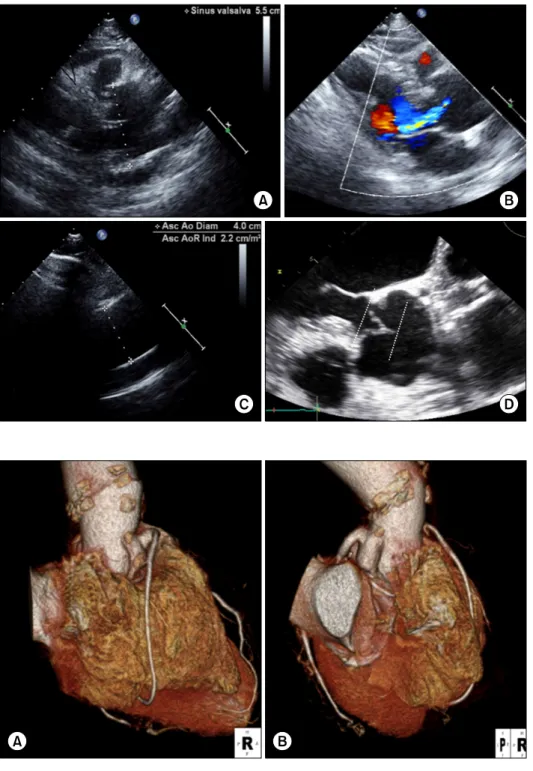
A 69-year-old man was referred for the surgical management of an aortic root aneurysm found by echocardiography during a preoperative work-up for spinal surgery. He did not have a family history or cardinal features of any heritable disorder of the connective tissue. A transthoracic echocardiographic examination showed an enlarged sinus of Valsalva (55 mm in diameter) with moderate aortic regur- gitation (Fig. 1A, B). The maximal diameters of the sinotubular junction and ascending aortic tubular portion were 44 and 40 mm, respectively (Fig. 1C).
The left ventricular ejection fraction was 65% with no regional wall motion abnormalities. A transesophageal echocardiographic examination showed aortic valve cusp coaptation at a level close to the middle of the
sinuses of Valsalva (Fig. 1D). The patient was sched- uled for aortic root remodeling with concomitant ex- ternal subvalvular ring annuloplasty.
After establishing cardiopulmonary bypass (CPB) and aortic clamping, the ascending aorta was trans- ected above the sinotubular junction. After external dissection down to the base of the aortic annulus, the wall of the aortic sinus was completely removed, leaving a fringe of the aortic wall, and the coronary artery buttons were individualized. The diameters of the Valsalva graft and the flexible ring were de- termined to be 26 and 25 mm, respectively, based on the internal aortic annular base diameter meas- ured with a 28-mm H egar dilator (Video 1). Five su- tures of 2-0 polyester fiber with pledgets were passed from the inside out as U stitches in the sub- valvular plane. A sixth suture without pledgeting was
Korean J Thorac Cardiovasc Surg 2016;49:489-492 □ HOW-TO-DO IT □
https://doi.org/10.5090/kjtcs.2016.49.6.489
Sang-Ho Cho, et al
− 490 −
Fig. 2. (A, B) Postoperative computed tomographic images (3-dimensional reconstruction) show reconstructed neo-sinuses, interleaflet triangles, and patent coronary artery button anastomoses.
Fig. 1. Preoperative echocardiogra- phy. (A) Transthoracic echocardiog- raphy shows an enlarged sinus of Valsalva (55 mm in diameter). (B) A parasternal long axis view shows moderate aortic regurgitation. (C) The maximal diameter of the as- cending aortic tubular portion was 40 mm. (D) Transesophageal echo- cardiography shows cusp coapta- tion at a level close to the middle of the sinuses of Valsalva.
placed externally at the level of the interleaflet tri- angle between the right and noncoronary sinus due to a lesion at the bundle of H is (Video 2). The first step of aortic cusp repair for the alignment of the coaptation margin was the placement of a central pli- cation stitch on the extended noncoronary cusp. The length of each hemi-cusp free edge between the right and noncoronary cusp was equalized (Video 3). The
remodeling technique was performed, using a 26 mm Gelweave Valsalva graft (Vascutek, Renfrewshire, Sco- tland), allowing a bulging aspect of the neo-sinuses of Valsalva and preserving the interleaflet triangles.
After remodeling the aortic root, the second step of aortic cusp repair was performed, involving the re- suspension of an effective height. Three plication stitches on the right coronary cusp and 1 plication
Aortic Root Remodeling with Annuloplasty
− 491 − stitch on the noncoronary cusp were added on the free edge until any residual or induced cusp prolapse was resolved and an effective height greater than 9 mm was obtained (Video 4). After reducing the ring length to 8 cm with a diameter of 25 mm, the six anchoring U stitches were passed through the inner aspect of the SJM Tailor flexible ring (St. Jude Medical, St. Paul, MN, USA). The ring was then brought downward around the remodeled aortic root and the U stitches were tied to fix the ring in a sub- valvular position (Video 5). The coronary ostia were re- implanted following the usual technique with a 6-0 prolene suture, and distal anastomosis was per- formed with the use of running 4-0 prolene sutures.
The total cross-clamp time was 210 minutes, and the patient was successfully weaned from CPB.
The patient had an uneventful postoperative course with regard to hemodynamic performance. Echocardi- ography on postoperative day 7 revealed a com- petent aortic valve with no regurgitation (Video 6) and a computed tomography scan showed a well-re- constructed aortic root (Fig. 2). H e was discharged from the hospital on postoperative day 13 and followed up at an outpatient clinic for 9 months. Follow-up echocardiography at 8 months was performed and showed no significant aortic regurgitation.
Discussion
In the past 2 decades, valve-sparing root replace- ments (VSRRs) have been developed as alternatives to the Bentall procedure for dystrophic aortic root aneurysms combined with aortic valve regurgitation.
The two most widely used techniques are the remod- eling technique, developed by Yacoub, and the reim- plantation technique, developed by David [1]. Aortic root remodeling does not correct the dilation of the aortic annulus that can lead to the failure of the pro- cedure and the recurrence of aortic insufficiency. As a result, many surgeons prefer the reimplantation technique. David et al. [2] recommended the reim- plantation technique when the aortic annulus is >27 mm in men and >25 mm in women, while the re- modeling technique is suitable for older patients with a normal aortic annulus. When selecting a technique, Hanke et al. suggested an annular diameter of 28–30 mm as a cutoff value, recommending the Yacoub technique for annuli with narrower diameters and
the David technique for wider diameters [3]. However, the classic remodeling technique provides more phys- iologic movements of the cusps within the three re- constructed neo-sinuses. This preserves root expan- sibility through the interleaflet triangles during the cardiac cycle. Recent dynamic studies have suggested that cusp motion and flow patterns across the re- constructed aortic root are more physiologic after re- modeling of the aortic root than after reimplantation of the aortic valve [1,4]. As a result, recent research has focused on modified remodeling techniques, such as a remodeling root reconstruction in association with an aortic annuloplasty to combine the advan- tages of both the remodeling and reimplantation techniques. In this case, we performed an approach standardized by Lansac that restored normal aortic cusp geometry (valve repair) and reduced the dilated aortic annulus using an external subvalvular ring an- nuloplasty device [1,5]. Valve repair is achieved in two steps. First, before root reconstruction to realign ad- jacent cusp free edges, the excess length of the cusp is corrected with central plication stitches along the nodule of Arantius or by triangular resection of this area. Second, after root reconstruction, residual or in- duced cusp prolapse is resuspended with central pli- cation stitches in order to obtain an equivalent effec- tive height of all cusps (distance between the free edge of the cusp to the aortic annular base) [1,5]. Prol- apse frequently occurs after VSRR irrespective of the preoperative degree of aortic regurgitation. An effec- tive height of less than 9 mm indicates prolapse, which in most instances should be corrected [5]. In addition to valve repair, Lansac et al. [1] advocated an external subvalvular annuloplasty using an ex- pansible flexible ring when the aortic annular base diameter is ≥25 mm. Compared with other techni- ques such as plication stitches, partial annuloplasty, and internal annuloplasty, an external ring annulo- plasty may ensure optimal reduction in the aortic an- nular base diameter, cusp mobility, and the preserva- tion of the conduction system. Moreover, subvalvular annuloplasty through an aortic ring partially in- creases the coaptation height and compensates for the induced symmetric prolapse that usually occurs after root replacement [1,5]. Six anchoring stitches are needed to implant the ring. Five sutures of 2-0 polyester fiber with pledgets are placed from the in- side out as U stitches circumferentially in the sub-
Sang-Ho Cho, et al
− 492 − valvular plane, below the nadirs of each cusp, and at the base of each interleaflet triangle except for one.
A sixth stitch is passed externally at the level of the interleaflet triangle between the right and noncoro- nary sinus in order to preserve the membranous sep- tum or the bundle of H is lesion [1].
This is a case report describing a standardized ap- proach to VSRR. We suggest that physiologic remod- eling of the aortic root can be safely combined with aortic valve repair and subvalvular annuloplasty us- ing an external ring for dystrophic aortic root aneurysms.
Conflict of interest
No potential conflict of interest relevant to this ar- ticle was reported.
Supplementary materials
Supplementary materials can be found via https://
doi.org/10.5090/kjtcs.2016.49.6.489. Video 1. Dissection of the subvalvular plane.
Supplementary materials can be found via https://
doi.org/10.5090/kjtcs.2016.49.6.489. Video 2. Placement of six anchoring sutures for ring annuloplasty. Five sutures with pledgets were passed from the inside out as U stitches circumferentially in the subvalvular plane, below the nadirs of each cusp, and at the base of each interleaflet triangle except for one. A sixth stitch without pledgeting was placed externally at the level of the interleaflet triangle between the right and noncoronary sinus.
Supplementary materials can be found via https://
doi.org/10.5090/kjtcs.2016.49.6.489. Video 3. The first step of aortic cusp repair for the alignment of the
coaptation margin. A polypropylene 6-0 stay suture was passed through each nodule of Arantius. The distance between the two stitches determined the area for the central plication stitches to equalize each hemi-cusp.
Supplementary materials can be found via https://
doi.org/10.5090/kjtcs.2016.49.6.489. Video 4. The sec- ond step of aortic cusp repair for the resuspension of an effective height. Central plication stitches were placed to obtain an equivalent effective height for all cusps. The effective height of all cusps should be greater than 9 mm.
Supplementary materials can be found via https://
doi.org/10.5090/kjtcs.2016.49.6.489. Video 5. Aortic annuloplasty using an external subvalvular ring.
Supplementary materials can be found via https://
doi.org/10.5090/kjtcs.2016.49.6.489. Video 6. Postope- rative echocardiography shows a competent aortic valve with no regurgitation.
References
1. Lansac E, Di Centa I, Vojacek J, et al. Valve sparing root replacement: the remodeling technique with external ring annuloplasty. Ann Cardiothorac Surg 2013;2:117-23.
2. David TE, Maganti M, Armstrong S. Aortic root aneurysm:
principles of repair and long-term follow-up. J Thorac Cardiovasc Surg 2010;140(6 Suppl):S14-9.
3. Hanke T, Charitos EI, Stierle U, et al. Factors associated with the development of aortic valve regurgitation over time after two different techniques of valve-sparing aortic root surgery. J Thorac Cardiovasc Surg 2009;137:314-9.
4. Soncini M, Votta E, Zinicchino S, et al. Aortic root perform- ance after valve sparing procedure: a comparative finite el- ement analysis. Med Eng Phys 2009;31:234-43.
5. Schafers HJ, Aicher D. Root remodeling for aortic root dilatation. Ann Cardiothorac Surg 2013;2:113-6.