INTRODUCTION
The vascular endothelial growth factor (VEGF) is known to be a vascular permeability factor and an endothelial cell growth factor secreted by both the smooth muscle and endo- thelial cells. VEGF is a secreted endothelial cell mitogen and play a major role in hypoxia-stimulated angiogenesis (1). Vas- cular endothelial cells have binding sites with a high affinity for VEGF and VEGF itself is thought to play a major role both in preserving vascular endothelial cells and vascular endothelial cell recovery (2). VEGF is regulated by the Flt-1 and Flk-1/KDR receptors, which are part of the tyrosine kinase family (3, 4). Flt-1, which has a higher affinity for VEGF than Flk-1/KDR, is thought to mainly regulate the formation of cellular architecture, while Flk-1/KDR is in- volved in cell proliferation (5-8).
However, the subsequent mechanism of signal transduc- tion following the VEGF receptor in vascular cells is uncer- tain. The effects of VEGF in permeability and vascular ten- sion are associated with nitric oxide (NO) formation (9, 10), and numerous studies have reported a correlation between the endothelial proliferating effect of VEGF with NO (11-
13). Angiogenesis associated with NO was investigated in mouse arteries in vitro or in vivo vascular injury experiments.
In experiments using arteries or arterioles, VEGF protein ex- pression is increased while the NO metabolite concentration initially decreases and then increases thereafter. However, in arteries injured with balloons, addition of VEGF protein or naked VEGF cDNA re-stimulates endothelial proliferation, and recovers the endothelial cell-dependent vascular response (14, 15). Such effects correlate VEGF with endothelial cell growth, and this new vascular formation is thought to be due to the continuous maintenance of NO formation. Also, the addition of NO synthase (NOS) inhibitors in endothelial cell cultures inhibited VEGF-induced cell growth in addition to the reticular formation of newly synthesized vascular tissues (12).
However, the exact mechanism of action of VEGF in NO formation is still unclear. Recently, several reports have de- monstrated that VEGF-stimulated NO production reguires activation of the phosphatidylinositol 3-kinase (PI-3K) and mitogen-activated protein kinase (MAPK) signal transduc- tion pathways (16, 17).
This study was designed to elucidate whether the changes Jong Seon Park, Gu Ru Hong,
Suk Whan Baek*, Dong Gu Shin, Young Jo Kim, Bong Sup Shim
Department of Internal Medicine, and Department of Biochemistry and Molecular Biology*, College of Medicine, Yeungnam University, Daegu, Korea
Address for correspondence Jong Seon Park, M.D.
Department of Internal Medicine, College of Medicine, Yeungnam University, 317-1 Daemyung-dong, Nam-gu, Daegu 705-717, Korea Tel : +82.53-620-3847, Fax : +82.53-654-8386 E-mail : [email protected]
161
Expression and Regulation of Endothelial Nitric Oxide Synthase by Vascular Endothelial Growth Factor in ECV 304 Cells
Nitric oxide (NO) seems to play a pivotal role in the vascular endothelial growth factor (VEGF)-induced endothelial cell proliferation. This study was designed to investigate the role and intracellular signal pathway of endothelial nitric oxide synthase (eNOS) activation induced by VEGF. ECV 304 cells were treated with VEGF165and then cell proliferation, eNOS protein and mRNA expression levels were analyzed to elucidate the functional role of eNOS in cell proliferation induced by VEGF. After exposure of cells to VEGF165, eNOS activity and cell growth were increased by approximately two-fold in the VEGF165-treated cells compared to the untreated cells. In addition, VEGF stimulated eNOS expression at both the mRNA and protein levels in a dose-dependent manner. Phosphatidylinositol-3 kinase (PI-3K) inhibitors were used to assess PI-3K involvement in eNOS regu- lation. LY294002 was found to attenuate VEGF-stimulated eNOS expression.
Wortmannin was not as effective as LY294002, but the reduction effect was de- tectable. Cells activated by VEGF showed increased ERK1/2 levels. Moreover, the VEGF-induced eNOS expression was reduced by the PD98059, MAPK path- way inhibitor. This suggests that eNOS expression might be regulated by PI-3K and the ERK1/2 signaling pathway. In conclusion, VEGF165induces ECV 304 cell proliferation via the NO produced by eNOS. In addition, eNOS may be regulated by the PI-3K or mitogen-activated protein kinase pathway.
Key Words : Angiogenesis; Nitric-Oxide Synthase
Received : 4 October 2001 Accepted : 11 January 2002
in eNOS activity by VEGF are simply due to variations in the eNOS activation rate, or due to the changes in the eNOS protein and mRNA levels. In addition, whether or not PI- 3K, a known crucial factor in cell growth, and extracellular regulated kinase 1/2 (ERK1/2), a MAPK family, are associ- ated with the eNOS protein expression by VEGF was ex- amined.
MATERIALS AND METHODS Materials
The recombinant VEGF protein, [3H] citrulline, [( -32P]
dCTP, [( -32P] ATP, and enhanced chemiluminescence (ECL) reagents were purchased from Amersham (Buckinghamshire, U.K.). M199 and the phosphate-buffered saline were obtained from Gibco BRL (Grand Island, NY, U.S.A.). The fetal calf serum was purchased from Hyclone (Logan, UT, U.S.A.). The rabbit polyclonal eNOS antibodies, and the anti-rabbit IgG peroxidase-conjugated secondary antibodies were purchased from Santa Cruz Biotechnology (Santa Cruz, CA, U.S.A.).
The phospho-ERK antibodies and phospho-p38 antibodies were purchased from New England BioLab (Beverly, MA, U.S.A.). The LY294002, wortmannin, and PD98059 were purchased from Biomol (Plymouth Meeting, PA, U.S.A.) and were dissolved in dimethyl sulfoxide (DMSO) prior to addi- tion to the cell cultures or enzyme assays; the final concentra- tions in DMSO were 0.1% or less. Controls using DMSO alone were run in parallel all cases.
Cell Culture
ECV 304 was obtained from the American Type Culture Collection. The cells were cultured in M199 supplemented with 2 mM L-glutamine, 100 U/mL penicillin, 100 mg/mL streptomycin, and 10% fetal calf serum. The cells were grown at 37℃, 5% CO2 in fully humidified air and subcultured twice weekly. The cells were seeded on 12-well plates at 1
×105 cells/well or 6-well plates at 5×10⁵ cells/well. The cells were stimulated for set times ranging from 1 hr to 24 hr in the presence of VEGF with or without the inhibitors.
The viability of the ECV 304 cells in the presence of the VEGF was assessed after different days of incubation by the Trypan Blue exclusion method.
Quantitation of Intracellular NO generation
The eNOS enzyme activity was quantified by L-[3H] cit- rulline formation from L-[3H] arginine. Briefly, the cells in a given well of the 6-well plate were incubated in 800 L of a buffer containing 25 mM HEPES, pH 7.3, 109 mM NaCl, 5.4 mM KCl, 0.9 mM CaCl2, 1 mM MgSO4, and 25 mM glucose at 37℃for 1 hr. The eNOS activity was assayed by
adding a mixture of unlabeled 10 M L-arginine, 10 Ci/mL L-[3H] arginine, and VEGF or the vehicle to the culture. Fol- lowing incubation at 37℃for 10 min, the cells were washed with ice-cold phosphate-buffered saline (PBS), scraped into 2 mL of a solution containing 20 mM sodium acetate, 1 mM L-citrulline, 2 mM EDTA, and 2 mM EGTA, pH 5.5, fol- lowed by sonication. An aliquot was withdrawn to determine the total protein concentration and the total amount of cel- lular 3H incorporation. The remaining sample was applied to the Dowex 50W-X8 400 column to separate L-[3H] cit- rulline. The flow-through fraction was analyzed by liquid scintillation counting.
Western blotting and MAPK activation
The ECV 304 cells were plated in a 6-well plate and treated with VEGF with various inhibitors. They were then washed with cold-PBS, scraped off and pelleted at 700×g and at 4
℃. The cell pellet was resuspended in lysis buffer (50 mM Tris-HCl, pH 8.0, 5 mM EDTA, 150 mM NaCl, 0.5% Non- idet P-40, 1 mM PMSF, and protease inhibitor cocktail). The preparation was then cleared by centrifugation and the super- natant saved as a whole-cell lysate. The proteins (50 g) were separated by 8% reducing SDS-PAGE and electroblotted in 20% methanol, 25 mM Tris, and 192 mM glycine onto a nitrocellulose membrane. The membrane was then blocked with 5% non-fat dry milk in TTBS (25 mM Tris-HCl, 150 mM NaCl, and 0.2% Tween-20) and subsequently incubated with the eNOS antibody for 4 hr. Subsequently, the mem- brane was washed and incubated for 1 hr with secondary anti- bodies conjugated to HRP. Finally, the membrane was washed and developed using an enhanced ECL system. The activa- tion of ERK and p38 was determined by Western blotting using antibodies specific for phosphorylated, active forms of the corresponding MAPKs. As loading controls, Western blots were also performed using antibody against total ERK in a 1:1,000 dilution.
RT-PCR
The ECV 304 cells (2×10⁶cells) were cultured and har- vested. The cells were then washed three times with PBS containing 2% bovine serum albumin (BSA), and the RNA was isolated using a Tri-Reagent kit (Molecular Research Cen- ter, Cincinnati, OH, U.S.A.). A modified reverse transcrip- tase-polymerase chain reaction (RT-PCR) technique was used to analyze eNOS at the mRNA level. Briefly, the total RNA was reverse transcribed into cDNA using the RT-PCR kit.
Oligonucleotide primers for the PCR were designed accord- ing to the published sequence for human eNOS (sense primer:
ATT ATC CAG AGC GAG AGC CT; antisense primer: TCT TGC CAC CAC TGT GTT GT). The PCR conditions for eNOS production was 24 cycles of denaturation (95℃/1 min), annealing (60℃/1 min), and extension (72℃/1 min) in the
presence of 2.5 mg MgCl2, followed by a final extension at 72℃for 20 min. Oligonucleotide primers for -actin were used as internal control for RT-PCR. The PCR products were separated by electrophoresis using a 2% agarose gel in TBE buffer containing 50 ng/mL of ethidium bromide.
Statistical analysis
Statistical analysis was performed using SPSS 10.0 for Win- dows. Data were given as mean±standard deviation and comparison of the two groups was made by unpaired Stu- dents t test. A p-value of <0.05 was considered significant.
RESULTS
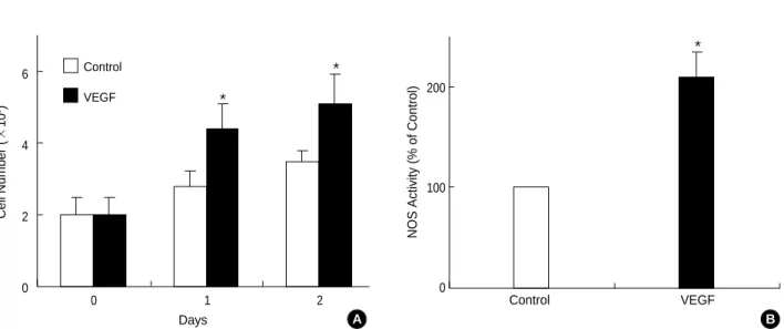
Initially, the ECV 304 cells were exposed to 20 ng/mL VEGF165 to determine the effect of VGEF on cell growth.
The VEGF165protein was found to significantly increase the number of cells by almost 2.2 times after 24 hr, and by 2.5 times after 48 hr (Fig. 1A).
To investigate the effect of VEGF165on eNOS activity, after treating 20 ng/mL VEGF165in an arginine substrate for 24 hr, the concentration of citrulline produced by the NOS en- zyme was measured. The results showed that NOS enzyme activation was increased by two-fold in the cells treated with the VEGF165protein, compared with the control group that was not treated with the VEGF165protein (Fig. 1B).
To determine whether the increase in NOS activity obser- ved in the ECV 304 group was the result of eNOS protein expression, the expression of the eNOS and inducible NOS (iNOS) protein was measured after exposing the ECV 304 cells to VEGF165 concentrations ranging from 5 ng/mL to
20 ng/mL for 24 hr. In the cells treated with the VEGF165 protein, eNOS protein expression (about 130 kDa) was in- creased in a dose-dependent manner, and eNOS protein ex- pression in the endothelial cells exposed to 20 ng/mL VEGF165 for 24 hr increased by approximately two-fold compared with the control cells (Fig. 2). However, iNOS expression was not
Cell Number (×105) 6
4
2
0
Fig. 1.Effect of VEGF on cell growth and NOS activity. (A) Confluent ECV 304 cells stimulated with the VEGF (20 ng/mL) for the different days. After treatment with VEGF, the cells were collected and counted the viable cells using Trypan Blue exclusion method. (B) ECV 304 cells were cultured for 24 hr at 37℃in serum free media in the absence or presence of VEGF (20 ng/mL). Afterward, cells were collect- ed and assayed for NOS activity. The results for NOS activities are average values±SD from three independent experiments. *p<0.05, compared with day 0 or control.
Days Control
VEGF
0 1 2
A
*
*
NOS Activity (% of Control)
200
100
0
Control VEGF
B
*
eNOS Expression (% of Control)
200
150
100
50
0
Fig. 2.Effect of VEGF on eNOS expression in ECV 304 cells. Con- fluent ECV 304 cells were incubated with the indicated concen- trations of VEGF for 24 hr. The cells were plated, and the expres- sion of eNOS was determined by Western blot analysis. The eNOS protein data are representative of three independent experiments.
The amounts of eNOS protein were quantified using scanning densitometry and expressed relative to the densities of control cells. *p<0.05 compared with control (0 ng/mL).
VEGF (ng/mL)
0 5 10 20
*
eNOS
detected.
To examine whether the increased eNOS protein and the enzyme activation was due to the increase in the mRNA, the amount of mRNA was determined by RT-PCR. After sepa- rating the total RNA from the cells harvested after VEGF165 protein treatment (0-20 ng/mL) for about 6 hr, RT-PCR was conducted using VEGF-specific primers. The result showed that the amount of -actin expression used as an internal con- trol was constant, while 296 bp bands that were thought to be the product of eNOS PCR increased by 1.1, 1.8, and 2.2 times when exposed to VEGF concentrations of 5, 10, and 20 ng/mL, respectively (Fig. 3).
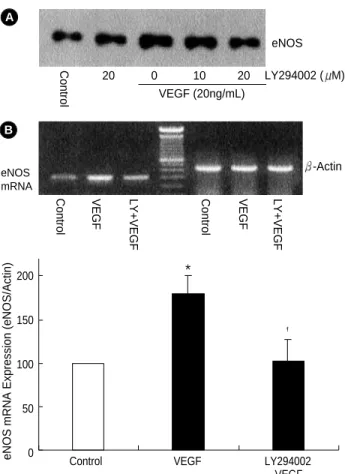
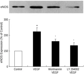
To examine the involvement of PI-3K in eNOS expression by VEGF, the endothelial cells were exposed to VEGF165after 1 hr pretreatment with LY294002 or wortmannin, which are well known as PI-3K inhibitors. In the cells pretreated with 10 M or 20 M of LY294002, the eNOS expression induced by VEGF165was suppressed depending on the con- centration of the inhibitor (Fig. 4A). In the group not pre- treated with LY294002, eNOS mRNA expression increased by 1.7 times, whereas in the group pretreated with 20 M of LY294002, it decreased to the same level as of the control group (Fig. 4B). This shows that PI-3K inhibitor inhibits eNOS mRNA expression by VEGF165. Furthermore, eNOS protein expression by VEGF165increased by 2.2 times, com- pared with 1.6 times when it was exposed to VEGF165after
pretreatment with wortmannin, the other PI-3K inhibitor (Fig. 5).
Because there are some reports showing that the eNOS activation in endothelial cells by VEGF165is controlled by ERK1/2, which is a downstream molecule and a type of MAPK, the effect of kinase phosphorylation on eNOS ex- pression was examined. eNOS protein expression by VEG- F165was measured after pretreatment with PD98059, which is a known ERK1/2 cascade inhibitor. As shown in Fig. 6, PD98059 pretreatment suppressed the VEGF-mediated eNOS expression (Fig. 6).
To examine whether or not eNOS expression by PD98059 correlates with ERK phosphorylation, immunoblotting was carried out using antibodies for phospho-ERK, an activated form of ERK. As a result, ERK phosphorylation was increased by the VEGF165. However, there was no significant change in p38 MAPK phosphorylation (Fig. 7). Furthermore, LY 294002 or PD9805 attenuated the ERK phosphorylation
eNOS Expression (eNOS/Actin)
300 250 200 150 100 50 0
Fig. 3.Effect of VEGF on induction of eNOS mRNA in ECV 304 cells. Confluent ECV 304 cells were treated with various concen- trations of VEGF for 24 hr. The mRNA levels of eNOS and -actin were determined by RT-PCR. The amounts of eNOS mRNA were quantified using scanning densitometry and expressed relative to the densities of -actin. *p<0.05 compared with control (0 ng/
mL).
VEGF (ng/mL)
0 5 10 20
*
eNOS mRNA
-Actin
0 5 10 20
VEGF (ng/mL) VEGF (ng/mL)
0 5 10 20
eNOS mRNA Expression (eNOS/Actin)
200
150
100
50
0
Fig. 4.Effect of LY294002 on VEGF-induced eNOS protein and mRNA expression. Confluent ECV 304 cells were pretreated with LY294002 (25 M) for 30 min and then stimulated with the indi- cated concentrations of VEGF for 24 hr. (A) eNOS expression as determined by Western blot analysis. (B) Total RNA was isolated and mRNA was determined by RT-PCR. The amounts of eNOS mRNA was quantified using scanning densitometry and expressed relative to the densities of -actin. *p<0.05 and �p<0.05 compared with control and VEGF, respectively.
VEGF
Control VEGF LY294002
*
�
eNOS mRNA
-Actin eNOS LY294002 ( M)
ControlControl VEGF LY+VEGF Control VEGF LY+VEGF
A
B
20 0 10 20
VEGF (20ng/mL)
by VEGF (Fig. 8).
DISCUSSION
Recent studies have shown that VEGF increases NO pro- duction in rabbit or human endothelial cells, and NO is thought to be an important mediator in VEGF-induced endothelial cell proliferation (16, 18). For the basic mecha- nism of the induction of NO by VEGF, some reports have shown the inhibition of VEGF-induced endothelial cell pro- liferation by the NOS inhibitor, the inhibition of reticular formation of endothelial cells in three-dimensional collagen
gel (12, 19), and that NO produced by substance P or VEGF stimulates bovine endothelial cell proliferation (12, 20).
Using ECV 304 endothelial cells, we observed the prolifera- tion of endothelial cells induced by VEGF and an increase in NOS activity. This suggests that NO is an important medi- ator in VEGF-induced endothelial proliferation.
The fact that the increase of NO through a brief exposure to VEGF is reduced by the tyrosine kinase inhibitor, calcium- chelating compounds, and PI-3K suggests that the NO in- crease is associated with an activation of tyrosine kinase and PI-3K and an increase in the cellular calcium concentration
eNOS Expression (% of Control)
300
200
150
0
Fig. 5.Effect of the phosphatidylinositol 3 kinase inhibitors on VEGF-induced eNOS expression. Confluent ECV 304 cells were pretreated with LY294002 (25 M) or wortmannin (100 nM) for 30 min and then stimulated with the VEGF (20 ng/mL) for 24 hr.
eNOS expression was determined by Western blot analysis. The eNOS protein data are representative of three independent exper- iments. *p<0.05 and �p<0.05 compared with control and VEGF, respectively.
VEGF VEGF
Control VEGF Wortmannin LY 294002
*
�
�
eNOS
eNOS Expression (% of Control)
250
200
150
100
50
0
Fig. 6.Effect of PD98059 on the VEGF-induced eNOS expression.
Confluent ECV 304 cells were incubated for 30 min in the absence or presence of PD98059 (50 M) prior to the addition of VEGF (20 ng/mL). eNOS expression was determined by Western blot anal- ysis. The eNOS protein data are representative of three indepen- dent experiments. *p<0.05 and �p<0.05 compared with control and VEGF, respectively.
VEGF
Control VEGF PD 98059
*
�
eNOS
Fig. 7.Effect of VEGF on ERK and p38 MAPK phosphorylation.
ECV 304 cells were stimulated with VEGF (20 ng/mL) for 10 min and harvested. Whole cell lysates were resolved by SDS-PAGE followed by Western blot analysis using a set of antibodies that recognize either phosphorylated ERK or phosphorylated p38 MAPK. The data are representative of three independent experi- ments.
B
Phospho-ERK
Control VEGF
A
Phospho-p38 MAPK
Fig. 8.Effect of inhibitors of VEGF-induced ERK phosphorylation.
ECV 304 cells were pretreated with LY294002 (25 M), wortman- nin (100 nM) or PD98059 (50 M) for 30 min and then stimulated with VEGF for 10 min. Whole cell lysates were resolved by SDS- PAGE and analyzed by Western blot analysis using phospho- ERK antibody. The data are representative of three independent experiments.
Phospho-ERK
Control VEGF LY294002+VEGF Wortmannin+VEGF PD98059+VEGF VEGF
(18, 19). Thus, the exposure of human umbilical vein endo- thelial cells to VEGF results in an increase in the cellular cal- cium concentration, while treatment with calcium-chelating compounds or calmidazolium results in a decrease of VEGF- induced NO production (9, 19). On the other hand, the chief mechanism of NO production through 24 hr or more of con- tinuous VEGF stimulation is thought to be due to an increase in NOS protein synthesis (7, 19, 21). It was thought that eNOS (type III NOS) is always activated in endothelial cells and its enzymatic activation is partially regulated by inter- cellular Ca2+, while iNOS (type II NOS) is regulated at the transcription level. However, recent studies have suggested that eNOS protein expression can also be regulated by phys- ical stress or hormone. The increase in eNOS expression by such stimuli as the transforming factor (22), a basic fibro- blast growth factor (23, 24), fluid shear stress (25), and hypox- ia (26) has been reported. Moreover, continuous physical exer- cise on dogs has been shown to increase eNOS expression and the consequent benefits of exercise on the vascular system has been reported (27). This study confirms that the increase of eNOS protein by VEGF at the mRNA level. The results also show that the increase in NO synthesis is through to be not only due to a simple increase in NOS activity, but also due to an increase in the NOS protein at the transcriptional level.
iNOS expression was not increased by VEGF in contrasts to Papapetropoulos et al. (19).
Tyrosine kinase is known to participate in the signal trans- duction and expression of eNOS induced by VEGF. In hu- man endothelial cells, tyrosine kinase inhibitors such as genis- tein and geldanamycin inhibit NO synthesis through VEGF.
At least two receptors, Flt-1 and Flk-1/KDR, are involved in the mechanism of action of VEGF and the cells express- ing Flk-1/KDR show more cell structural changes and cell proliferation than those with Flt-1 (5). However, Flk-1/KDR receptor activation through VEGF shows increases in eNOS and iNOS protein expression, but Flt-1 receptor stimulation does not increase NOS protein expression (21). Each one of these receptors has endogenous tyrosine kinase activity. In addition to these receptors, at least 11 proteins, such as phos- pholipase C- , PI-3K, Ras GTPase activating protein (GAP), and Nck (oncogenic adaptor protein), are activated through tyrosine phosphorylation (28). Among the tyrosine phos- phorylated proteins, there is also ERK1/2, a number of the MAPK family. In human umbilical vein endothelial cells, VEGF increases tyrosine phosphorylation of the Flk-1/KDR receptors and both receptors phosphorylate and activate the p85, a regulating unit of PI-3K. It then activates MAPK (29). ERK1/2 plays a central role in the growth and prolif- eration of various cells, in the increase of eNOS expression through VEGF treatment, and consequently in the neovas- cular formation by VEGF. Studies have shown that the pre- treatment with PI-3K inhibitors in human umbilical vein endothelial cells inhibits NO formation through VEGF (19).
However, the actual mechanism of PI-3K-associated NO
synthesis is still unknown. It is also unclear whether this is due to a simple increase in NOS activity or due to changes in NOS protein synthesis. The present study revealed that the pre-treatment with PI-3K inhibitor, LY294002 or wortman- nin, attenuated the eNOS expression in response to VEGF.
LY294002 showed a higher degree of inhibition than wort- mannin. Furthermore, the pre-treatment with the ERK1/2 inhibitor, PD98059, decreased eNOS expression by VEGF and phosphorylated ERK1/2. Both PI-3K inhibitors, LY 294002 and wortmannin, inhibited ERK1/2 phosphoryla- tion by VEGF. These results suggest that both PI-3K and ERK1/2 pathways play a major role in NOS protein synthe- sis by VEGF.
In conclusion, NOS protein synthesis is one of the key me- chanisms responsible for the increase in NOS activity indu- ced by VEGF and PI-3K and ERK1/2 are deeply involved in VEGF-induced eNOS expression in ECV 304 cells.
ACKNOWLEDGMENT
This study was supported by a research grant (1999) from the Institute of Basic Medicine and Research Institute of Clin- ical Medicine, Yeungnam University.
REFERENCES
1. Shweiki D, Itin A, Soffer D, Keshet E. Vascular endothelial growth factor induced by hypoxia may mediate hypoxia-initiated angiogen- esis. Nature 1992; 359: 843-5.
2. Leung DW, Cachianes G, Kuang WJ, Goeddel DV, Ferrara N. Vas- cular endothelial growth factor is a secreted angiogenic mitogen.
Science 1989; 246: 1306-9.
3. de Vries C, Escobedo JA, Ueno H, Houck K, Ferrara N, Williams LT. The fms-like tyrosine kinase, a receptor for vascular endothelial growth factor. Science 1992; 255: 989-91.
4. Terman BI, Dougher-Vermazen M, Carrion ME, Dimitrov D, Armel- lino DC, Gospodarowicz D, Bohlen P. Identification of the KDR tyro- sine kinase as a receptor for vascular endothelial cell growth factor.
Biochem Biophys Res Commun 1992; 187: 1579-86.
5. Fong GH, Rossant J, Gertsenstein M, Breitman ML. Role of the Flt- 1 receptor tyrosine kinase in regulating the assembly of vascular endothelium. Nature 1995; 376: 66-70.
6. Shalaby F, Rossant J, Yamaguchi TP, Gertsenstein M, Wu XF, Bre- itman ML, Schuh AC. Failure of blood-island formation and vascu- logenesis in Flk-1-deficient mice. Nature 1995; 376: 62-6.
7. Waltenberger J, Claesson-Welsh L, Siegbahn A, Shibuya M, Heldin CH. Different signal transduction properties of KDR and Flt1, two receptors for vascular endothelial growth factor. J Biol Chem 1994;
269: 26988-95.
8. Keyt BA, Nguyen HV, Berleau LT, Duarte CM, Park J, Chen H, Ferrara N. Identification of vascular endothelial growth factor deter- minants for binding KDR and FLT-1 receptors. Generation of recep-
tor-selective VEGF variants by site-directed mutagenesis. J Biol Chem 1996; 271: 5638-46.
9. Brock TA, Dvorak HF, Senger DR. Tumor-secreted vascular per- meability factor increases cytosolic Ca2+and von Willebrand factor release in human endothelial cells. Am J Pathol 1991; 138: 213-21.
10. Wu HM, Huang Q, Yuan Y, Granger HJ. VEGF induces NO-depen- dent hyperpermeability in coronary venules. Am J Physiol 1996; 271:
2735-9.
11. Ku DD, Zaleski JK, Liu S, Brock TA. Vascular endothelial growth factor induces EDRF-dependent relaxation in coronary arteries. Am J Physiol 1993; 265: 586-92.
12. Morbidelli L, Chang CH, Douglas JG, Granger HJ, Ledda F, Ziche M. Nitric oxide mediates mitogenic effect of VEGF on coronary venu- lar endothelium. Am J pysiol 1996; 270: 411-5.
13. Ziche M, Morbidelli L, Choudhuri R, Zhang HT, Donnini S, Granger HJ, Bicknell R. Nitric oxide synthase lies downstream from vascular endothelial growth factor-induced but not basic fibroblast growth factor-induced angiogenesis. J Clin Invest I 1997; 99: 2625-34.
14. Asahara T, Bauters C, Pastore C, Kearney M, Rossow S, Bunting S, Ferrara N, Symes JF, Isner JM. Local delivery of vascular endothe- lial growth factor accelerates reendothelialization and attenuates intimal hyperplasia in balloon-injured rat carotid artery. Circula- tion 1995; 91: 2793-801.
15. Asahara T, Chen D, Tsurumi Y, Kearney M, Rossow S, Passeri J, Symes JF, Isner JM. Accelerated restitution of endothelial integrity and endothelium-dependent function after phVEGF165gene transfer.
Circulation 1996; 94: 3291-302.
16. Parenti A, Morbidelli L, Cui XL, Douglas JG, Hood JD, Granger HJ, Ledda F, Ziche M. Nitric oxide is an upstream signal of vascular endothelial growth factor-induced extracellular singal-regulated kinase1/2 activation in postcapillary endothelium. J Biol Chem 1998; 73: 4220-6.
17. Yu Y, Sato Y. MAP kinases, phosphatidylinositol 3-kinase, and p70 S6 kinase mediate the mitogenic response of human endothelial cells to vascular endothelial growth factor. J Cell Physiol 1999;
178: 235-46.
18. Bouloumie A, Schini-Kerth VB, Busse R. Vascular endothelial growth factor up-regulates nitric oxide synthase expression in endo- thelial cells. Cardiovasc Res 1999; 41: 773-80.
19. Papapetropoulos A, Garcia-Cardena G, Madri JA, Sessa WC. Nitric
oxide production contributes to the angiogenic properties of vascu- lar endothelial growth factor in human endothelial cell. J Clin Invest 1997; 100: 3131-9.
20. van der Zee R, Murohara T, Luo Z, Zollmann F, Passeri J, Lekutat C, Isner JM. Vascular endothelial growth factor/vascular perme- ability factor augments nitric oxide release from quiescent rabbit and human vascular endothelium. Circulation 1997; 95: 1030-7.
21. Kroll J, Waltenberger J. VEGF-A induces expression of eNOS and iNOS in endothelial cells via VEGF receptor-2 (KDR). Biochem Biophys Res Commun 1998; 27: 743-6.
22. Inoue N, Venema RC, Sayegh HS, Ohara Y, Murphy TJ, Harrison DG. Molecular regulation of the bovine endothelial cell nitric oxide synthase by transforming growth factor-beta 1. Arterioscler Thromb Vasc Biol 1995; 15: 1255-61.
23. Kostyk SK, Kourembanas S, Wheeler EL, Medeiros D, McQuillan LP, D’Amore PA, Braunhut SJ. Basic fibroblast growth factor in- creases nitric oxide synthase production in bovine endothelial cells.
Am J Physiol 1995; 269: 1583-9.
24. Arnal JF, Yamin J, Dockery S, Harrison DG. Regulation of endothe- lial nitric oxide synthase mRNA, protein, and activity during cell growth. Am J Physiol 1994; 267: 1381-8.
25. Uematsu M, Ohara Y, Navas JP, Nishida K, Murphy TJ, Alexander RW, Nerem RM. Regulation of endothelial cell nitric oxide synthase mRNA expression by shear stress. Am J Physiol 1995; 269: 1371-8.
26. Xu XP, Pollock JS, Tanner MA, Myers PR. Hypoxia activates nitric oxide synthase and stimulates nitric oxide production in porcine coronary resistance arteriolar endothelial cells. Cardiovasc Res 1995; 30: 841-7.
27. Sessa WC, Pritchard K, Seyedi N, Wang J, Hintze TH. Chronic exer- cise in dogs increases coronary vascular nitric oxide production and endothelial cell nitric oxide synthase gene expression. Circ Res 1994;
74: 349-53.
28. Guo D, Jia Q, Song HY, Warren RS, Donner DB. Vascular endothe- lial cell growth factor promotes tyrosine phosphorylation of media- tors of signal transduction that contain SH2 domains. Association with endothelial cell proliferation. J Biol Chem 1995; 270: 6729-33.
29. Thakker GD, Hajjar DP, Muller WA, Rosengart TK. The role of phosphatidylinositol 3-kinase in vascular endothelial growth factor signaling. J Biol Chem 1999; 274: 10002-7.