Preventive Effects of Zoledronic Acid on Bone Metastasis in Mice Injected with Human Breast Cancer Cells
Bisphosphonates are used routinely to reduce bone-related events in breast cancer patients with bone metastasis. We evaluated the effects of zoledronic acid, a third generation, nitrogen-containing bisphosphonate, to prevent bone metastasis in breast cancer.
Zoledronic acid or vehicle alone was administered to nude mice either simultaneously or after intracardiac injection of human breast cancer MDA-MB-231 cells. Nude mice treated with zoledronic acid at early time points showed a lower incidence of bone metastases than did vehicle-treated nude mice, but these differences were not statistically significant.
Only 37.5% of mice treated with zoledronic acid at the time of tumor cell inoculation developed bone metastases compared to over 51.8% of mice receiving vehicle alone (P = 0.304). Cell count of apoptosis confirmed by immunohistochemical staining in metastatic bone tissue significantly increased in the zoledronic acid-treated groups compared to non-treated group (1,018.3 vs 282.0; P = 0.046). However, metastatic tumor cells, which invade soft tissue around the bone, did not show extensive apoptosis;
there were no differences between the zoledronic acid-treated and control groups. These results suggest that zoledronic acid increases apoptosis of metastatic breast tumor cells in the bone and could therefore reduce metastatic tumor burden. These results support the use of zoledronic acid to reduce the incidence of bone metastasis in breast cancer.
Key Words: Zoledronic Acid; Breast Neoplasms; Bone Metastasis; Prevention Joon Jeong1, Kyung Sun Lee2,
Yang-Kyu Choi2, Young Ju Oh1 and Hy-De Lee1
1Department of Surgery, College of Medicine, Yonsei University, Seoul; 2Department of Veterinary Medicine, Konkuk University, Seoul, Korea Received: 27 May 2011
Accepted: 19 October 2011 Address for Correspondence:
Hy-De Lee, MD
Department of Surgery, Gangnam Severance Hospital, Yonsei University, 712 Eonjuro, Gangnam-gu, Seoul 135-720, Korea Tel: +82.2-2019-3370, Fax: +82.2-3462-5994 E-mail: [email protected]
The present research has been partly supported by Korean Breast Cancer Foundation (2005).
http://dx.doi.org/10.3346/jkms.2011.26.12.1569 • J Korean Med Sci 2011; 26: 1569-1575 Oncology & Hematology
INTRODUCTION
Breast cancer is the most common malignancy among women, and bone metastases occur in approximately 80% of cases of ad- vanced breast cancer. Bone metastasis in breast cancer is large- ly osteolytic metastasis coupled with complications such as bone pain, pathologic fractures, hypercalcemia, spinal cord compres- sion, and bone marrow suppression (1-3), which in turn lead to increased morbidity and mortality rates in breast cancer patients.
Osteolytic destruction in breast cancer patients with bone me- tastasis is caused by the activation of osteoclasts secondary to numerous factors secreted by breast cancer cells and the stimu- lation of osteoclast production by osteoblasts through the recep- tor activator of nuclear factor-κB ligand (4, 5). Bisphosphonates are potent inhibitors of bone resorption that suppress both the activities of mature osteoclasts and the formation of new osteo- clasts (6-9). Therefore, bisphosphonates are being widely used in Paget’s disease, osteoporosis, malignancy with hypercalce- mia, and osteolytic bone metastases (7, 10-13).
Zoledronic acid, a third generation nitrogen-containing bis- phosphonate, is one of the most potent bisphosphonates clini- cally used at present. It was approved for the treatment of com-
plications associated with osteolytic lesions of metastatic breast cancer. As shown in several clinical studies, zoledronic acid re- duces the incidence of skeletal complications in breast cancer patients with confirmed bone metastases (14-16). However, it has not yet been used for the primary prevention of bone me- tastasis in high-risk breast cancer patients. There have been an- imal studies demonstrating that certain bisphosphonates re- duce bone metastasis, and it was observed that high-dose pami- dronate exerts preventive effects in nude mice with osteolytic lesions where human breast cancer cells were injected into the heart (17-20). Yet, these studies primarily cited a reduction in the magnitude of bone metastasis, but did not mention the ef- fects of bisphosphonates on the development of bone metasta- sis. If zoledronic acid can be administered safely without caus- ing serious side effects to reduce bone metastasis in breast can- cer, it would be clinically meaningful. Given the mechanism of action of zoledronic acid on osteoclasts, zoledronic acid should have a great potential for the prevention of bone metastasis. Thus, preclinical research of zoledronic acid is warranted to explore the development of bone metastases in breast cancer.
The majority of bone metastases that occur in breast cancer are osteolytic metastases, which can be diagnosed with radiog-
raphy. However, when small animals are used, detection of bone metastasis is difficult. In addition, very early lesions may be un- likely viewed on radiography. Thus, in this study, a breast can- cer cell line, transfected with the luciferase gene, was used; to detect metastasized cells, the IVISTM imaging system, which en- ables the detection of early metastatic sites, was used. Further- more, by comparing the preventive effects of zoledronic acid administered at different times, an appropriate administrative dosing schedule was explored.
MATERIALS AND METHODS
MDA-MB-231 cell line transfected with luciferase
MDA-MB-231 is a breast cancer cell line that does not express estrogen receptors. Cells were incubated in RPMI 1640 (Gibco, Grand Island, NY, USA) supplemented with 10% fetal bovine serum (FBS; Cansera, Toronto, Ontario, Canada) and antibiotic- antimycotic (Gibco) in a 5% CO2 incubator. Using lipofectamine 2000 (Invitrogen, Carlsbad, CA, USA), the cells were cotrans- fected with GL3-control (Promega, Madison, WI, USA) and TK- Hygro (Clontech, Mountain View, CA, USA) at a ratio of 1:4. Trans- fected cells were incubated in hygromycin (200 µg/mL, Invitro- gen)-containing medium. Resistant colonies were isolated by ring cloning. Using Promega Bright-Glo reagent, verification of hygromycin-resistant colonies expressing luciferase was per- formed, with levels expressed as RLU/µg protein. After verifica- tion, subclones with high luciferase activity were isolated and named MDA-MB-231/Luc, which were then incubated in RPMI 1640 containing 10% FBS.
Intracardiac injection of MDA-MB-231/Luc and administration of zoledronic acid
These studies (Protocol No. 2011-239) were approved by the In- stitutional Animal Care and Use Committees of Yonsei Univer- sity College of Medicine (Seoul, Korea). Five-week-old female BALB/c-Foxn1nu mice (Japan SLC, Shizuoka, Japan) were used.
For administration of the bisphosphonate, zoledronic acid [1- hydroxy-2-(1H-imidazole-1-yl)ethylidene-bisphosphonic acid], ZOMETA injection (Novartis Pharmaceuticals Ltd., Basel, Swit- zerland) was used. Zoledronic acid was stored at 4°C and dilut- ed with PBS (pH 7.4) prior to use.
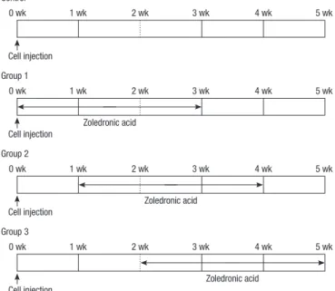
Cells were incubated in fresh medium for 24 hr before injec- tion. MDA-MB-231/Luc cells were prepared in phosphate-buff- ered saline (PBS; 5 × 10⁵ cells/0.1 mL PBS) and injected into the left ventricle of anesthetized mice using 27-gauge needle syringes (Fig. 1). Beginning at different times after the injection of cancer cells, 120 µg zoledronic acid/kg was administered twice weekly for 3 weeks, and all nude mice were sacrificed at week 5 (day 35). The experiment was undertaken using three different meth- ods of zoledronic acid administration: administration of zole-
dronic acid for 3 weeks beginning at the time of tumor cell in- jection (group 1), administration of zoledronic acid for 3 weeks beginning 1 week after tumor cell injection (group 2), and ad- ministration of zoledronic acid for 3 weeks beginning 2 weeks after tumor cell injection (group 3). All three corresponding con- trol groups with no administration of zoledronic acid were ad- ministered vehicle (PBS) in the same way (Fig. 1).
Detection of MDA-MB-231 cell/Luc in nude mice
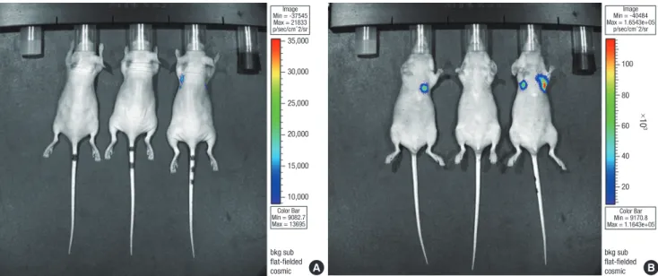
MDA-MB-231 cell/Luc was measured at week 5 after injection of the tumor cell line. After 5-min anesthesia with ketamine/xy- lazine, 150 mg D-luciferin/kg (Xenogen Corp., Hopkinton, MA, USA) was given to mice by intraperitoneal (IP) injection. After 5 min, light emitted from bioluminescent tumor cells was mea- sured using the IVISTM imaging system (Xenogen Corp). Mice were imaged in both ventral and dorsal views (Fig. 2). After im- aging at week 5, several sites with bone metastasis were isolated from the sacrificed mice, fixed, dehydrated, and hematoxylin and eosin (H&E)-dyed for histopathological confirmation of bone metastasis.
Terminal deoxynucleotidyl transferase dUTP nick end labeling (TUNEL) assay
The detection of apoptosis was performed using ApopTag, ac- cording to the instructions of the manufacturer (Chemicon In- ternational, Inc., Temecula, CA, USA). Briefly, the sections were deparaffined, hydrated, treated with proteinase K (20 µg/mL) in PBS (pH 7.5) at 37°C for 20 min, and then treated with 3% hy- droperoxidase in PBS to block endogenous peroxidase. After washing with PBS, the slides were incubated with terminal de- oxynucleotidyl transferase and a nucleotide mixture at 37°C for
0 wk
Cell injection Control
2 wk 4 wk
1 wk 3 wk 5 wk
0 wk
Cell injection
Zoledronic acid Group 2
2 wk 4 wk
1 wk 3 wk 5 wk
0 wk
Cell injection
Zoledronic acid Group 1
2 wk 4 wk
1 wk 3 wk 5 wk
0 wk
Cell injection
Zoledronic acid Group 3
2 wk 4 wk
1 wk 3 wk 5 wk
Fig. 1. Experimental schedule of zoledronic acid administration.
1 hr and then incubated for 30 min with anti-digoxigenin con- jugate. The slides were treated with DAB before counterstaining with hematoxylin. After dehydration and washing, the sections were placed on coverslips and examined by microscopy (Olym- pus BX 51, Tokyo, Japan).
Images of slides processed for apoptosis detection were quan- titatively analyzed using a computerized image analyzer (Meta- Morph 7.5, Molecular Devices, Sunnyvale, CA, USA). Immu- nopositive cells in bone from three inconsecutive sections per three mice of the zoledronic acid-treated and untreated groups were blindly counted with the image analyzer at a magnifica- tion of × 400 by an experienced pathologist and expressed as the number of cells per 1 mm3. All values are expressed as the means ± standard deviation, and statistical significance was evaluated by one-way analysis of variance using SPSS for Win- dows (version 14.0 SPSS Inc., Chicago, IL, USA)
RESULTS
Effects of zoledronic acid on the prevention of bone metastasis
At 35 days after intracardiac injection using the Xenogen IVIS- 200 imaging system, bioluminescence imaging was performed;
a single hot spot, was considered to have bone metastasis. Fig. 2 shows an example of bioluminescence imaging. Imaging results showed bone metastasis in 14 of 27 mice (51.8%) in the control group, 9 of 24 mice in test group 1 (37.5%), 7 of 15 mice in test group 2 (46.6%), and 9 of 14 mice in test group 3 (64.2%; Fig. 3).
Differences between group 1 and control (P = 0.304), between group 2 and control (P = 0.747), and between group 3 and con- trol (P = 0.447) were not statistically significant. The differences between each test group were also not statistically significant.
The number of mice tested in each group differed because some mice died during intracardiac injection; other mice showed mas- sive spillage of injected cancer cells into the pericardial space and were therefore excluded. Although results were not statisti- cally significant, test group 1 treated prophylactically with zole- dronic acid demonstrated a trend toward a decreased incidence of bone metastases.
Apoptosis of metastatic sites
After identifying sites in which bone metastases occurred in zole- dronic acid-treated groups and the control group and staining with ApopTag peroxidase in situ (Chemicon), the number of apoptotic metastatic cells per mm3 were counted. The apopto- sis of tumor cells with bone metastasis occurred regardless of the timing of zoledronic acid administration. Among the mice that
A B
35,000
30,000
25,000
20,000
15,000
10,000
bkg sub flat-fielded cosmic
bkg sub flat-fielded cosmic Color Bar
Min = 9082.7 Max = 13695 Image Min = -37545 Max = 21833 p/sec/cm^2/sr
Image Min = -40484 Max = 1.6543e+05
p/sec/cm^2/sr
Color Bar Min = 9170.8 Max = 1.1643e+05
100
×103
80
60
40
20
Fig. 2. Metastatic activities visualized using bioluminescent imaging in the control group (A; dorsal side, B; ventral side). First and third mice show hot spots on their ventral sides, confirmed as rib metastases.
Bone metastasis (%)
Group
61.8% (14/27)
37.6% (9/24)
46.6% (7/15)
64.2% (9/14)
Control Group 1 Group 2 Group 3 100
90 80 70 60 50 40 30 20 10 0
Fig. 3. Incidence of bone metastases in each group. Differences between group 1 and control (P = 0.304), between group 2 and control (P = 0.747), and between group 3 and control (P = 0.447) were not statistically significant. Additionally, the differences between each test group were not statistically significant.
A B
50.0 µM 50.0 µM
Fig. 4. Immunohistochemical staining results for apoptosis in metastatic tumor cells in the bone (× 400). ApopTag peroxidase in situ results of metastatic tumor tissues of bone in test (A) and control (B) groups. Many apoptotic tumor cells were positively stained in the test groups.
A B
50.0 µM 50.0 µM
Fig. 5. Immunohistochemical staining results for apoptosis in metastatic tumor cells outside the bone (× 400). ApopTag peroxidase in situ results of metastatic tumor tissues outside the bone in the test (A) and control (B) groups. No differences were seen between the groups.
Apoptotic cell
Apoptotic cell
Fig. 6. Number of apoptotic tumor cells inside and outside of the bone in each group. Apoptosis of metastatic cells increased on average by 1,018.3 in the zoledronic acid- treated groups (zol) and on average by 282.0 in the control groups (con), indicating a significant increase in apoptosis in the zoledronic acid-treated groups (P = 0.046). On the other hand, apoptosis of cells metastasized to the outside of the bone was 173.6 in the zoledronic acid-treated groups and 217.3 in the control groups, suggesting no increase in any of the groups and no differences between groups (P = 0.86).
1,750 1,500 1,250 1,000 750 500 250 0
500450 400350 300250 200150 10050 0
Apoptotic tumor cell (cell/mm2) inside bone Apoptotic tumor cell (cell/mm2) inside bone
Zol Con
Zol Con
Average SD
Zol 173.6 217.4
Con 217.3 236.9
had bone metastases, we selected three mice from the treated (regardless of timing) and control groups, respectively, and apop-
tosis of tumor cells were evaluated as mentioned above (Fig. 4, 5).
At certain metastatic sites, tumor cells extended to the outside
Average SD
Zol 1,018.3 421.2
Con 282.0 147.8
of the bone tissue. Apoptosis of metastatic cells inside the bone increased on average by 1,018.3 in the zoledronic acid-treated groups and on average by 282.0 in the controls, indicating a sig- nificant increase in apoptosis in the zoledronic acid-treated groups (P = 0.046, Fig. 6). Apoptosis of cells that metastasized to the outside of the bone was 173.6 in the zoledronic acid-treat- ed groups and 217.3 in the controls, suggesting no increase in any of the groups and no difference between groups (P = 0.86, Fig. 6).
DISCUSSION
Zoledronic acid, a nitrogen-containing bisphosphonate, exerts its pharmacologic activity by inducing apoptosis of osteoclasts;
it inhibits the mevalonate pathway of osteoclasts and prevents the prenylation of small GTP proteins, such as Ras, Rho, and Rab, thereby leading to apoptosis (21, 22). The inhibitory effects of zoledronic acid on osteolytic metastasis in breast cancer can be explained by two mechanisms of action. One mechanism is that zoledronic acid reduces the release of growth factors, such as insulin-like growth factor and transforming growth factor beta (TGF-β), which are abundant in bone matrix by inhibiting osteo- clasts, which are considered to contribute to the inhibition of bone metastasis in breast cancer. The other mechanism is the direct apoptotic effects of zoledronic acid on breast cancer cells with bone metastasis. High-concentration bisphosphonates have been reported to induce apoptosis of cancer cells in the labora- tory (23-25), and high concentrations of zoledronic acid were reported to induce apoptosis in an experiment using 4T1 mouse breast cancer cell lines by inhibiting the mevalonate pathway (26). In this study, relatively high concentrations of zoledronic acid were also used. In general, the dose in mice equivalent to the 4 mg intravenous dose in humans is known to be 98-100 µg/kg (27); in this study, zoledronic acid was administered twice a week at 120 µg/kg per dose. Thus, the dose given should be taken into consideration for any attempt to apply the results of this study to clinical settings.
In this study, the increased apoptosis of bone metastasis in breast cancer in zoledronic acid-treated groups was confirmed, and no apoptotic effects of zoledronic acid were observed on areas that metastasized to the outside of the bone. This result is in line with previous studies citing that bisphosphonates did not induce apoptosis in breast cancer cells prior to metastases to other organs or on the primary lesions (18). Bisphosphonates accumulate primarily in bone when administered intravenous- ly, thus resulting in higher concentrations in bone than other organs. Additionally, bisphosphonates mainly work on bone metastasis other than the primary sites or lesions of other organs.
The apoptotic effects of zoledronic acid on bone metastasis in breast cancer can be explained by the aforementioned two mech- anisms. In this study, zoledronic acid effects on the growth fac-
tors were not fully evaluated. We checked serum TGF-β levels, both pre- and post-zoledronic acid treatment in some of mice in the zoledronic acid treated groups but there was no definite change after zoledronic acid treatment (data not shown). How- ever, no absolute conclusions can be drawn due to the extremely small number of samples and limited number of animals used.
In addition, it cannot be assumed that changes in TGF-β in the areas of bone metastasis would be reflected by serum changes.
This study aimed to elucidate whether zoledronic acid could reduce the incidence of bone metastasis. Most previous study results confirmed that bisphosphonates conferred preventive effects on bone metastasis by decreasing the volume of bone metastasis (20, 26). However, in a clinical setting, treatments begin when bone metastasis is viewed in even one area, which considers the presence of bone metastasis. Thus, the incidence of bone metastasis may be more important than the volume of metastasis. The results of this study showed a trend toward the reduction in the incidence of metastasis, although not statisti- cally significant, in groups in which zoledronic acid was preven- tively administered. Yet, when zoledronic acid administration was delayed, the effects of such decreased incidence were dimin- ished. Therefore, it may be concluded that the preventive effects of zoledronic acid on bone metastasis may no longer exist when bone metastasis has already begun.
In previous studies, X-rays were used to diagnose bone me- tastasis. However, X-rays have a low sensitivity for diagnosis of bone metastasis in mice, and bone micrometastases may not be discovered on X-rays. In fact, it was reported that certain cases that were not found to have bone metastasis on X-rays revealed to have metastasis by histological examination (20). To overcome these limitations, bioluminescence imaging was used to identify bone metastasis in this study. The use of green fluorescent pro- tein was reported to allow a diagnosis 1 week earlier than the use of X-rays (28).
A more accurate diagnostic method was used to elucidate the effects of zoledronic acid on the prevention of bone metastasis in this study. However, the mechanism of action of zoledronic acid on bone metastasis is associated with the apoptosis of met- astatic breast cancer cells. Additionally, zoledronic acid does not prevent the colonization of breast cancer cells in the bone. There- fore, the effect of zoledronic acid on the prevention of metasta- sis involves a decrease in the burden of lesions rather than inci- dence of metastasis. If the effect on the burden of metastatic le- sions is large, it may result in a decreased incidence of metasta- sis, whereas the incidence may not change if the effect on the burden of metastatic lesions is small. Several clinical studies of the preventive effects on bone metastasis in breast cancer pa- tients using other bisphosphonates, clodronate or pamidronate, have shown both the presence and absence of the preventive effects in addition to the presence of preventive effects with no statistical significance (29, 30). These contradictory results can
be explained by the reasons discussed above.
In summary, the reduced incidence of bone metastasis with zoledronic acid, although not statistically significant, was con- firmed in mice in which breast cancer with bone metastases was induced. Apoptosis of tumor cells inside the bone occurred to a larger degree in the zoledronic acid-treated group than in the control group but was statistically significant. The effects of zoledronic acid on the prevention of bone metastasis are asso- ciated with apoptosis of breast cancer cells at metastatic sites inside the bone.
REFERENCES
1. Rubens RD. Bone metastases: the clinical problem. Eur J Cancer 1998;
34: 210-3.
2. Yoneda T, Sasaki A, Mundy GR. Osteolytic bone metastasis in breast can- cer. Breast Cancer Res Treat 1994; 32: 73-84.
3. Kanis JA. Bone and cancer: pathophysiology and treatment of metasta- ses. Bone 1995; 17: 101S-5S.
4. Body JJ. Bisphosphonates. Eur J Cancer 1998; 34: 263-9.
5. Mundy GR. Bisphosphonates as cancer drugs. Hosp Pract (Minneap) 1999; 34: 81-4, 88-9, 93-4.
6. Boonekamp PM, van der Wee-Pals LJ, van Wijk-van Lennep MM, Thesing CW, Bijvoet OL. Two modes of action of bisphosphonates on osteoclastic resorption of mineralized matrix. Bone Miner 1986; 1: 27-39.
7. Fleisch H. Bisphosphonates. Pharmacology and use in the treatment of tumor-induced hypercalcaemic and metastatic bone disease. Drugs 1991;
42: 919-44.
8. Carano A, Teitelbaum SL, Konset JD, Schlesinger PH, Blair HC. Bisphos- phonates directly inhibit the bone resorption activity of isolated avian osteoclasts in vitro. J Clin Invest 1990; 85: 456-61.
9. Sato M, Grasser W, Endo N, Akins R, Simmons H, Thompson DD, Golub E, Rodan GA. Bisphosphonate action. Alendronate localization in rat bone and effects on osteoclast ultrastructure. J Clin Invest 1991; 88: 2095-105.
10. Frijlink WB, Bijvoet OL, Te Velde J, Heynen G. Treatment of Paget’s dis- ease with (3-amino-1-hydroxypropylidene)-1, 1-bisphosphonate (A.P.D.).
Lancet 1979; 1: 799-803.
11. Ryan PJ, Sherry M, Gibson T, Fogelman I. Treatment of Paget’s disease by weekly infusions of 3-aminohydroxy-propylidene-1, 1-bisphospho- nate (APD). Br J Rheumatol 1992; 31: 97-101.
12. Kanis JA, O’Rourke N, McCloskey E. Consequences of neoplasia induced bone-resorption and the use of clodronate. Int J Oncol 1994; 5: 713-31.
13. Rizzoli R, Buchs B, Bonjour JP. Effect of a single infusion of alendronate in malignant hypercalcaemia: dose dependency and comparison with clodronate. Int J Cancer 1992; 50: 706-12.
14. Rosen LS, Gordon D, Tchekmedyian S, Yanagihara R, Hirsh V, Krzakows- ki M, Pawlicki M, de Souza P, Zheng M, Urbanowitz G, Reitsma D, Sea- man JJ. Zoledronic acid versus placebo in the treatment of skeletal me- tastases in patients with lung cancer and other solid tumors: a phase III, double-blind, randomized trial. The Zoledronic Acid Lung Cancer and Other Solid Tumors Study Group. J Clin Oncol 2003; 21: 3150-7.
15. Rosen LS, Gordon D, Kaminski M, Howell A, Belch A, Mackey J, Apffel- staedt J, Hussein M, Coleman RE, Reitsma DJ, Seaman JJ, Chen BL, Am- bros Y. Zoledronic acid versus pamidronate in the treatment of skeletal
metastases in patients with breast cancer or osteolytic lesions of multiple myeloma: a phase III. double-blind, comparative trial. Cancer J 2001; 7:
377-87.
16. Rosen LS, Gordon D, Kaminski M, Howell A, Belch A, Mackey J, Apffel- staedt J, Hussein MA, Coleman RE, Reitsma DJ, Chen BL, Seaman JJ.
Long-term efficacy and safety of zoledronic acid compared with pami- dronate disodium in the treatment of skeletal complications in patients with advanced multiple myeloma or breast carcinoma. Cancer 2003; 98:
1735-44.
17. El Abdaimi K, Dion N, Papavasiliou V, Cardinal PE, Binderup L, Goltzman D, Ste-Marie LG, Kremer R. The vitamin D analogue EB 1089 prevents skeletal metastasis and prolongs survival time in nude mice transplant- ed with human breast cancer cells. Cancer Res 2000; 60: 4412-8.
18. Sasaki A, Boyce BF, Story B, Wright KR, Chapman M, Boyce R, Mundy GR, Yoneda T. Bisphosphonate risedronate reduces metastatic human breast cancer burden in bone in nude mice. Cancer Res 1995; 55: 3551-7.
19. Sasaki A, Kitamura K, Alcalde RE, Tanaka T, Suzuki A, Etoh Y, Matsumu- ra T. Effect of a newly developed bisphosphonate, YH529, on osteolytic bone metastases in nude mice. Int J Cancer 1998; 77: 279-85.
20. El-Abdaimi K, Ste-Marie L, Papavasiliou V, Dion N, Cardinal PE, Huang D, Kremer R. Pamidronate prevents the development of skeletal metas- tasis in nude mice transplanted with human breast cancer cells by reduc- ing tumor burden within bone. Int J Oncol 2003; 22: 883-90.
21. Fisher JE, Rogers MJ, Halasy JM, Luckman SP, Hughes DE, Masarachia PJ, Wesolowski G, Russell RG, Rodan GA, Reszka AA. Alendronate mechanism of action: geranylgeraniol, an intermediate in the mevalon- ate pathway, prevents inhibition of osteoclast formation, bone resorp- tion, and kinase activation in vitro. Proc Natl Acad Sci U S A 1999; 96:
133-8.
22. Luckman SP, Hughes DE, Coxon FP, Russell G, Rogers MJ. Nitrogen-con- taining bisphosphonates inhibit the mevalonate pathway and prevent post-transitional prenylation of GTP-binding proteins, including Ras. J Bone Miner Res 1998; 13: 581-9.
23. Shipman CM, Rogers MJ, Apperley JF, Russell RG, Croucher PI. Bisphos- phonate induces apoptosis in human myeloma cell lines: a novel anti- tumor activity. Br J Haematol 1997; 98: 665-72.
24. Shipman CM, Croucher PI, Russell RG, Helfrich MH, Rogers MJ. The bisphosphonate incadronate (YM175) causes apoptosis of human my- eloma cells in vitro by inhibiting the mevalonate pathway. Cancer Res 1998; 58: 5294-7.
25. Aparicio A, Cardner A, Tu Y, Savage A, Berenson J, Lichtenstein A. In vitro cytoreductive effects on multiple myeloma cells induced by bisphos- phonates. Leukemia 1998; 12: 220-9.
26. Hiraga T, Williams PJ, Ueda A, Tamura D, Yoneda T. Zoledronic acid in- hibits visceral metastases in the 4T1/luc mouse breast cancer model. Clin Cancer Res 2004; 10: 4559-67.
27. Daubiné F, Le Gall C, Gasser J, Green J, Clézardin P. Antitumor effects of clinical dosing regimens of bisphosphonates in experimental breast can- cer bone metastasis. J Natl Cancer Inst 2007; 99: 322-30.
28. Peyruchaud O, Winding B, Pécheur I, Serre C, Delmas P, Clézardin P.
Early detection of bone metastases in a murine model using fluorescent human breast cancer cells: application to the use of the bisphosphonate zoledronic acid in the treatment of osteolytic lesions. J Bone Miner Res 2001; 16: 2027-34.
29. Diel IJ, Solomayer EF, Costa SD, Gollan C, Goerner R, Wallwiener D,
Kaufmann M, Bastert G. Reduction in new metastases in breast cancer with adjuvant clodronate treatment. N Engl J Med 1998; 339: 357-63.
30. Saarto T, Vehmanen L, Virkkunen P, Blomqvist C. Ten-year follow-up of
a randomized controlled trial of adjuvant clodronate treatment in node- positive breast cancer patients. Acta Oncol 2004; 43: 650-6.
AUTHOR SUMMARY
Preventive Effects of Zoledronic Acid on Bone Metastasis in Mice Injected with Human Breast Cancer Cells
Joon Jeong, Kyung Sun Lee, Yang-Kyu Choi, Young Ju Oh and Hy-De Lee
Bisphosphonates are used routinely to reduce bone-related events in breast cancer patients with bone metastasis. We evaluated the effects of zoledronic acid, a third generation, nitrogen-containing bisphosphonate, to prevent bone metastasis in breast cancer, using mouse xenograft model. Zoledronic acid decreased incidence of bone metastasis comparing with control, although the results were not statistically significant. Apoptosis in metastatic bone tissue significantly increased in the zoledronic acid- treated groups compared to non-treated group. However, metastatic tumor cells, which invade soft tissue around the bone, did not show extensive apoptosis; there were no differences between the zoledronic acid-treated and control groups. These results suggest that zoledronic acid increases apoptosis of metastatic breast tumor cells in the bone and could therefore reduce metastatic tumor burden. These results support the use of zoledronic acid to reduce the incidence of bone metastasis in breast cancer.