Introduction
Scapular dyskinesis is characterized by altered scapular position and motion (Kibler et al, 2013;
Kibler and McMullen, 2003; Ludewig et al, 2009). This dyskinesis can change the roles of the scapula in the shoulder kinematics and scapula-humeral rhythm (Kibler et al, 2013). It presents an abnormal scapula
medial border and inferior angle prominence in the static position or dynamic motion, early scapula ele- vation or shrugging on arm elevation, as well as excessive/insufficient upward and downward rotation of the scapula during arm elevation and lowering (McClure et al, 2009). Once scapular dyskinesis have been confirmed, it is related to muscle imbalance, leading to excessive activity of the upper trapezius Corresponding author: Heon-seock Cynn [email protected]
Does the Use of Sling Influence Scapular Stabilizers’ Activity During Push Up Plus Exercises in Subjects With Scapular Dyskinesis?
Dong-hun Lee1, BHSc, PT, Heon-seock Cynn2,3, PhD, PT, Tae-lim Yoon4, PhD, PT, Ji-hyun Lee1, MSc, PT
1Dept. of Physical Therapy, The Graduate School, Yonsei University
2Dept. of Physical Therapy, College of Health Science, Yonsei University
3Dept. of Ergonomic Therapy, The Graduate School of Health and Environment, Yonsei University
4Department of Physical Therapy, College of Health Science, Cheongju University
Abstract
1)Background: Scapular dyskinesis is characterized by altered scapular position and motion. Specifically, excess activation of the Upper trapezius (UT) combined with decreased Lower trapezius (LT) and Serratus anterior (SA) have been observed. The Standard push-up plus exercise (SPP) is considered as a therapeutic exercise for increasing SA activity and maintaining the scapular kinematics. In addition, Using the Sling surface can lead to higher muscle activity. However, the advantage of an unstable surface has been uncertatin.
Objects: To compare the activation of the UT, LT, and lower serratus anterior (LSA) muscles during various push-up plus exercises with and without sling in subjects with scapular dyskinesis.
Methods: Total 18 male subjects with scapular dyskinesis were recruited. The UT, LT, and LSA electromyographic activities and the UT/LSA and UT/LT EMG activity ratios were measured during three push-up plus exercises with and without sling. Two-way repeated of analysis of variance was used to determine the statistical significance.
Results: The UT activity was significantly lower in all postures without sling than that with sling. In addition, the LSA activity was significantly greater without than with sling, and significantly large in SPP, Low back supported push-up plus (LSPP), and Quadruped push-up plus. Additionally, the UT/LSA and UT/LT activity ratios were lower in SPP and LSPP without sling than with the other four push-up plus exercises.
Conclusion: The push-up plus without sling were considered to decrease UT and increase LSA activity compared with exercises with sling. Furthermore, SPP without sling seems to be a more effective exercise for increasing LSA activity and lowering the UT/LSA and UT/LT activity ratios in scapular dyskinesis subjects.
Key Words: Push-up plus exercise; Scapular dyskinesis; Sling.
(UT) muscle, combined with reduced activity of the lower trapezius (LT) and serratus anterior (SA) muscles (Cools et al, 2003; Ludewig and Cook, 2000).
The SA plays a significant role in stabilizing the scapula (Kibler and Sciascia, 2010). In the presence of cooperation with the LT, the SA also maintains the scapula to keep the thorax aligned and to retain dy- namic stabilization (Maenhout et al, 2010). Specifically, the selective activation of the lower parts of the ser- ratus anterior (LSA) results in a higher synergistic activity of the LT than that of the UT (Holtermann et al, 2010). Thus, exercises for the scapular muscles in the treatment of dysfunction related to scapular dyskinesis should focus on LT and SA activation, rather than UT activation (Cools et al, 2007; Kibler and McMullen, 2003; Ludewig and Cook, 2000), in order to reduce the scapular muscule imbalance.
The standard push-up plus exercise (SPP) is con- sidered as a therapeutic exercise approach for main- taining the scapular kinematics of patients with shoulder pathologies (Lunden et al, 2010). This ex- ercise involves eliciting high SA activity along with relatively high LT activity (Decker et al, 1999) and the lowest UT/SA activity ratio (Ludewig et al, 2004). However, it is difficult for patients to sustain their weights through their hands and wrists only (Huang et al, 2011). Therefore, modifications to the SPP are commonly used clinically, including the quadruped push-up plus (QPP) and knee push-up plus (Maenhout et al, 2010). These modified push-up plus exercises are considered to be less demanding and thus should be operated in rehabilitation pro- grams because patients may be able to repetitively perform the modified push-up plus than SPP (Ludewig et al, 2004).
Sling-based exercises have been proposed to pro- vide an unstable environment under control, which allows subjects to perform a number of exercises at various levels of difficulty (Huang et al, 2011).
Because the closed kinetic chain exercise as push-up plus stimulates mechanoreceptors and activates sta- bility muscles around shoulder girdle (Martins et al,
2008), some researchers suggest that this stimulus can be enhanced by adding an unstable surface, ulti- mately leading to higher muscle activity (Behm and Anderson, 2006). Naughton et al (2005) reported that push up posture using unstable surfaces led to im- prove in shoulder joint proprioceptive balance in sub- jects with shoulder instability. However, this advant- age of an unstable surface, especially in the scap- ulothoracic musculature, has been inefficient (de Oliveira et al, 2008; Lehman et al, 2008) because some researchers demonstrated that exercises on an unstable surface increase the UT/SA activity ratio (De Mey et al, 2014; Tucker et al, 2010).
Most studies (de Oliveira et al, 2008; Lehman et al, 2008; Ludewig et al, 2004) examined the upper part of the serratus anterior muscle of subjects without scapular dyskinesis. Park and Yoo (2011) proposed that the Lower serratus anterior (LSA) is mainly responsible for stabilizing the scapula.
Moreover, the advantages of push-up plus on an unstable surface with a supporting novel training device, the sling, in subjects with scapular dyskine- sis have not been sufficiently investigated. The purpose of this study was to compare the activation of the UT, LT, and LSA muscles; the UT/LSA ac- tivity ratio; and the UT/LT activity ratio during various push-up plus exercises with and without sling in subjects with scapular dyskinesis. The hy- pothesis was that the UT, LT, LSA muscle activa- tion; the UT/LSA activity ratio; and the UT/LT activity ratio would be different among different exercises and surfaces in subjects with scapular dyskinesis.
Methods
Subjects
Eighteen male subjects with scapular dyskinesis participated in this study (age=27.7 years, height=173.4
㎝, weight=70.7 ㎏). The subjects were included if they (1) were from 20 to 40 years old; and (2) had
a positive diagnosis of scapular dyskinesis during dynamic observation and palpation (patternⅠ: Inferior medial scapular angle prominence; pattern Ⅱ: Entire medial scapular border prominence; pattern Ⅲ: Early scapular elevation or excessive/insufficient scapular upward rotation; and mixed pattern) (6 subjects as Pattern Ⅱ, 2 subjects as Pattern Ⅲ, 5 subjects as pattern Ⅰ+Ⅱ, 5 subjects as pattern Ⅱ+Ⅲ) (Huang et al, 2014). The subjects were excluded if they (1) were unable to perform the required exercises in the study; (2) had musculoskeletal problems within the last 1 year; (3) had more than mild pain (visual an- alogue scale; VAS score: >3) during the arm ele- vation in the scapular plane (Pirauá et al, 2014). The subjects provided written informed consent to partic- ipate in this study. This investigation was approved by Yonsei University Wonju Institutional Review Board (approval number: 1041849-201512-BM-086-01).
Scapular dyskinesis assessment
Scapular dyskinesis was assessed by using the test proposed by Huang et al (2014), which consists of dynamic clinical examination. The assessment in- cluded visual observation combined palpation of the medial scapular border during 12 trials of arm ele- vation and lowering with 2.3 ㎏ (5 lb) dumbbells (Huang et al, 2014). Two blinded clinicians, one with 10 years of musculoskeletal clinical experience and the other with 8 years of experience in the field, performed cross-rating assessments and categorized the scapular motion into one of the 5 categories (4-pattern and mixed pattern), as previously de- scribed (Huang et al, 2014). The interrater reliability of the pattern classification in our study (κ=.636;
agreement, 72.2%) was similar to that previously re- ported by Huang et al (2014) (κ=.49~.64; agreement, 68~83%). Subsequently, the evaluators categorized the subjects into only two types (yes or no). All dyskinesis categories (patternsⅠ to Ⅲ and mixed patterns) were classified into a single category of
“yes” (scapular dyskinesis) and pattern Ⅳ was relabeled
“no” (normal scapular motion). According to Uhl TL
et al. (2009), the yes/no assessment method has sat- isfactory levels of interrater agreement (79%), sensi- tivity (78%), and positive predictive value (74%) (Pirauá et al, 2014).
Surface electromyography (EMG)
Surface EMG was used to obtain the EMG sig- nals of the UT, LT, and LSA by using a Wave wireless EMG system (Cometa srl, Milan, Italy), which were analyzed with the Noraxon MyoResearch 1.06 XP software (Noraxon USA Inc., Scottsdale, AZ, USA). The sampling rate was 1000
㎐. Raw EMG data were digitally filtered at a fre- quency bandwidth between 20 and 500 ㎐ and the root mean square value was calculated in sliding windows of 100 ㎳. Skin was shaved at the elec- trode sites, gently abraded, and cleaned with alcohol to reduce skin impedance. Three surface electrodes were placed on the following muscles on the scap- ular dyskinesis side: UT—at midway between the spinous process of the seventh cervical vertebra and the posterior tip of the acromion process, along the line of the trapezius; LT-at obliquely upward and laterally along a line between the intersection of the spine of the scapula and the vertebral border of the scapula and the seventh thoracic spinous process (Cools et al, 2007); LSA-at the belly of the muscle branched to the seventh rib (Hermens et al, 2000;
Park and Yoo, 2011). Maximal voluntary isometric contractions (MVICs) were performed in a random sequence, according to the recommendations of Ekstrom et al. (2005), for each muscle: UT-with the shoulder abducted to 90° with the neck first side-bent to the same side, rotated to the opposite side, and then extended as resistance was applied at the head and above the elbow with the subject sit- ting in an erect posture with no back support;
LT-with the arm raised above the head in line with the LT muscle fibers as resistance was applied above the elbow with the subject in the prone posi- tion; LSA-with the scapula protracted at 90° of shoulder flexion as resistance was applied over the
hand and at the elbow with the subject in the su- pine position. Three repetitions of 5 seconds each were performed with 5 seconds rest between contractions. Between MVIC measurements of the different muscles, 2 minutes rest was provided to avoid fatigue (Cools et al, 2007). The first and last second of the EMG data from each MVIC trial were discarded, and the remaining 3 seconds of data were used. We obtained the mean value of 3 seconds in three trials. Then, the mean of the three trials was calculated for data analysis. The gathered EMG amplitudes during the exercises are expressed as a percentage of the mean MVIC (%MVIC). The ratio of muscle activity between UT/LT and UT/LSA in each task could be assessed by calcu- lating the EMG ratio. The normalized value of the UT was divided by the normalized value of the LT and LSA.
Experimental procedure
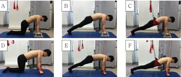
A single instructor educated the subjects on how to perform each exercise and allowed them to practice for 10 minutes for accustomization so that the proper motion would be achieved. After 10 mi- nutes, each subject randomly performed one of six variations of the push-up plus exercise (Figure 1).
The SPP is a standard push-up with the addition
of full scapular protraction (the “plus”) after ob- taining full elbow extension at the end of the usual push-up with the feet together. The subjects were asked to keep their body in a neutral position with the hip and legs extended. The locations of the hands and feet were determined with tape by calcu- lating the distance as 75% of the subject’s height (Pirauá et al, 2014). The QPP was performed with the subjects on their knees with 90° hip flexion. The Low back supported push-up plus (LSPP) was used clinically as an alternative to SPP in patients with weak core stability, because according to Beach et al. (2008), the SPP requires a high level of trunk core muscle activity.
The LSPP was the same as the SPP except that the lower back is supported (12043 Redcord wide sling) under the abdomen at the height of the ante- rior superior iliac spine (ASIS), by using an black elastic cord attachment supported 20 ㎏; the elastic cords should always be perpendicular to the ASIS.
To provide a stable surface during push-up plus, handles (pole: 4 ㎝×16 ㎝, bottom: 14.5 ㎝×30 ㎝×10
㎝) were used. In addition, a sling device (Redcord sling with 12130 Redcord power grip) was used to provide an unstable surface. All trials were com- pleted in the standardized exercise positions: neu- tral head position with the chin tucked, 90°
A B C
D E F
Figure 1. Three variations of the push-up plus exercise with and without sling. (A) Quadruped push-up plus without sling, (B) Standard push-up plus without sling, (C) Low back supported push-up plus without sling, (D) Quadruped push-up plus with sling, (E) Standard push-up plus with sling, and (F) Low back supported push-up plus with sling.
shoulder flexion with full elbow extension, neutral forearms, neutral wrist position, and hands beneath the acromioclavicular joint. Furthermore, the han- dles were placed 10 ㎝ above the ground with the participants positioned. A bar was placed at the T4 level where the scapula was fully protracted. The starting phase of the push-up plus exercises was protracting the scapula by translating the thorax posteriorly until the spinous process of the thoracic vertebra touched the target bar for 2 seconds. The holding phase of the exercise was holding the posi- tion for 5 seconds (Park et al, 2014). The partic- ipants performed three repetitions of each exercise with 5 seconds of intermediate rest, whereas a resting period of 2 minutes was held between exercises. If the subjects could not maintain the standard position, the holding phase data were not collected.
Statistical analysis
The PASW Statistics ver. 18.0 (SPSS Inc., Chicago, IL, USA) was used for all statistical analyses. The test-retest reliability of the EMG measurements in six interventions was assessed by using the in- tra-class correlation coefficient and 95% confidence interval. Two-way repeated analyses of variance (ANOVA) with two within-subject factors (surface condition: with and without sling; exercise: QPP, SPP, and LSPP) and Bonferroni’s post-hoc test were used to assess the statistical significance of the UT, LT, and LSA EMG activity and the UT/LT and UT/LSA EMG activity ratio. The level of significance was set
at α<.05. If a significant surface condition×exercise interaction was not revealed from the two-way re- peated ANOVA, the main effects of surface con- dition and exercise were determined. If a sig- nificant main effect of exercise was observed, the Bonferroni correction was used (α=.05/3=.017). If a significant surface condition×exercise interaction was found, pairwise comparison with Bonferroni correction was used to determine the simple effect for the post-hoc t-test, to reduce the type I error rate (α=.05/6=.008).
Results
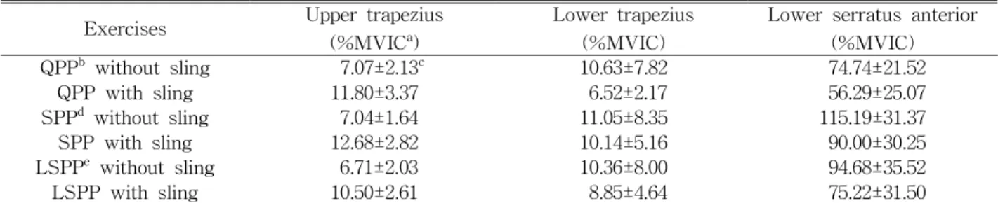
The mean and standard deviation (SD) for EMG measurement of the UT, LT, and LSA during six interventions are depicted in Table 1. For the UT activity, a significant surface condition×exercise in- teraction was found (F2,34=3.884, p=.030). The UT activity was significantly lower in all exercises without sling than in those with sling (p<.001).
The UT activity was significantly lower in the LSPP with sling than in SPP with sling (p<.001) (Figure 2A). In the LT activity, no significant sur- face condition×exercise interaction was found (F2,34=2.44, p=.103). No significant main effects of surface condition (F1,17=1.69, p=.211) and exercise (F2,34=2.50, p=.097) were found (Figure 2B). In the LSA activity, no significant surface condition×exercise interaction was found (F2,34=.32, p=.727). Significant main effects of surface condition (F1,17=17.77, p<.001)
Exercises Upper trapezius
(%MVICa)
Lower trapezius (%MVIC)
Lower serratus anterior (%MVIC)
QPPb without sling 7.07±2.13c 10.63±7.82 74.74±21.52
QPP with sling 11.80±3.37 6.52±2.17 56.29±25.07
SPPd without sling 7.04±1.64 11.05±8.35 115.19±31.37
SPP with sling 12.68±2.82 10.14±5.16 90.00±30.25
LSPPe without sling 6.71±2.03 10.36±8.00 94.68±35.52
LSPP with sling 10.50±2.61 8.85±4.64 75.22±31.50
apercentage of the mean maximal voluntary isometric contractions, bquadruped push-up plus, cmean±standard deviation,
dstandard push-up plus, elow back supported push-up plus.
Table 1. Mean muscle activity for three push-up plus exercise with and without sling
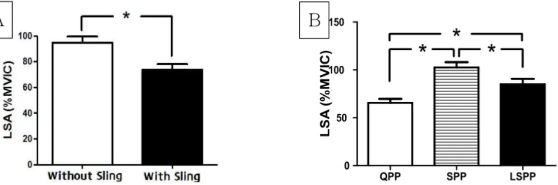
and exercise (F2,34=25.49, p<.001) were found. The LSA activity was significantly greater without than with sling (p=.001) (Figure 3A). The LSA activity was significantly decreased in the order of SPP, LSPP, iand QPP (p<.017) (Figure 3B).
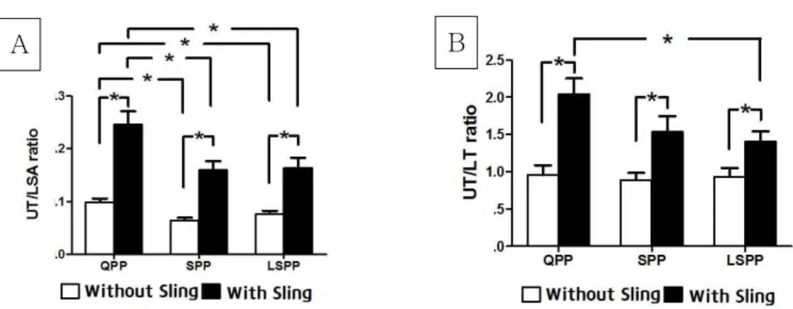
In addition, a significant surface condition×exercise interaction was observed in UT/LSA activity ratio (F2,34=5.48, p=.009). The UT/LSA activity was sig- nificantly lower in all exercises without sling than in those with sling. The UT/LSA activity ratio was significantly lower in SPP and LSPP than in QPP with or without sling (p<.001) (Figure 4A). In the UT/LT activity ratio, a significant surface con- dition×exercise interaction was observed (F2,34=5.87, p=.006). The UT/LT was significantly lower in all ex- ercises without sling than in those with sling (p≤.002).
The UT/LT activity ratio was significantly lower in
LSPP than in QPP with sling (p=.004) (Figure 4B).
Discussion
Our study found that UT activity was sig- nificantly lower in all exercises without sling than in all exercises with sling. This finding is consistent with that of a previous research that recruited sub- jects with scapular dyskinesis (Pirauá et al. 2014).
However, our results differ from those of previous studies with healthy subjects (Lehman et al, 2008;
Maenhout et al, 2010; Park and Yoo, 2011), which showed no significant difference. Such differences may have resulted from differences in subject char- acteristics; that is, whether or not the subjects had scapular dyskinesis. Because of scapular dyskinesis,
A
QPP SPP LSPP
0 50 100 150
QPP SPP LSPP
* *
*
LSA (%MVIC)
B
Figure 3. Comparison of LSA EMG activity during three push-up plus exercise with and without sling. significant difference in main effect (A) LSA for condition, *p<.05; (B) LSA for exercise,
*p<.017 (LSA: Lower serratus anterior, QPP: Quadruped push-up plus, SPP: Standard push-up plus, LSPP: Low back supported push-up plus, MVIC: maximal voluntary isometric contractions).
A B
Figure 2. Comparison EMG activity during three push-up plus exercise with and without sling.
*p<.008, significant simple effect by pairwise comparison with Bonferroni post-hoc test (A) UT (B) LT (UT: upper trapezius, LT: lower trapezius, QPP: quadruped push-up plus, SPP: standard push-up plus, LSPP: low back supported push-up plus, MVIC: maximal voluntary isometric contractions).
altered muscle activities such as a decreased SA and increased UT activity, and abnormal kinematics would be expected to appear (Cools et al, 2003;
Kibler and Sciascia, 2010; Ludewig and Cook, 2000;
Tucker et al, 2010). Park et al. (2014) also demon- strated that reduced SA activity can cause less scapular protraction and increased pectoralis major activity during push-up plus. Considering all of these muscle imbalances and the findings of this study, push-up plus without sling would be a better option for decreasing UT activity. In addition, the UT ac- tivity was significantly lower in LSPP with sling than in SPP with sling. Because the low-back sup- port was designed to provide a stable condition for easily performing LSPP, LSPP with sling would be less influenced by an unstable surface than SPP with sling. Consequently, LSPP would be recommended over SPP in case of using a sling.
Our results of LT activity did not show significant differences according to the surface and exercise.
This result differs from that of a previous study (Pirauá et al, 2014) that showed a significant in- crease on an unstable surface. Such difference may be related to the dyskinesis pattern. Unlike our study, in the study by Pirauá et al. (2014), only sub- jects with patterns I and II dyskinesis participated, and these subjects may show excessive LT activa- tion during the performance of push-up plus.
Patterns I and II mainly caused SA weakness and fatigue (Huang et al, 2015), and inhibited or delayed the LT activity, whereas pattern III is a result of excessive UT activity (Kibler and Sciascia, 2010). As mentioned above, the LT activity would be un- affected by the push-up plus exercise used in our study.
In this study, the LSA activity was significantly greater without than with sling. Pirauá et al. (2014) indicated that subjects with scapular dyskinesis pre- sented increased LSA activity on a stable surface compared with on an unstable surface. And, some other studies with healthy subjects also reported similar results (De Mey et al, 2014; Pontillo et al, 2007). However, some previous studies showed no significant differences between stable and unstable surfaces (de Oliveira et al, 2008; Lear and Gross, 1998; Lehman et al, 2008). Such results may be re- lated to the different conditions in an unstable surface. Pontillo et al. (2007), by using Thera-Band stability trainer, demonstrated that the SA activity was reduced and triceps activity was increased with the increase in the difficulty of balancing the upper extremity during a one-arm push-up posture. This result may have shown the need for more stability at the elbow rather than the shoulder on unstable surface. Because the sling creates a much more un- stable surface in multiple directions as there was no
A B
Figure 4. Comparison EMG ratio during three push-up plus exercise with and without sling.
*p<.008, significant simple effect by pairwise comparison with Bonferroni post-hoc test (A) UT/LSA (B) UT/LT (UT: Upper trapezius, LT: Lower trapezius, LSA: Lower serratus anterior, QPP: Quadruped push-up plus, SPP: Standard push-up plus, LSPP: Low back supported push-up plus, MVIC: maximal voluntary isometric contractions).
contact with the ground (De Mey et al, 2014), it would have more influence on the stability of the el- bow than that of the shoulder. Consequently, the ex- ercise with sling would be ineffective in increasing the LSA muscle activity.
The findings of this study indicated that LSA ac- tivity was significantly increased in the order of QPP, LSPP, and SPP. Previous studies have shown similar findings of significantly greater LSA activity in SPP than in QPP (Decker et al, 1999; Herrington et al, 2015; Ludewig et al, 2004). These results can be explained by the effect of increased upper-ex- tremity weight-bearing load (Lehman et al, 2008 Uhl et al, 2009). To perform SPP, the upper-extremity weight-bearing load applied on the dominant arm should be about 35% of the body weight, whereas QPP requires about 19% of the body weight (Uhl et al, 2003). In addition, the LSA activity in LSPP was lower than that in SPP and higher than that in QPP in our study. This finding can also be explained by the upper-extremity weight-bearing load, because the low-back support in LSPP would have influenced to reduce the load of the body weight on the upper extremity. Consequently, SPP was the best exercise to increase LSA activity. Furthermore, SPP and LSPP could be used to increase SA strength because ludewig et al. (2004) stated that loads >66% of the MVIC would be sufficient to increase SA strength.
This means that LSPP is also recommended to in- crease strength for subjects with difficulty in per- forming SPP owing to weak core stability
In our study, the UT/LSA activity ratio was sig- nificantly lower in all three exercises without sling (<.1) than in those with sling (range: .15~.25). Our study result supports those of previous researches that used a stable surface to decrease the UT/LSA in subjects with scapular dyskinesis during quad- ruped push-up or standard push-up (Batista et al, 2013; Pirauá et al, 2014). These results may be re- lated to scapular dyskinesis which showed a de- creased ratio (Batista et al, 2013). However, unlike our study, both studies demonstrated a higher UT/LSA on
the stable surface (.52 and .52, respectively) and un- stable surface (.78 and .87, respectively). For re- habilitation of the scapula stabilizer, the UT/LSA ac- tivity ratio was considered low if it was nearly or less than .3 (Ludewig et al, 2004; Martins et al, 2008). The reason for the low UT/LSA in our study may be explained by the presence of the plus phase.
Ludewig et al. (2004) showed that UT/SA for the SPP tended to be lower in the push-up plus phase (<.2) than in the push-up without plus phase (<.5).
Although UT/LSA in all exercises was <.3 in our study, push-up plus without sling would be more recommended for decreasing UT/LSA. In addition, in our study, we found that SPP and LSPP produced significantly lower UT/LSA than QPP with and without sling. It can also be explained by the up- per-extremity weight-bearing load. QPP may present significantly lower UT/LSA than do SPP and LSPP as the distal base of support (Ludewig et al, 2004).
Overall, SPP and LSPP without sling are recom- mended for the producing the lowest UT/LSA activ- ity ratio in subjects with scapular dyskinesis. This means that LSPP is recommended for a subjects with difficulty in performing SPP.
Our results showed that the UT/LT activity ratio was significantly lower in all exercises without sling (<1.0) than in those with sling (about 1.5~2.0). The results differ from those of previous studies (De Mey et al, 2014; Maenhout et al, 2010; Pirauá et al, 2014) that showed no significant difference between stable and unstable surfaces. These differences may be ex- plained by the differences in subjects. De Mey et al.
(2014) and Maenhout et al. (2010) included healthy subjects and Pirauá et al. (2014) excluded subjects with pattern III scapular dyskinesis. Because pattern III causes excessive UT activity, and inhibits or de- lays LT activity (Kibler and Sciascia, 2010), the in- cluded pattern III subjects in our study may have more altered muscle activity. Therefore, the use of a sling is not recommend for decreasing UT/LT in subjects with scapular dyskinesis. In addition, the UT/LT activity ratio was significantly lower in
LSPP with sling than in QPP with sling. The reason for the lower UT/LT in LSPP with sling would be both the effect of low-back support and the hip flexion angle. The UT activity would be decreased by the stable condition with low-back support, and LT activity would be increased to stimulate the glu- teus maximus, inducing a myofascial connection by decreasing the hip flexion angle (Kibler and Sciascia, 2010). For the rehabilitation of the scapula stabilizer, UT/LT was considered low if it was nearly or less than 1.0 (Pirauá et al, 2014). As a result, exercises without sling are recommended for increasing the LT activity with a decrease in UT activity to <1.0.
The present study has some limitations. First, only young man subjects were recruited into our study and the number of subjects is relatively small. Thus, our study cannot be generalized to subject with scapular dyskinesis. Second, altered muscle activity, such as that of the pectoralis major, that can affect scapular control was not considered in this study.
Third, we did not analyze the kinematic data to quantify the scapular position during the exercises.
However, with the visual observation by using the target bar, there would be a possibility to present abnormal scapular motion such as scapular winging.
Finally, the dyskinesis patterns need to be classified more specifically because they were observed in dif- ferent kinematics through the influence of scapular muscles.
Conclusion
This study was performed to compare the activa- tion of the UT, LT, and LSA muscles; the UT/LSA activity ratio; and the UT/LT activity ratio during various push-up plus exercises with and without sling in subjects with scapular dyskinesis. The push-up plus exercises without sling were considered to decrease UT and increase LSA activity compared with exercises with sling. Moreover, the UT/LSA and UT/LT ratios were lower in SPP and LSPP
without sling than in the other four push-up plus exercises. Consequently, SPP without sling seems to be the most effective exercise for increasing LSA activity and lowering the UT/LSA and UT/LT ratios in subjects with scapular dyskinesis. In addition, LSPP without sling might be an alternative method to SPP without sling for subjects with difficulty in performing SPP.
References
Batista L, Oliveira V, Piraua A, et al.
Electromyographic activity of scapular stabilizers muscles during push up exercise variations in subjects with and without shoulder impingement syndrome. Motricidade. 2013;9(3):70-82.
Beach TA, Howarth SJ, Callaghan JP. Muscular contribution to low-back loading and stiffness during standard and suspended push-ups. Hum Mov Sci. 2008;27(3):457-472. https://doi.org/10.1016/
j.humov.2007.12.002
Behm DG, Anderson KG. The role of instability with resistance training. J Strength Cond Res.
2006;20(3):716-722.
Cools AM, Dewitte V, Lanszweert F, et al.
Rehabilitation of scapular muscle balance which exercises to prescribe? Am J Sports Med.
2007;35(10):1744-1751.
Cools AM, Witvrouw EE, Declercq GA, et al.
Scapular muscle recruitment patterns: Trapezius muscle latency with and without impingement symptoms. Am J Sports Med. 2003;31(4):542-549.
De Mey K, Danneels L, Cagnie B, et al. Shoulder muscle activation levels during four closed kinetic chain exercises with and without redcord slings.
J Strength Cond Res. 2014;28(6):1626-1635.
https://doi.org/10.1519/JSC.0000000000000292 de Oliveira AS, de Morais Carvalho M, de Brum DP.
Activation of the shoulder and arm muscles during axial load exercises on a stable base of support and on a medicine ball. J Electromyogr
Kinesiol. 2008;18(3):472-479.
Decker MJ, Hintermeister RA, Faber KJ, et al.
Serratus anterior muscle activity during selected rehabilitation exercises. Am J Sports Med.
1999;27(6):784-791.
Ekstrom RA, Soderberg GL, Donatelli RA.
Normalization procedures using maximum volun- tary isometric contractions for the serratus ante- rior and trapezius muscles during surface emg analysis. J Electromyogr Kinesiol. 2005;15(4):418-428.
Hermens HJ, Freriks B, Disselhorst-Klug C, et al.
Development of recommendations for semg sen- sors and sensor placement procedures. J Electromyogr Kinesiol. 2000;10(5):361-374.
Herrington L, Waterman R, Smith L. Electromyographic analysis of shoulder muscles during press-up var- iations and progressions. J Electromyogr Kinesiol.
2015;25(1):100-106. https://doi.org/10.1016/j.jelekin.
2014.10.002
Holtermann A, Mork P, Andersen L, et al. The use of emg biofeedback for learning of selective activation of intra-muscular parts within the serratus anterior muscle: A novel approach for rehabilitation of scapular muscle imbalance. J Electromyogr Kinesiol. 2010;20(2):359-365.
https://doi.org/10.1016/j.jelekin.2009.02.009
Huang JS, Pietrosimone BG, Ingersoll CD, et al. Sling exercise and traditional warm-up have similar effects on the velocity and accuracy of throwing.
J Strength Cond Res. 2011;25(6):1673-1679.
https://doi.org/10.1519/JSC.0b013e3181da7845 Huang TS, Huang HY, Wang TG, et al. Comprehensive
classification test of scapular dyskinesis: A reli- ability study. Man Ther. 2014;20(3):427-432.
https://doi.org/10.1016/j.math.2014.10.017
Huang TS, Ou HL, Huang CY, et al. Specific kine- matics and associated muscle activation in in- dividuals with scapular dyskinesis. J Shoulder Elbow Surg. 2015;24(8):1227-1234. https://doi.org/
10.1016/j.jse.2014.12.022
Kibler W, Ludewig P, McClure P, et al. Clinical im- plications of scapular dyskinesis in shoulder in-
jury: The 2013 consensus statement from the
‘scapular summit’. Br J Sports Med. 2013;47(14):
877-885. https://doi.org/10.1136/bjsports-2013-092425 Kibler WB, McMullen J. Scapular dyskinesis and its
relation to shoulder pain. J Am Acad Orthop Surg. 2003;11(2):142-151.
Kibler WB, Sciascia A. Current concepts: Scapular dyskinesis. Br J Sports Med. 2010;44(5):300-305.
https://doi.org/10.1136/bjsm.2009.058834
Lear LJ, Gross MT. An electromyographical analysis of the scapular stabilizing synergists during a push-up progression. J Orthop Sports Phys Ther. 1998;28(3):146-157.
Lehman GJ, Gilas D, Patel U. An unstable support surface does not increase scapulothoracic stabi- lizing muscle activity during push up and push up plus exercises. Man Ther. 2008;13(6):500-506.
Ludewig PM, Hoff MS, Osowski EE, et al. Relative balance of serratus anterior and upper trapezius muscle activity during push-up exercises. Am J Sports Med. 2004;32(2):484-493.
Ludewig PM, Cook TM. Alterations in shoulder kin- ematics and associated muscle activity in people with symptoms of shoulder impingement. Phys Ther. 2000;80(3):276-291.
Ludewig PM, Phadke V, Braman JP, et al. Motion of the shoulder complex during multiplanar humeral elevation. J Bone Joint Surg. 2009;91(2):378-389.
Lunden JB, Braman JP, Laprade RF, et al. Shoulder kinematics during the wall push-up plus exercise.
J Shoulder Elbow Surg. 2010;19(2):216-223.
https://doi.org/10.1016/j.jse.2009.06.003
Maenhout A, Van Praet K, Pizzi L, et al.
Electromyographic analysis of knee push up plus variations: What is the influence of the kinetic chain on scapular muscle activity? Br J Sports Med. 2010;44(14):1010-1015. https://doi.org/10.1136/
bjsm.2009.062810
Martins J, Tucci HT, Andrade R, et al.
Electromyographic amplitude ratio of serratus anterior and upper trapezius muscles during modi- fied push-ups and bench press exercises. J Strength
Cond Res. 2008;22(2):477-484. http://doi.org/
10.1519/JSC.0b013e3181660748
McClure P, Tate AR, Kareha S, et al. A clinical method for identifying scapular dyskinesis, part 1: Reliability. J Athl Train. 2009;44(2):160-164.
https://doi.org/10.4085/1062-6050-44.2.160
Naughton J, Adams R, Mahr C. Upper-body wobble- board training effects on the post-dislocation shoulder. Phys Ther Sport. 2005;6(1):31-37.
Park KM, Cynn HS, Kwon OY, et al. Comparison of pectoralis major and serratus anterior muscle activities during different push-up plus exercises in subjects with and without scapular winging.
J Strength Cond Res. 2014;28(9):2546-2551.
https://doi.org/10.1519/JSC.0000000000000443 Park SY, Yoo WG. Differential activation of parts of
the serratus anterior muscle during push-up var- iations on stable and unstable bases of support. J Electromyogr Kinesiol. 2011;21(5):861-867.
https://doi.org/10.1016/j.jelekin.2011.07.001
Pirauá ALT, Pitangui ACR, Silva JP, et al.
Electromyographic analysis of the serratus ante- rior and trapezius muscles during push-ups on stable and unstable bases in subjects with scapular dyskinesis. J Electromyogr Kinesiol.
2014;24(5):675-681. https://doi.org/10.1016/j.jelekin.
2014.05.009
Pontillo M, Orishimo KF, Kremenic IJ, et al.
Shoulder musculature activity and stabilization during upper extremity weight-bearing activities.
Am J Sports Phys Ther: 2007;2(2):90-96.
Tucker WS, Armstrong CW, Gribble PA, et al.
Scapular muscle activity in overhead athletes with symptoms of secondary shoulder impinge- ment during closed chain exercises. Arch Phys Med Rehabil. 2010;91(4):550-556. https://doi.org/
10.1016/j.apmr.2009.12.021
Uhl TL, Carver TJ, Mattacola CG, et al. Shoulder musculature activation during upper extremity weight-bearing exercise. J Orthop Sports Phys Ther. 2003;33(3):109-117.
Uhl TL, Kibler WB, Gecewich B, et al. Evaluation of clinical assessment methods for scapular dyskinesis. Arthroscopy. 2009;25(11):1240-1248.
https://doi.org/10.1016/j.arthro.2009.06.007
Weon JH, Kwon OY, Cynn HS, et al. Real-time vis- ual feedback can be used to activate scapular upward rotators in people with scapular wing- ing: An experimental study. J Physiother.
2011;57(2):101-107. https://doi.org/10.1016/S1836 -9553(11)70020-0
This article was received November 22, 2016, was reviewed November 22, 2016, and was accepted January 10, 2017.