Intramyocardial Injection of Stem Cells in Pig Myocardial Infarction Model: The First Trial in Korea
Although cell therapy is emerged for cardiac repair, its efficacy is modest by intracoronary infusion. Therefore, we established the intramyocardial delivery technique using a left ventricular (LV) mapping system (NOGA® XP) using 18 pigs. After adipose tissue-derived mesenchymal stem cells (ATSCs) were delivered intramyocardially to porcine infarcted heart, LV ejection fraction (EF) was increased, and LV chamber size was decreased. We proved the therapeutic effect of intramyocardial injection of ATSC through a LV mapping system in the porcine model for the first time in Korea. The adoption of this technique may accelerate the translation into a clinical application in the near future.
Keywords: Mesenchymal Stem Cell Transplantation; Heart Failure; Myocardial Infarction Min Chul Kim,1* Yong Sook Kim,2,3*
Wan Seok Kang,2 Ki Hong Lee,1 Meeyoung Cho,3 Moon Hwa Hong,3 Kyung Seob Lim,2 Myung Ho Jeong,1 and Youngkeun Ahn1,2
1Department of Cardiology, Chonnam National University Hospital, Gwangju, Korea; 2Biomedical Research Institute, Chonnam National University Hospital, Gwangju, Korea; 3Cell Regeneration Research Center, Chonnam National University Hospital, Gwangju, Korea
* Min Chul Kim and Yong Sook Kim contributed equally to this work.
Received: 19 March 2017 Accepted: 1 July 2017 Address for Correspondence:
Youngkeun Ahn, MD, PhD
Department of Cardiology, Chonnam National University Hospital, 42 Jebong-ro, Dong-gu, Gwangju 61469, Republic of Korea
E-mail: [email protected]
Funding: This study was supported by a grant of the Korea Health Technology R & D Project through the Korea Health Industry Development Institute (HI15C0498), grants of National Research Foundation of Korea (NRF) funded by the Korea government (2015M3A9B4051063, 2015M3A9B4066496, and 2016R1D1A1A09917796), the Bio & Medical Technology Development Program of the NRF funded by the Korean Ministry of Science, ICT and Future Planning (MSIP) (2017M3A9E8023001), and grant of Chonnam National University Hospital Biomedical Research Institute (CRI13901- 22.1).
https://doi.org/10.3346/jkms.2017.32.10.1708 • J Korean Med Sci 2017; 32: 1708-1712
Ischemic heart failure (HF) after myocardial infarction (MI) is the most common cause of HF and its incidence is 52% in Korea (1). Although infarct size is the main determi- nant of long-term mortality in patients with MI and reducing infarct size is essential for suppressing chronic ischemic HF (2), the effects of many pharmacological or non-phar- macological interventions with percutaneous coronary intervention were modest and the impact on clinical outcomes have not been well-studied (3). Stem cell therapy has emerged as a complement to these aforementioned modalities for reducing ischemic HF in patients with MI (4), and various routes can be used to deliver stem cells. Among these, percutaneous intramyocardial delivery is theoretically more effective than the intracoronary or surgical intramyocardial route for targeting the injection site, because it can use myocardial mapping system for the injection (5). Cell type may also be an is- sue. Mesenchymal stem cells (MSCs) are emerging as an extremely promising thera- peutic agent for tissue regeneration (6). Despite the myocardial protective role of MSCs, the therapeutic capacity is limited after intravenous or intracoronary delivery (7). There- fore, this study investigated the effect of delivering MSCs by the percutaneous intramyo- cardial route, using a novel myocardial mapping system, on left ventricular (LV) systol- ic function recovery in a porcine MI model.
This study included 18 pigs (MI was induced by occluding the left anterior descend- ing [LAD] coronary artery: 8 pigs received MSC treatment and 10 did not) from the ani- mal catheterization laboratory of Chonnam National University Hospital. Yorkshire × Landrace F1 crossbred castrated male pigs (25 kg) were observed from the Laboratory Animal Center of Chonnam National University Medical Institute 7 days before the ex- periment. On the day of the procedure, the pigs were anesthetized and the left carotid artery was surgically exposed, and MI was induced with occlusion of LAD using a poly- ethylene terephthalate (PET) occlude. Porcine adipose tissue-derived mesenchymal stem cells (ATSCs) were isolated from abdominal subcutaneous white adipose tissue.
To verify stemness of the porcine MSCs, adipogenic, chondrogenic, and osteogenic differentiation was induced using Stem Cell Differentiation Kits (Invitrogen Life Tech- nologies, Carlsbad, CA, USA). These cells were used in the experiment at passages 3–4 and 1 × 10⁷ cells were injected into each animal. After 1 week of MI, a total of 8 acute myocardial infarction (AMI) pigs received ATSC injections via intramyocardial route after LV mapping. The transthoracic echocardiography (TTE) was performed before BRIEF COMMUNICATION
Cell Therapy & Organ Transplantation
2017-03-16 https://crossmark-cdn.crossref.org/widget/v2.0/logos/CROSSMARK_Color_square.svg
injecting the ATSCs, and again after 4 weeks, in 8 MI pigs to ex- amine the LV ejection fraction (EF) which represents LV systolic function. TTE was also done at the same time in 10 control MI pigs. Images were taken using a conventional cardiac ultrasound system (Vivid S3; GE Healthcare, Schenectady, NY, USA) under general anesthesia while the pigs were in the supine position.
LV end-systolic volume (LVESV) and end-diastolic dimensions (LV end-diastolic volume [LVEDV]) were determined from two- dimensional imaging, and LVEF was calculated using the mod- ified biplane method (8). After anesthesia, the right carotid ar- tery was surgically exposed and an 8F introducer sheath with a side port was placed in the right carotid artery. Then, a LV map- ping catheter (NOGASTAR®; Biosense Webster, Irwindale, CA, USA) was inserted into the left ventricle and connected to a map- ping system (NOGA® XP; Biosense Webster). The mapping cath- eter was manipulated in every part of the left ventricle, and the catheter recognized the voltage in the myocardium and sent a signal to the mapping system. The mapping system showed an image of the mapping results which were defined by the bound- ary of different colors (red: infarcted myocardium, purple: heal- thy myocardium, and green: borderline infarcted myocardium).
An intramyocardial injection catheter with a protruding needle (MyoStar®; Biosense Webster) was used for the ATSC injections.
The borderline infarction zone was the site for the ATSCs injec- tions which is represented as a green color in the mapping sys- tem. The length of the needle was adjusted to no more than 50%
of the target tissue depth (5–10 mm) and the rate of injection for each 0.1 mL was 30 seconds, followed by a 5 seconds hold at the same pressure at the injection site before removing the nee- dle (9). A total of 2 mL of ATSCs were injected, and the injection points were spaced at least 10 mm apart according to the man- ufacturer’s guidelines.
Statistical analysis was performed using the Statistical Pack- age for Social Sciences (SPSS) for Windows, version 21.0 (IBM, Armonk, NY, USA). The data are presented as means ± standard deviation. The Mann-Whitney test was used to compare the echo- cardiographic data between the ATSC injection and control groups. The baseline and 4-week follow-up echocardiographic data were compared using the Wilcoxon signed-rank test. A P value < 0.05 was considered significant.
Table 1 showed a comparison of the echocardiographic data at baseline and 4 weeks after MI between the ATSCs and control groups. LVEF (46.1% ± 6.1% vs. 48.2% ± 2.4%, P = 0.829), LVEDV (52.0% ± 11.5% vs. 45.1% ± 6.2%, P = 0.360), and LVESV (28.8 ± 8.0 vs. 23.9 ± 4.2 mL, P = 0.146) were comparable between the two groups at baseline. However, LVEF was higher in the MSC group than in the control group (56.9% ± 8.1% vs. 45.1% ± 7.3%, P = 0.012) 4 weeks post-MI. There was a tendency toward a small- er LVEDV (41.2 ± 9.4 vs. 50.8 ± 12.2 mL, P = 0.055) in the ATSCs group, and LVESV was also smaller (18.1 ± 6.7 vs. 27.5 ± 8.5 mL, P = 0.012) in the ATSCs group at follow-up compared with that
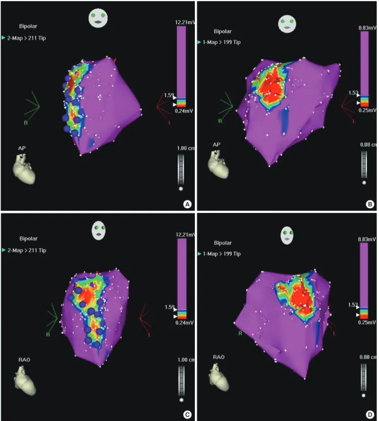
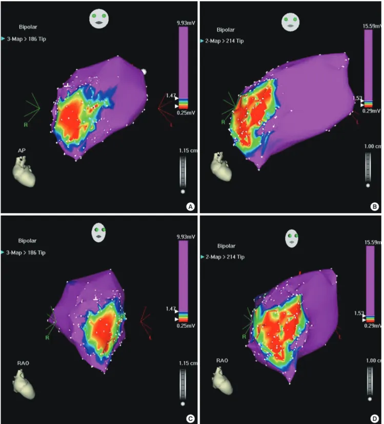
in the control group. On comparisons of echocardiography data between baseline and 4 weeks after MI, LVEF increased signifi- cantly (P = 0.039) in the ATSCs group, and the LVEDV (P = 0.013) and LVESV (P = 0.001) values decreased significantly from base- line to follow-up. No differences in LVEF (P = 0.228), LVEDV (P = 0.153), and LVESV (P = 0.225) were observed in the two groups. Follow-up LV mapping was performed to compare the infarct sizes between 1 and 4 weeks after MI. Visual estimation showed that infarct size decreased in cell-injected pigs (Fig. 1) on the bipolar voltage map at 4 weeks after MI: anteroposterior (1 week and 4 weeks; Fig. 1A and 1B) and right anterior oblique (1 week and 4 weeks; Fig. 1C and 1D) projections. On the other hand, infarct size was extended in the control pig on the same projections (Fig. 2).
Intramyocardial injection of ATSCs into the accurate sites fol- lowed by LV mapping improved LV systolic function, and de- creased infarct size on follow-up mapping. This is the first pre- clinical study in Korea to investigate the efficacy of intramyocar- dial injection of MSCs using a novel LV mapping system (NOGA® XP) and catheter (NOGASTAR®) in an MI model. Although many preclinical and clinical studies have evaluated the efficacy of stem cell therapy in cases of MI and ischemic HF, the impact of cell therapy remained questionable and they reported only mod- est success with a mean increase of 2%–8% in LVEF by intracor- onary route (9). The percutaneous intramyocardial route is more easily accessible and results in a better engraftment of injected cells compared to the direct injection method. However, special equipment and a catheter system are required for mapping the left ventricle prior to and during cell injections (10).
Other preclinical studies showed the efficacy of percutane- ous intramyocardial injections of stem cells in terms of reduced infarct size and improved LV systolic function (11-16). Although the present study did not evaluate clinical outcomes, LV remod- eling represented as LVEDV improved and LV remodeling is the main component of ischemic HF. Therefore, its clinical useful- ness is encouraging in patients with MI, although a further large- Table 1. Comparison of echocardiography data at baseline and 4 weeks post-MI be- tween cell injection group and control group
Echocardiography findings ATSCs group (n = 8)
Control group
(n = 10) P value Baseline (1 wk after MI)
LVEF, % 46.1 ± 6.1 48.2 ± 2.4 0.829
LVEDV, mL 52.0 ± 11.5 45.1 ± 6.2 0.360
LVESV, mL 28.8 ± 8.0 23.9 ± 4.2 0.146
Follow-up (4 wk after MI)
LVEF, % 56.9 ± 8.1 45.1 ± 7.3 0.012
LVEDV, mL 41.2 ± 9.4 50.8 ± 12.2 0.055
LVESV, mL 18.1 ± 6.7 27.5 ± 8.5 0.012
Data are presented as mean ± SD.
MI = myocardial infarction, ATSCs = adipose tissue-derived mesenchymal stem cells, LVEF = left ventricular ejection fraction, LVEDV = left ventricular end-diastolic volume, LVESV = left ventricular end-systolic volume, SD = standard deviation.
Fig. 1. Comparisons of infarct size by visual estimation between 1 week and 4 weeks post-AMI in cell injected pig (purple dots). (A, B) Anteroposterior (1 week and 4 weeks) and (C, D) right anterior oblique (1 week and 4 weeks) projections. Infarct size was reduced on bipolar voltage map 4 weeks after AMI.
AMI = acute myocardial infarction.
A B
C D
scaled clinical study is needed. To date, no previous trial to our knowledge has applied stem cell injection via intramyocardial route in Korea.
The limitations of this study include the lack of a sham group,
the small sample size, and the loss of histological analyses. Fur- ther studies should be performed to investigate the action mech- anisms of ATSCs.
In conclusion, we successfully established the intramyocar-
Fig. 2. Comparisons of infarct size by visual estimation between 1 week and 4 weeks post-AMI in control pig. (A, B) Anteroposterior (1 week and 4 weeks) and (C, D) right an- terior oblique (1 week and 4 weeks) projections. There was an extension of infarct size in control pig.
AMI = acute myocardial infarction.
A B
C D
dial injection system and stably delivered stem cells into por- cine infarcted myocardium. This is the first pre-clinical trial in Korea, and ATSCs injections using LV mapping system can be useful to recover LV systolic function and to reduce LV remod-
eling in porcine MI.
Ethics statement
The animal experiment for this study was approved by the In-
stitutional Animal Care and Use Committee of Chonnam Na- tional University Medical School and Chonnam National Uni- versity Hospital (CNU IACUC-H-2015-38), and conformed to the Guide for the Care and Use of Laboratory Animals published by the US National Institutes of Health (NIH Publication No. 85- 23, revised 1996).
DISCLOSURE
The authors have no potential conflicts of interest to disclose.
AUTHOR CONTRIBUTION
Conceptualization: Kim MC, Kim YS, Cho M, Lee KH, Jeong MH, Ahn Y. Investigation: Kim MC, Kim YS, Kang WS, Lee KH, Cho M, Lim KS, Hong MH. Writing - original draft: Kim MC, Kim YS, Ahn Y. Writing - review & editing: Kim MC, Kim YS, Ahn Y.
ORCID
Min Chul Kim https://orcid.org/0000-0001-6026-1702 Yong Sook Kim https://orcid.org/0000-0001-9483-5639 Wan Seok Kang https://orcid.org/0000-0002-8041-3109 Ki Hong Lee https://orcid.org/0000-0002-9938-3464 Meeyoung Cho https://orcid.org/0000-0003-0697-6867 Kyung Seob Lim https://orcid.org/0000-0002-9117-887X Moon Hwa Hong https://orcid.org/0000-0003-4608-9859 Myung Ho Jeong https://orcid.org/0000-0003-4173-1494 Youngkeun Ahn https://orcid.org/0000-0003-2022-9366 REFERENCES
1. Choi DJ, Han S, Jeon ES, Cho MC, Kim JJ, Yoo BS, Shin MS, Seong IW, Ahn Y, Kang SM, et al. Registry. Characteristics, outcomes and predictors of long-term mortality for patients hospitalized for acute heart failure: a re- port from the Korean heart failure registry. Korean Circ J 2011;41:363-71.
2. McMurray JJ. Clinical practice. Systolic heart failure. N Engl J Med 2010;
362: 228-38.
3. Ibáñez B, Heusch G, Ovize M, Van de Werf F. Evolving therapies for myo- cardial ischemia/reperfusion injury. J Am Coll Cardiol 2015; 65: 1454-71.
4. Gyöngyösi M, Wojakowski W, Lemarchand P, Lunde K, Tendera M, Bar- tunek J, Marban E, Assmus B, Henry TD, Traverse JH, et al. ACCRUE In- vestigators. Meta-Analysis of Cell-based CaRdiac stUdiEs (ACCRUE) in patients with acute myocardial infarction based on individual patient data.
Circ Res 2015; 116: 1346-60.
5. Charwat S, Lang I, Dettke M, Graf S, Nyolczas N, Hemetsberger R, Zamini S, Khorsand A, Sochor H, Maurer G, et al. Effect of intramyocardial deliv- ery of autologous bone marrow mononuclear stem cells on the regional myocardial perfusion. NOGA-guided subanalysis of the MYSTAR prospec-
tive randomised study. Thromb Haemost 2010; 103: 564-71.
6. Williams AR, Hare JM. Mesenchymal stem cells: biology, pathophysiolo- gy, translational findings, and therapeutic implications for cardiac disease.
Circ Res 2011; 109: 923-40.
7. Kim YS, Kwon JS, Hong MH, Kang WS, Jeong HY, Kang HJ, Jeong M, Ahn Y. Restoration of angiogenic capacity of diabetes-insulted mesenchymal stem cells by oxytocin. BMC Cell Biol 2013; 14: 38.
8. Lang RM, Bierig M, Devereux RB, Flachskampf FA, Foster E, Pellikka PA, Picard MH, Roman MJ, Seward J, Shanewise JS, et al. Recommendations for chamber quantification: a report from the American Society of Echo- cardiography’s Guidelines and Standards Committee and the Chamber Quantification Writing Group, developed in conjunction with the Euro- pean Association of Echocardiography, a branch of the European Society of Cardiology. J Am Soc Echocardiogr 2005;18:1440-63.
9. Clifford DM, Fisher SA, Brunskill SJ, Doree C, Mathur A, Clarke MJ, Watt SM, Martin-Rendon E. Long-term effects of autologous bone marrow stem cell treatment in acute myocardial infarction: factors that may influence outcomes. PLoS One 2012; 7: e37373.
10. Gyöngyösi M, Dib N. Diagnostic and prognostic value of 3D NOGA map- ping in ischemic heart disease. Nat Rev Cardiol 2011; 8: 393-404.
11. Gyöngyösi M, Lang I, Dettke M, Beran G, Graf S, Sochor H, Nyolczas N, Charwat S, Hemetsberger R, Christ G, et al. Combined delivery approach of bone marrow mononuclear stem cells early and late after myocardial infarction: the MYSTAR prospective, randomized study. Nat Clin Pract Cardiovasc Med 2009; 6: 70-81.
12. Heldman AW, DiFede DL, Fishman JE, Zambrano JP, Trachtenberg BH, Karantalis V, Mushtaq M, Williams AR, Suncion VY, McNiece IK, et al. Tran- sendocardial mesenchymal stem cells and mononuclear bone marrow cells for ischemic cardiomyopathy: the TAC-HFT randomized trial. JAMA 2014; 311: 62-73.
13. Trachtenberg B, Velazquez DL, Williams AR, McNiece I, Fishman J, Nguyen K, Rouy D, Altman P, Schwarz R, Mendizabal A, et al. Rationale and de- sign of the Transendocardial Injection of Autologous Human Cells (bone marrow or mesenchymal) in Chronic Ischemic Left Ventricular Dysfunc- tion and Heart Failure Secondary to Myocardial Infarction (TAC-HFT) trial: a randomized, double-blind, placebo-controlled study of safety and efficacy. Am Heart J 2011; 161: 487-93.
14. Williams AR, Trachtenberg B, Velazquez DL, McNiece I, Altman P, Rouy D, Mendizabal AM, Pattany PM, Lopera GA, Fishman J, et al. Intramyocar- dial stem cell injection in patients with ischemic cardiomyopathy: func- tional recovery and reverse remodeling. Circ Res 2011; 108: 792-6.
15. Pokushalov E, Romanov A, Chernyavsky A, Larionov P, Terekhov I, Artyo- menko S, Poveshenko O, Kliver E, Shirokova N, Karaskov A, et al. Efficien- cy of intramyocardial injections of autologous bone marrow mononucle- ar cells in patients with ischemic heart failure: a randomized study. J Car- diovasc Transl Res 2010; 3: 160-8.
16. Bartunek J, Behfar A, Dolatabadi D, Vanderheyden M, Ostojic M, Dens J, El Nakadi B, Banovic M, Beleslin B, Vrolix M, et al. Cardiopoietic stem cell therapy in heart failure: the C-CURE (Car diopoietic stem Cell therapy in heart failURE) multicenter randomized trial with lineage-specified bio- logics. J Am Coll Cardiol 2013; 61: 2329-38.