Brief Report
Vol. 30, No. 4, 2018 491
Received July 19, 2017, Revised August 23, 2017, Accepted for publication August 28, 2017
*These authors contributed equally to this work and should be considered co-corresponding authors.
Corresponding author: Chun Wook Park, Department of Dermatology, Kangnam Sacred Heart Hospital, Hallym University College of Medicine, 1 Singil-ro, Yeongdeungpo-gu, Seoul 07441, Korea. Tel: 82-2-829-5221, Fax: 82-2-832-3237, E-mail: [email protected]
ORCID: https://orcid.org/0000-0003-4512-8668
Hye One Kim, Department of Dermatology, Kangnam Sacred Heart Hospital, Hallym University College of Medicine, 1 Singil-ro, Yeongdeungpo-gu, Seoul 07441, Korea. Tel: 82-2-829-5221, Fax: 82-2-832-3237, E-mail: [email protected]
ORCID: https://orcid.org/0000-0001-5846-0008
This is an Open Access article distributed under the terms of the Creative Commons Attribution Non-Commercial License (http://creativecommons.org/
licenses/by-nc/4.0) which permits unrestricted non-commercial use, distribution, and reproduction in any medium, provided the original work is properly cited.
Copyright © The Korean Dermatological Association and The Korean Society for Investigative Dermatology
https://doi.org/10.5021/ad.2018.30.4.491
A Case of Vascular Leiomyoma on the Heel: A Rarely Seen Benign Soft Tissue Tumor with Brief Reviews
Jee Hee Son, Hyun Ji Kim, Min Je Jung, Yong Won Choi, Bo Young Chung, Hye One Kim*, Chun Wook Park*
Department of Dermatology, Hallym University Kangnam Sacred Heart Hospital, Hallym University College of Medicine, Seoul, Korea
Dear Editor:
A 33-year-old male visited our clinic for painful mass on the heel of his right foot. The patient did not know when occurred. A slight blue to skin-colored nodule about 1×1 cm in size was found on his right heel (Fig. 1A). Ultraso- nography was performed and a well-demarcated oval hy- perechoic mass in the subcutaneous fat layer, with prom- inent vascularity was indicated (Fig. 1B). A vascular leio- myoma, hemangioma, glomus tumor or complicated epi- dermoid cyst was suspected. The tumor was completely resected and the microscopic examination exposed nu- merous tortuous vascular channels with proliferation of spindle-shaped cells displaying an interlacing band-like pattern. Elongated spindle cells with abundant brightly eo- sinophilic cytoplasm were observed without necrosis, pleomorphism, mitosis or nuclear atypia (Fig. 2A; H&E,
×40). With these results, fibrous components with edema- tous stroma were seen (Fig. 2B; H&E, ×200). Immunohis- tochemistry revealed the spindle-shaped tumor cells were diffusely positive with desmine (Fig. 2C, ×100). Tissue were stained with Masson’s trichrome (Fig. 2D, ×100).
S-100 stain was negative (Fig. 2E, ×100). The final diag- nosis appeared to be vascular leiomyoma.
Leiomyoma is a benign smooth muscle tumor which usu- ally occurs in the extra-skeletal area, such as ovaries, ute- rus, bladder, lung and gastrointestinal tract1. Occasionally
skin and subcutaneous soft tissues are involved2. Enzinger and Weiss explained that leiomyomas can be classified in- to three types; vascular, cutaneous and deep soft tissue3. Vascular leiomyomas, featured by a painful solitary tumor occurring most frequently in the extremities, originate from the tunica media layer of a vein2,3. Pain is accom- panied in approximately 60% of patients whether there is tenderness4. It typically affects middle aged females in the third and fourth decades but may occur at any age1,2,4. It usually presents as a solitary mass. Its differential diagnosis includes tender tumors such as neuroma, neurilemmoma, and eccrine spiradenoma because pain is the most charac- teristic subjective symptom. Etiology is still unknown.
Morimoto suggested that pain may be caused by the con- traction of vessels which lead to local ischemia5. X-ray findings are mostly normal, however, rarely, dystrophic calcifications might be seen3. The essential histologic fea- tures include bundles and masses of smooth muscle fibers, irregularly separated by strands of collagen fibers1. A vari- ety of immunohistochemical stainings such as desmin, vi- mentin, S-100 protein, neuron specific enolase, actin and factor VIII-related protein could be performed for differ- entiation or an indication of muscle origin and endothel- ical cell1,3. The most satisfactory treatment is complete ex- cision1. In the Korean dermatology literature, there have been several reported cases on lower extremities, cheek,
Brief Report
492 Ann Dermatol
Fig. 1. (A) A slightly bluish to skin-colored nodule about 1×1 cm in size was found on his right heel.
(B) By ultrasonography, a well-de- marcated oval hyperechoic mass in the subcutaneous fat layer, with prominent vascularity, was deter- mined in the subcutaneous fat layer on the right heel.
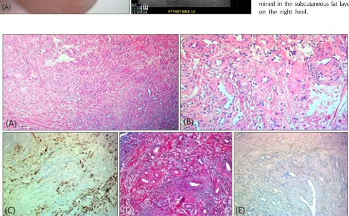
Fig. 2. (A) Numerous tortuous vascular channels with proliferation of spindle-shaped cells displaying an interlacing band-like pattern.
The angioleiomyoma composed of rich vascular channels with thick vessel walls and smooth muscle bundles with elongated nuclei.
Elongated spindle cells with abundant brightly eosinophilic cytoplasm were observed without necrosis, pleomorphism, mitosis or nuclear atypia (H&E, ×40). (B) Fibrous components and edematous stroma were observed. The mitotic activity was low and no necrosis was seen. (H&E, ×200). (C) The tumor cells show positivity for desmin (×100). (D) Masson’s trichrome stain demonstrating smooth muscle fibers in red, erytherocytes in blue, collagen fibers in blue and nuclei in black-blue (×100). (E) S-100 stain was negative (×100).
lip and multiple locations involving shoulders, upper chest, upper back, arm, and neck1. Few studies analyzing vascular leiomyoma have been conducted in Korea1. Little has been reported on the heel in young adult male pa- tient, just like our case. Vascular leiomyomas are rarely di- agnosed clinically before biopsy, but can occur anywhere in various ages. Although vascular leiomyoma is an in- frequent soft tissue tumor, it is worth consideration as the differential diagnosis of nodule with pain on the heel.
ACKNOWLEDGMENT
This study was supported by grants from the National Research Foundation of Korea (NRF), funded by the Ministry of Science, ICT & Future Planning (NRF-2017- R1A2B4006252), the Korea Healthcare technology R&D Project, funded by the Ministry of Health & Welfare, Republic of Korea (HI17C0597), and the Hallym Univer- sity Research Fund (HURF-2017-35, HURF-2017-52).
Brief Report
Vol. 30, No. 4, 2018 493
Received July 6, 2017, Revised August 22, 2017, Accepted for publication September 4, 2017
*These authors have equally contributed to the article.
Corresponding author: Min Kyung Shin, Department of Dermatology, School of Medicine, Kyung Hee University, 23 Kyungheedae-ro, Dongdaemun-gu, Seoul 02447, Korea. Tel: 82-2-958-8300, Fax: 82-2-969-6538, E-mail: [email protected]
ORCID: https://orcid.org/0000-0001-9834-7931
This is an Open Access article distributed under the terms of the Creative Commons Attribution Non-Commercial License (http://creativecommons.org/
licenses/by-nc/4.0) which permits unrestricted non-commercial use, distribution, and reproduction in any medium, provided the original work is properly cited.
Copyright © The Korean Dermatological Association and The Korean Society for Investigative Dermatology
CONFLICT OF INTEREST
The authors have nothing to disclose.
REFERENCES
1. Kim MS, Kwon WJ, Cho EB, Park EJ, Kim KH, Kim KJ.
Angioleiomyoma: a clinicopathological study of 27 cases.
Korean J Dermatol 2016;54:91-97.
2. Gajanthodi S, Rai R, Chaudhry RK. Vascular leiomyoma of
foot. J Clin Diagn Res 2013;7:571-572.
3. Sayit E, Sayit AT, Zan E, Bakirtas M, Akpinar H, Gunbey HP.
Vascular leiomyoma of an extremity: Report of two cases with MRI and histopathologic correlation. J Clin Orthop Trauma 2014;5:110-114.
4. Kinoshita T, Ishii K, Abe Y, Naganuma H. Angiomyoma of the lower extremity: MR findings. Skeletal Radiol 1997;26:
443-445.
5. Morimoto N. Angiomyoma (vascular leiomyoma): a clinico- pathologic study. Med J Kagoshima Univ 1973;24:663-688.
https://doi.org/10.5021/ad.2018.30.4.493
Clinical Factors Influencing Outcomes of 1064 nm Neodymium-Doped Yttrium Aluminum Garnet (Nd:YAG) Laser Treatment for Onychomycosis
Hyun Joo Kim*, Hyung-jin Park
1,*, Dong Hye Suh, Sang Jun Lee, Ki-Heon Jeong
1, Mu-Hyoung Lee
1, Min Kyung Shin
1Arumdaun Nara Dermatologic Clinic, 1Department of Dermatology, School of Medicine, Kyung Hee University, Seoul, Korea
Dear Editor:
Onychomycosis is a common fungal infection of the nails, approximately 2% to 13% of the general population is af- fected by onychomycosis1. There are several therapeutic modalities, among which oral antifungal agents are con- sidered the gold standard. While these agents are gen- erally well tolerated, they carry a treatment failure risk of approximately 25% to 40% primarily due to poor patient compliance and drug interactions. There are also several contraindications for oral antifungal drugs including liver disease and congestive cardiac failure. Apart from these medical causes, some patients are reluctant to take oral
antifungal drugs due to concerns and misconceptions about the risks associated with systematic antifungals.
Thus, there is a growing need for new therapeutic options that are safe and beneficial to a wider population without contraindications. Recently, a number of different devices have been developed, most of which utilize a 1064 nm neodymium-doped yttrium aluminum garnet (Nd:YAG) la- ser3. Although not included in recent guidelines2, several studies have shown that 1064 nm Nd:YAG lasers are ef- fective in treating onychomycosis, while other studies have reported conflicting results4-6. The Food and Drug Administration has also approved the 1064 nm Nd:YAG