Corresponding author:
Sae-Ock Oh
Address: Beomeo-Ri, Mulgeum-eup, Yangsan, Korea [626-870]
Tel: +82-51-510-8045, Fax: +82-51-510-8049, E-mail: hedgehog@pusan.
ac.kr
Copyright © 2010. Anatomy and Cell Biology
pISSN 2093-3665 eISSN 2093-3673
CDH3/P-Cadherin regulates migration of HuCCT1 cholangiocarcinoma cells
Sungmin Baek
1,2, Yong-Whan Lee
1, Sik Yoon
1, Sun-Yong Baek
1, Bong-Seon Kim
1, Sae-Ock Oh
1,21Department of Anatomy, School of Medicine, Pusan National University, 2Medical Research Center for Ischemic Tissue Regeneration, Pusan National University, Yangsan, Korea
Abstract: Intrahepatic cholangiocarcinoma is the second most common subtype of primary hepatobilliary cancer. Despite advances in surgical and medical therapy, its survival rate remains poor. Compared to hepatocellular carcinoma (HCC), the most common liver malignancy, the underlying mechanisms of cholangiocarcinoma carcinogenesis are poorly characterized.
P-cadherin (CDH3) is a cadherin super family member. Although CDH3 is frequently over-expressed in cholangiocarcinoma tissues, its roles have never been characterized. To determine the roles of CDH3 in cholangiocarcinoma, we investigated CDH3 function in HuCCT1 cells using specifi c siRNA. Transfection with CDH3 siRNA did not aff ect proliferation of HuCCT1 cells.
However, cell migration and invasion were signifi cantly reduced when CDH3 was down-regulated. In addition, expressions of several biomarkers for epithelial-mesenchymal transition (EMT) were not changed by CDH3 down-regulation. Th ese results suggest that CDH3 regulates cell migration independent of EMT in cholangiocarcinoma cells.
Key words: Cholangiocarcinoma, CDH3, migration
Received April 4, 2010; Revised April 22, 2010; Accepted May 12, 2010
geographic variation in the incidence of cholangiocarcinoma, with the highest incidence in East Asia. Th is high incidence is likely due to regional risk factors, such as heptolithiasis and liver fl uke infection. Despite advances in surgical and medical therapy, its survival rate is still poor. The main reasons of poor prognosis are diagnostic diffi culty, extensive local tumor invasion at diagnosis and multi-drug resistance.
An important prognostic factor for cholangiocarcinoma is metastasis, which precludes curative surgical resection.
Prognosis is dependent on the presence of free margins in resected tissues and the absence of lymph nodes metastasis (Olnes & Erlich, 2004). Increased cell invasion and migration is a key phenotypic advantage of malignant cells favoring metastasis. Several distinct steps have been described in the process of metastasis: detachment of tumor cells from the primary tumor, invasion into surrounding tissue, intravasation into blood or lymphatic vessels, dissemination in the blood stream or the lymphatic system, extravasation and outgrowth at a secondary site (Yilmaz & Christofori, 2009). Each of these steps requires a distinct molecular
Introduction
Cholangiocarcinoma is a malignant tumor originated from bile duct epithelial cells (Olnes & Erlich, 2004). Intrahepatic cholangiocarcinoma is the second most common subtype of primary hepatobilliary cancer (Kato et al., 1990; Taylor- Robinson et al., 1997; Olnes & Erlich, 2004; Shaib et al., 2004). Between 1973 and 1997, the incidence and mortality rates of intraheptic cholangiocarcinoma in the United States was increased by approximately 9%, and is the most common primary liver cancer-related cause of death in the United Kingdom. The mortality rate of intrahepatic cholangiocarcinoma has increased in Japan, Western Europe, and Australia between 1979 and 1998. There is significant
program. To detach from the primary tumor and invade into the surrounding tissue, tumor cells have to break down cell- cell contacts and the remodel cell-matrix adhesion sites. Th ese processes are known as epithelial-mesenchymal transition (EMT). During EMT, non-motile and polarized epithelial cells that are usually embedded via cell-cell junctions in a cell collective, dissolve their cell-cell junctions and convert into individual, non-polarized, motile and invasive mesenchymal cells (Yilmaz & Christofori, 2009).
Many EMT biomarkers have been reported (Zeisberg &
Neilson, 2009). One of most important biomarkers is down- regulation of E-cadherin. E-cadherin is a cell-cell adhesion molecule expressed on the membrane of epithelial cells.
Moreover, cancer cells undergoing EMT gain mesenchymal markers, including N-cadherin, P-cadherin, vimentin, collagen 1 and 2. EMT can be prompted by various intrinsic signals (e.g. gene mutations) as well as extrinsic signals (e.g.
growth factor signaling) (Yilmaz & Christofori, 2009). Among the growth factors known to induce EMT are transforming growth factor β (TGFβ), hepatocyte growth factor (HGF), members of the epidermal growth factor (EGF) family, insulin-like growth factor (IGF) and fi broblast growth factor (FGF).
Cadherins (Calcium dependent adhesion molecules) are transmembrane proteins which play a role in cell to cell adhesion and junction (Takeichi 1995). The majority of studies on cadherins have focused on E-cadherin (CDH1) and N-cadherin (CDH2). E-cadherin is the prototype family member of classical cadherins, single-span transmembrane glycoproteins that interact in a calcium-dependent, hemophilic manner with E-cadherins on neighboring cells.
E-cadherin-mediated cell-cell adhesion complexes are anchored to the actin cytoskeleton via its cytoplasmic domain, β-catenin and α-catenin. N-cadherin also forms hemophilic cell-cell adhesion junctions. It is normally expressed in nervous tissue, vascular endothelial cells and in skeletal and cardiac muscle cells. It is found to be localized in the lamellipodia and fi llopodia. P-cadherin (CDH3) is one of the cadherin super family members and fi rst identifi ed in mouse placenta (Nose & Takeichi, 1986). In human its expression is not detectable in the placenta but is present in a few organs, such as mammary gland and prostate (Taniuchi et al., 2005).
Unlike the E- and N-cadherins, CDH3 has not fully been investigated and its roles remain unclear. Although previous studies have shown that CDH3 was related to various cancers, such as breast cancer (Paredes et al., 2005; Paredes et al., 2008;
Gorski et al., 2009), colorectal cancer (Milicic et al., 2008;
Hibi et al., 2009a), gastric cancer (Hibi et al., 2009b), head and neck cancer (Dasgupta et al., 2006), ovarian cancer (Cheung et al., 2010), and pancreatic cancer (Taniuchi et al., 2005; Imai et al., 2008), the pathological roles played by CDH3 remain poorly investigated.
Compared to HCC, the underlying mechanisms of cholangiocarcinoma carcinogenesis are poorly characterized.
A previous study showed that CDH3 is frequently over- expressed in cholangiocarcinoma cells (Obama et al., 2005).
However its roles in cholangiocarcinoma cells have not yet been characterized. Th e aim of this study was to demonstrate the roles CDH3 play in cholangiocarcinoma cells.
Materials and Methods
Cell culture and transfection
HuCCT1 cell line was purchased from the Health Science Research Resources Bank (Osaka, Japan). HuCCT1 cells were cultured with RPMI1640, 10% FBS and 1x penicillin/
streptomycin at 37oC and 5% CO2 incubator. CDH3 siRNA (Bioneer, Daejeon, Korea) and scrambled (SCR) siRNA (Dhamacon, Lafayette, CO, USA) were purchased. Cells were transfected with 100 nM of CDH3 siRNA, or SCR siRNA with Dhamafect reagent (Dhamacon, Lafayette, CO, USA) in accordance with the manufacturer’s protocol. SCR siRNA was used as negative control. Th e sequences of CDH3 siRNA duplex were as follows: 5'-CUC UCU GGA AUG GAA CCU U-3', 5'-GAC UGA CCU ACA GUG GAC U-3', and 5'-GUG ACA ACG UCU UCU ACU A-3'
Proliferation assay
Four days aft er transfection of CDH3 siRNA into HuCCT1 cells in a 96-well plate, 10 ul of Ez-Cytox (ITSBIO, Seoul, Korea) were added and incubated for 2 h under normal cell culture conditions. Cell viability was measured by absorbance at 450 nm using an ELISA reader (TECAN, Mannedorf, Switzerland).
Wound-healing assay
A day after transfection of CDH3 siRNA into HuCCT1 cells in a 6-well plate, cells were transferred and cultured to a 48-well plate until confl uent. Th ree hours following treatment with mitomycin C at 5 μg/ml (Sigma-Aldrich, St. Louis, MO, USA), cells were scratched using 200 ul yellow tips and
changed fresh media to incubate for 22 h.
Matrigel invasion assay
As described by Jeon et al. (Jeon et al., 2010), following a day of transfection with SCR or CDH3 siRNA into HuCCT1 cells, transfected cells were seeded to a 24-well BioCoatTM MatrigelTM chamber inserts (BD Biosciences, San Jose, CA, USA). After 36 h, cells on the inside of the inserts were removed with cotton tips, and the invaded cells on the outside of the inserts were visualized using hematoxylin/eosin staining.
Real-time RT-PCR
Total RNA was extracted using RNeasy Mini kit (Qiagen, Valencia, CA, USA) in accordance with the manufacturer’s protocol. cDNA was synthesized with MMLV reverse transcriptase (Promega, Madison, WI, USA), dNTP and oligo- dT primers. Real-time RT-PCR was carried out using Power SYBR Green PCR Master Mix (Applied Biosystems, Foster city, CA, USA) in the ABI Prism 7500 sequence detector (Applied Biosystems, Foster city, CA, USA) in accordance with the manufacturer’s protocol. Th e primer sequences were as follows: CDH1 (F: 5'-TGG GCC AGG AAA TCA CAT CC- 3', R: 5'-CTC AGC CCG AGT GGA AAT GG-3'), CDH3 (F:
5'-CCC CCA GAA GTA CGA GGC CCA-3', R: 5'-ACG CCA CGC TGG TGA GTT GG-3'), Fibronectin (F: 5'-GAG CTG CAC ATG TCT TGG GAA C-3', R: 5'-GGA GCA AAT GGC ACC GAG ATA-3'), SNAI1 (F: 5'-GGA CCC ACA CTG GCG AGA AG-3', R: 5'-ATT CGG GAG AAG GTC CGA GC-3'), SNAI2 (F: 5'-TTG CAA GAT CTG CGG CAA GG-3', R: 5'- AAT GCT CTG TTG CAG TGA GGG C-3'), Vimentin (F: 5'- TGA GTA CCG GAG ACA GGT GCA G-3', R: 5'-TAG CAG CTT CAA CGG CAA AGT TC-3') and β-actin (F: 5'-CAA GAG ATG GCC ACG GCT GC-3', R: 5'-TCC TTC TGC ATC CTG TCG GC-3'). β-actin was used as a loading control and all signals were normalized to β-actin.
Western blotting
Transfected cell lysates were run onto 10% SDS-PAGE gel and transferred to a PVDF membrane. Blocking was carried out with 3% BSA in PBS for 1 h at room temperature. CDH3 antibody (BD Biosciences, CA, USA) was diluted to 1 : 500 in 3% BSA in PBS, and incubated overnight at 4oC. β-actin antibody (Abcam, Cambridge, MA, USA) was diluted to 1 : 2000 in 3% BSA in PBS. HRP-conjugated secondary antibody (Jackson ImmunoResearch Laboratories, West Grove, PA,
USA) was diluted to 1 : 2,000 in PBST and incubated for 2 h at room temperature. Blots were visualized by enhanced chemiluminescence (Amersham Bioscience, Freiburg, Germany).
Data analysis
All data are presented as means±SEM. All experiments were repeated at least 4 times. The difference between the mean values of two groups was evaluated using the Student’s t-test (unpaired comparison). For comparison of more than three groups, we used one-way analysis of variance (ANOVA) test followed by Tukey’s multiple comparison. A P value of
<0.05 was considered statistically signifi cant.
Results
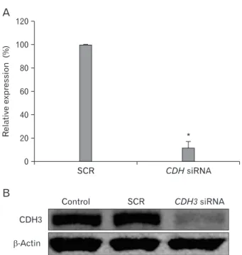
CDH3 is knockdowned through the siRNA
Prior to investigating CDH3 functions in cholangio- carcinoma cells using siRNA, we checked whether CDH3 siRNA specifically down-regulate its expression level. Cells
Fig. 1. CDH3 siRNA specifically knock-downed CDH3 expression level. 48 h following transfection, CDH3 and SCR siRNA-transfected HuCCT1 cells were collected and RNA and protein were purified.
Real-time RT-PCR (A) and Western blotting (B) were performed with CDH3 specific primers and antibody. β-actin was used for loading control. Data are expressed as percent change (means±SEM) compared to control. *P<0.01 (Student’s t-test).
were harvested two days after transfection with CDH3 or scrambled siRNA. Real-time RT-PCR showed transfection of CDH3 siRNA into HuCCT1 cells dramatically decreased its mRNA expression level while scrambled control siRNA made no impact (Fig. 1A). CDH3 protein level was measured by Western blotting using CDH3 antibody. CDH3 siRNA significantly reduced its protein level, whereas scrambled siRNA did not (Fig. 1B). These results indicate that CDH3 siRNA could specifically down-regulate CDH3 expression
level in HuCCT1 cells.
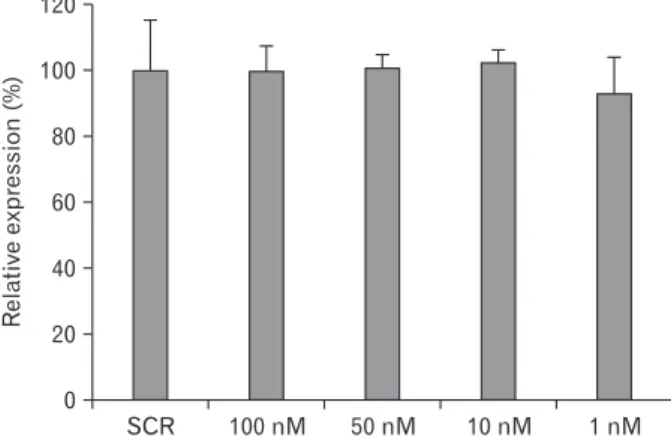
CDH3 has no eff ect on cell proliferation
Previous reports showed that CDH3 promoter is hypomethylated and highly expressed in various cancer cells and tissues (Milicic et al., 2008; Hibi et al., 2009a; Hibi et al., 2009b). However, the roles of CDH3 in cell proliferation are poorly characterized, especially in cholangiocarcinoma cells. We therefore carried out cell proliferation assay to determine whether CDH3 affects cell proliferation in cholangiocarcinoma cells. Different concentration series of CDH3 siRNA were transfected into HuCCT1 cells 1 d after cell seeding. Following 4 d of siRNA transfection, cell proliferation assay was performed as described in the Materials and Methods section. As shown in Fig. 2, down- regulation of CDH3 did not aff ect cell proliferation, consistent with a previous report (Cheung et al., 2010). Th is data suggest that CDH3 plays in other cellular events rather than cell proliferation in cholangiocarcinoma cells.
Cell migration is decreased by CDH3 knockdown Previous reports showed that CDH3 aff ects cell migration in pancreatic cancer cells (Taniuchi et al., 2005) and ovarian cancer cells (Cheung et al., 2010). We hypothesize that CDH3 regulates cell migration in cholangiocarcinoma cells. In order to verify this hypothesis, CDH3 siRNA or scrambled siRNA Fig. 2. Cell proliferation is not aff ected by down-regulation of CDH3
in HuCCT1 cells. SCR siRNA and diff erent concentration series (100 nM, 50 nM, 10 nM, and 1 nM) of CDH3 siRNA-transfected HuCCT1 cells in a 96-well plate were incubated for 4 d, and MTT assay was performed. Data are expressed as percent change (means±SEM) compared to control.
Fig. 3. Down-regulation of CDH3 in HuCCT1 cells reduced migration in wound healing assay. Aft er transfection as described in Materials and Methods, HuCCT1 cells were scratched and allowed to migrate up to 22 h. Control = no transfected, SCR = Scrambled siRNA transfected.
was transfected into HuCCT1 cells, and cell migration was examined using wound-healing assay. Fig. 3 shows that down- regulation of CDH3 decreased cell migration rate in HuCCT1, compared to control and scrambled siRNA. To confi rm this data, we conducted matrigel invasion assay, which is a gold standard cancer cell migration and invasion assay. As shown in Fig. 4, invasion of HuCCT1 cells through the matrigel was signifi cantly reduced when CDH3 was knock-downed. Th ese results are consistent with previous reports (Taniuchi et al., 2005; Cheung et al., 2010), and suggest that CDH3 positively aff ect cell migration in HuCCT1 cells.
CDH3 is not related with EMT
To reveal how CDH3 regulates cell migration in HuCCT1 cells, we decided to study EMT because it can affect cancer cell migration and invasion (Yilmaz & Christofori, 2009). To determine whether EMT is involved in the eff ects of CDH3 on cell migration, change in expression level of EMT-related genes was measured, using real time RT-PCR after CDH3 knock-down. Fig. 5 shows that the mRNA expression levels of mesenchymal markers (snail1, snail2, and vimentin) were not signifi cantly changed. Although the expression level of CDH1 was increased, there was no change in CDH2 expression
level during CDH3 knockdown (data not shown). Th is result implies that CDH3-dependent cell migration may not involve EMT.
Fig. 4. Down-regulation of CDH3 in HuCCT1 cells reduced invasion in Matrigel invasion assay. After trans- fection as described in Materials and Methods, HuCCT1 cells were allowed to invade through the matrigel up to 36 h. Invaded cells were stained with hematoxylin/eosin (A) and counted to quantif y (B). Data are expressed a s p ercent chang e (means±S E M) compared to control. *P<0.01 (Student’s t-test).
Fig. 5. Expression of EMT-related genes in CDH3-knockdowned HuCCT1 cells. RNA was purified from SCR or CDH3 siRNA- transfected cells and real-time RT-PCR was carried out. After normalization to β-actin, data are expressed as percent change (means±SEM) compared to control.
Discussion
The carcinogenesis of cholangiocarcinoma has been poorly characterized compared to HCC. Over-expression of CDH3 in patients with cholangiocarcinoma has been shown in a previous report (Obama et al., 2005). However, its roles in carcinogenesis of cholangiocarcinoma have never been examined. In the present study, we show that CDH3 regulates migration rather than proliferation in cholangiocarcinoma.
Metastatic cancers can preclude curative surgical resection and results in poor prognosis. Over-expression of CDH3 was reported to be associated with aggressive character and poor prognosis in breast and endometrial cancers (Peralta Soler et al., 1999; Gamallo et al., 2001; Stefansson et al., 2004).
Although over-expression of CDH3 in cholangiocarcinoma tissues was reported in an immunohistochemistry study (Obama et al., 2005), associations between its expression and lymph node metastasis, distant metastasis or gross appearance were not observed. It must be emphasized that only 23 tumors specimens from patients were analyzed. Further studies including more tissues specimens should be conducted.
Th ere are controversies surrounding the roles of CDH3 in cancer cells migration. CDH3 is known to promote migration in pancreatic and ovarian cancer (Taniuchi et al., 2005;
Cheung et al., 2010). In contrast, in mammary epithelial cells, CDH3 inhibited migration (Simpson et al., 2008). When Panc-1 cells were stably transfected with full-length CDH3 cDNA, cell migration was significantly enhanced although N-cadherin expression was significantly reduced (Taniuchi et al., 2005). In CDH3-overexpressing Panc-1 cells, the activities of Rac1 and Cdc42 were significantly increased (Taniuchi et al., 2005). Moreover, when OVCAR-3 cells and Caov-3 cells were stably transfected with CDH3 cDNA, cell migration and invasion were significantly enhanced (Cheung et al., 2010). When CDH3 expression was down- regulated using CDH3 siRNA in these cells, cell migration and invasion was significantly reduced. In contrast to these results, when CDH3 expression was down-regulated using CDH3 siRNA in MCF-10A cells (mammary epithelial cells), cell migration was significantly enhanced (Simpson et al., 2008). Th is discrepancy may be due to diff erent roles CDH3 play, depending on cell type. Another explanation may be that MCF-10A cells are non-cancerous cells, whereas Panc- 1, Caov-3, and OVCAR-3 cells are cancerous cells. As such, the roles of CDH3 in cancerous cells and non-cancerous cells may be diff erent. In the present study, CDH3 siRNA inhibited
migration of cholangiocarcinoma cells in two experiments:
wound healing assay, and matrigel invasion assay. Th e results suggest that CDH3 in cancerous cells supports cell migration.
Previous reports suggested possible mechanisms on how CDH3 regulates migration of cancer cells. Taniuchi et al.
showed that p120-catenin was involved in the regulation of motility by CDH3 (Taniuchi et al., 2005). P120-catenin is found in two forms, one bound to cadherins under the plasma membrane and the other in the cytoplasm (Kinch et al., 1995; Staddon et al., 1995; Anastasiadis & Reynolds, 2001). Different types of cadherins regulate cell movement by controlling the levels of p120-catenin present in the cytoplasmic pool. Taniuchi et al. showed that cytoplasmic accumulation of p120-catenin significantly correlated with CDH3 levels but not with the levels of E-cadherin or N-cadherin (Taniuchi et al., 2005). Diff erent distribution may be due to diff erent affi nity of p120 for each classic cadherin.
Because of the low affi nity for CDH3, more p120-catenin is distributed in the cytoplasm when CDH1 is replaced with CDH3 by EMT. In addition, Taniuchi et al. showed that activation of Rac1 and Cdc42 by CDH3 was inhibited by p120-catenin siRNA, which suggested that p120-catenin activated Rac1 and Cdc42 GTPases, which are known modulators of actin dynamics essential for cell migration and invasion (Yilmaz & Christofori, 2009).
Another mechanism on how p120-catenin regulates cell migration and invasion may involve transcriptional repressor Kaiso (van Roy & McCrea, 2005). P120-catenin can transfer to the nucleus where it binds to Kaiso. In contrast to β-catenin/Tcf-mediated transcriptions, where β-catenin acts as transactivator, p120-catenin has no transactivation domain, but rather release Kasio from its promoter binding sites, thereby activates gene expression by de-repression. However, the nature of p120/Kaiso target genes is still poorly defined (Yilmaz & Christofori, 2009).
Cadherins have been shown to play roles in signal transduction in addition to their structural roles in adhesion and migration (Pece & Gutkind, 2000; Suyama et al., 2002;
Qian et al., 2004). For example, E-cadherin is associated with epidermal growth factor receptor (EGFR), thus activating the mitogen-activated protein kinase pathway. N-cadherin has also been found to interact with fibroblast growth factor receptor (FGFr). Moreover, interaction between CDH3 and insulin-like growth factor receptor (IGFRr) has been suggested. Furthermore, EMT is regulated by various growth factors, including EGF, FGF, HGF and IGF, whose
signaling is fi nally transmitted into the nucleus where the key transcription factors, such as SNAI1, SNAI2, ZEB1 and ZEB2 are induced (Th iery et al., 2009; Yilmaz & Christofori, 2009).
Th ese results suggest that a change in CDH3 expression level may modulate EMT induced by a certain growth factor. To test this hypothesis, we examined change in the expression of EMT marker genes following transfection with CDH3 siRNA.
However, we did not observe consistent changes in EMT marker genes expression (Fig. 5).
Although other EMT marker genes including SNAI1, SNAI2, and vimentin were not significantly changed after transfection with CDH3 siRNA, the expression of CDH1 was increased, which may suggest redundancy in the level of cadherins (Fig. 5). During EMT in carcinogenesis, cadherin switch has been well documented (Zeisberg & Neilson, 2009).
However, effects of change in the expression of CDH3 on other cadherins expression were different in different cells.
Th e expression level of CDH1 or CDH2 was not changed by over-expression or knock-down of CDH3 in ovarian cancer cells (Cheung et al., 2010). Th e expression level of CDH2 was reduced however that of CDH1 was not changed by over- expression of CDH3 in pancreatic cancer cells (Taniuchi et al., 2005).
The roles of cadherins in signal transduction pathway indicate that they can regulate proliferation. N-cadherin negatively controls osteoblast proliferation and survival via inhibition of autocrine/paracrine Wnt3a ligand expression and attenuation of Wnt, ERK and PI3K/Akt signaling (Haÿ et al., 2009). E-cadherin can negatively regulate, in an adhesion- dependent manner, ligand-dependent activation of divergent classes of receptor tyrosine kinases (RTKs), by inhibiting their ligand-dependent activation in association with decreases in receptor mobility and in ligand-binding affinity (Qian et al., 2004). As such, E-cadherin neutralizing antibody inhibited proliferation of MDCK cells. However, according to published reports, CDH3 does not regulate cell proliferation, although CDH3 is highly expressed in numerous cancer tissues (Taniuchi et al., 2005; Milicic et al., 2008; Cheung et al., 2010). In Panc-1 cells, neither over-expression of CDH3 nor functional-blocking of CDH3 regulated cell proliferation (Taniuchi et al., 2005). Moreover, when OVCAR-3 cells and Caov-3 cells were stably transfected with CDH3 cDNA, cell proliferation was not affected (Cheung et al., 2010).
Furthermore, the crypt fission rate was not significantly increased in CDH3 transgenic mouse that harbored CDH3 over-expression in the intestinal and colonic epithelium
(Milicic et al., 2008). Consistent with these results, the present study showed that down-regulation of CDH3 did not affect proliferation in cholangiocarcinoma cells.
In summary, CDH3 regulates cell migration through EMT- independent signaling in cholangiocarcinoma cells. Our data will contribute to the development of diagnostic molecular markers and therapies against cholangiocarcinoma.
Acknowledgements
This work was supported for two years by the Pusan National University Research Grant.
References
Anastasiadis PZ, Reynolds AB. (2001). Regulation of Rho GTPases by p120-catenin. Curr Opin Cell Biol 13: 604- 610
Cheung LW, Leung PC, Wong AS. (2010). Cadherin switching and activation of p120 catenin signaling are mediators of gonadotropin-releasing hormone to promote tumor cell migration and invasion in ovarian cancer. Oncogene 29:
2427-2440
Dasgupta S, Tripathi PK, Qin H, Bhattacharya-Chatterjee M, Valentino J, Chatterjee SK. (2006). Identification of molecular targets for immunotherapy of patients with head and neck squamous cell carcinoma. Oral Oncol 42:
306-316
Gamallo C, Moreno-Bueno G, Sarrió D, Calero F, Hardisson D, Palacios J. (2001). The prognostic significance of P-cadherin in infiltrating ductal breast carcinoma. Mod Pathol 14: 650-654
Gorski JJ, James CR, Quinn JE, et al. (2009). BRCA1 trans- criptionally regulates genes associated with the basal-like phenotype in breast cancer. Breast Cancer Res Treat [Epub ahead of print]
Haÿ E, Nouraud A, Marie PJ. (2009). N-cadherin negatively regulates osteoblast proliferation and survival by antagonizing Wnt, ERK and PI3K/Akt signalling. PLoS One 4: e8284
Hibi K, Goto T, Mizukami H, et al. (2009a). Demethylation of the CDH3 gene is frequently detected in advanced colorectal cancer. Anticancer Res 29: 2215-2217
Hibi K, Kitamura YH, Mizukami H, et al. (2009b). Frequent
CDH3 demethylation in advanced gastric carcinoma.
Anticancer Res 29: 3945-3947
Imai K, Hirata S, Irie A, et al. (2008). Identifi cation of a novel tumor-associated antigen, cadherin 3/P-cadherin, as a possible target for immunotherapy of pancreatic, gastric, and colorectal cancers. Clin Cancer Res 14: 6487-6495 Jeon TY, Han ME, Lee YW, et al. (2010). Overexpression of
stathmin1 in the diff use type of gastric cancer and its roles in proliferation and migration of gastric cancer cells. Br J Cancer 102: 710-718
Kato I, Kuroishi T, Tominaga S. (1990). Descriptive epide- miology of subsites of cancers of the liver, biliary tract and pancreas in Japan. Jpn J Clin Oncol 20: 232-237
Kinch MS, Clark GJ, Der CJ, Burridge K. (1995). Tyrosine phosphor ylation regulates the adhesions of ras- transformed breast epithelia. J Cell Biol 130: 461-471 Milicic A, Harrison LA, Goodlad RA, et al. (2008). Ectopic
expression of P-cadherin correlates with promoter hypomethylation early in colorectal carcinogenesis and enhanced intestinal crypt fi ssion in vivo. Cancer Res 68:
7760-7768
Nose A, Takeichi M. (1986). A novel cadherin cell adhesion molecule: its expression patterns associated with implantation and organogenesis of mouse embryos. J Cell Biol 103: 2649-2658
Obama K, Ura K, Li M, et al. (2005). Genome-wide anal ysis of gene expression in human intrahepatic cholangiocarcinoma. Hepatology 41: 1339-1348
Olnes MJ, Erlich R. (2004). A review and update on chola- ngiocarcinoma. Oncology 66: 167-179
Paredes J, Albergaria A, Oliveira JT, JerÓnimo C, Milanezi F, Schmitt FC. (2005). P-cadherin overexpression is an indicator of clinical outcome in invasive breast carcinomas and is associated with CDH3 promoter hypomethylation.
Clin Cancer Res 11: 5869-5877
Paredes J, Correia AL, Ribeiro AS, Milanezi F, Cameselle- Teijeiro J, Schmitt FC. (2008). Breast carcinomas that co-express E- and P-cadherin are associated with p120- catenin cytoplasmic localisation and poor patient survival.
J Clin Pathol 61: 856-862
Pece S, Gutkind JS. (2000). Signaling from E-cadherins to the MAPK pathway by the recruitment and activation of epidermal growth factor receptors upon cell-cell contact
formation. J Biol Chem 275: 41227-41233
Peralta Soler A, Knudsen KA, Salazar H, Han AC, Keshgegian AA. (1999). P-cadherin expression in breast carcinoma indicates poor survival. Cancer 86: 1263-1272
Qian X, Karpova T, Sheppard AM, McNally J, Lowy DR.
(2004). E-cadherin-mediated adhesion inhibits ligand- dependent activation of diverse receptor tyrosine kinases.
Embo J 23: 1739-1748
Shaib YH, Davila JA, McGlynn K, El-Serag HB. (2004). Rising incidence of intrahepatic cholangiocarcinoma in the United States: a true increase? J Hepatol 40: 472-477 Simpson KJ, Selfors LM, Bui J, et al. (2008). Identification
of genes that regulate epithelial cell migration using an siRNA screening approach. Nat Cell Biol 10: 1027-1038 Staddon JM, Smales C, Schulze C, Esch FS, Rubin LL. (1995).
p120, a p120-related protein (p100), and the cadherin/
catenin complex. J Cell Biol 130: 369-381
Stefansson IM, Salvesen HB, Akslen LA. (2004). Prognostic impact of alterations in P-cadherin expression and related cell adhesion markers in endometrial cancer. J Clin Oncol 22: 1242-1252
Suyama K, Shapiro I, Guttman M, Hazan RB. (2002). A signaling pathway leading to metastasis is controlled by N-cadherin and the FGF receptor. Cancer Cell 2: 301-314 Takeichi M. (1995). Morphogenetic roles of classic cadherins.
Curr Opin Cell Biol 7: 619-627
Taniuchi K, Nakagawa H, Hosokawa M, et al. (2005).
Overexpressed P-cadherin/CDH3 promotes motility of pancreatic cancer cells by interacting with p120ctn and activating rho-family GTPases. Cancer Res 65: 3092-3099 Taylor-Robinson SD, Foster GR, Arora S, Hargreaves S,
Thomas HC. (1997). Increase in primary liver cancer in the UK, 1979-94. Lancet 350: 1142-1143
Thiery JP, Acloque H, Huang RY, Nieto MA. (2009).
Epithelial-mesenchymal transitions in development and disease. Cell 139: 871-890
van Roy FM, McCrea PD. (2005). A role for Kaiso-p120ctn complexes in cancer? Nat Rev Cancer 5: 956-964
Yilmaz M, Christofori G. (2009). EMT, the cytoskeleton, and cancer cell invasion. Cancer Metastasis Rev 28: 15-33 Zeisberg M, Neilson EG. (2009). Biomarkers for epithelial-
mesenchymal transitions. J Clin Invest 119: 1429-1437