INTRODUCTION
Atopic dermatitis and allergen
Atopic dermatitis (AD) is a chronic, inflammatory skin disease with intractable pruritus. As one of the leading skin diseases in Westernized countries, its prevalence is increasing steadily world-wide,1-3 AD affects approximately 20% of pediatrics and 1%-3% of adults,4 and 40%-60% of pediatric AD patients contin- ue on as adult-forms later in their lives.1,5,6 Its pathogenesis is multifactorial with roots in a combination of genetic, environ- mental, skin barrier, and other immunological factors. Although there is no single gene responsible for onset of the disease, fam- ily history contributes in predicting prognosis of AD along with interplays between environmental and individual factors. Fur- thermore, abnormalities of the skin barrier have been exten- sively studied in the pathogenesis of AD in several studies,7-12 and these barrier dysfunctions lead to dry and rough surfaced skin of AD patients. Consequently disrupted barrier leads to in- creased rate of secondary infection and penetration of foreign antigens through damaged stratum corneum.
AD can be classified into either intrinsic or extrinsic AD de- pending on co-existence with allergic features; barrier dysfunc- tion and increased penetration of foreign allergens of food and environment are closely associated with aggravation of extrinsic AD. In an acute stage, allergen penetrates through damaged
skin barrier and binds with an epidermal dendritic cell (DC) ex- pressing FcεRI which plays a role in recruiting cutaneous lym- phocyte antigen-bearing T cell to initiate cutaneous inflamma- tion13 and activate Th2 polarization.14,15 Also, interleukin (IL)-16 and monocyte chemotactic protein 1 (MCP-1) produced by these epidermal DCs induce differentiation of monocytes into inflammatory dendritic epidermal cells (IDECs), which produce IL-1, IL-6, and tumor necrosis factor α (TNF- α). Other cytokines in AD pathogenesis such as IL-12 and IL-18 aid in transforma- tion of inflammatory responses from Th2 to Th1/0 and enter chronic phase.16 Through above mechanisms, allergens in envi- ronment are important in both acute phase from repetitive ex- posure and also in chronic status of disease; hence, it is impera- tive for extrinsic patients who have elevated serum and specific IgE for allergens to avoid possible exacerbating factors. And one of the most frequently noted allergens for AD exacerbation is a house dust mite (HDM).
Specific Immunotherapy in Atopic Dermatitis
Jungsoo Lee, Chang Ook Park, Kwang Hoon Lee*
Department of Dermatology, Severance Hospital, Cutaneous Biology Research Institute, Yonsei University College of Medicine, Seoul, Korea
This is an Open Access article distributed under the terms of the Creative Commons Attribution Non-Commercial License (http://creativecommons.org/licenses/by-nc/3.0/) which permits unrestricted non-commercial use, distribution, and reproduction in any medium, provided the original work is properly cited.
Allergen specific immunotherapy (SIT) using house dust mite (HDM) extracts has been performed mainly with patients of asthma and allergic rhini- tis. In the meanwhile, there has been a long debate on the efficacy of SIT in atopic dermatitis (AD) with only a few double-blind placebo-controlled trials. However, several randomized controlled trials of SIT in AD revealed significant improvement of clinical symptoms and also, positive result was shown by a following meta-analysis study of these trials. In order to predict and evaluate the treatment outcome, finding a biomarker that can predict treatment responses and treatment end-points is critical but it is very challenging at the same time due to the complexity of causes and mechanisms of AD. Other considerations including standardization of the easiest and safest treatment protocol and optimizing the treatment prepa- rations should be studied as well. This review summarizes the basics of SIT in AD including the brief mechanisms, treatment methods and sched- ules, and also highlights the clinical efficacy of SIT in AD along with mild, controllable adverse reactions. Immunologic effects and studies of various biomarkers are also introduced and finally, future considerations with upcoming studies on SIT were discussed.
Key Words: Specific immunotherapy; subcutaneous immunotherapy; atopic dermatitis; clinical efficacy; biomarker
Correspondence to: Kwang Hoon Lee, MD, PhD, Department of
Dermatology, Severance Hospital, Cutaneous Biology Research Institute, Yonsei University College of Medicine, 50-1 Yonsei-ro, Seodaemun-gu, Seoul 120-752, Korea.
Tel: +82-2-2228-2080; Fax: +82-2-393-9157; E-mail: [email protected] Received: February 3, 2014; Revised: April 30, 2014; Accepted: June 30, 2014
•There are no financial or other issues that might lead to conflict of interest.
Allergy Asthma Immunol Res. 2015 May;7(3):221-229.
http://dx.doi.org/10.4168/aair.2015.7.3.221 pISSN 2092-7355 • eISSN 2092-7363
Pyroglyphidae Dermatophagoides farinae (Der f), Derma- tophagoides pteronyssinus (Der p) and Euroglyphus maynei are the most common types of HDM. The antigenically active parti- cles contain high enzymatic activity and act through destroying tight junction of epidermis, enhancing penetration of allergens deep into the skin.17,18 One of enzymes that HDM possesses is serine cysteine proteinase, and these enzymes are able to acti- vate proteinase-activated receptors (PARs). Among many PARs, PAR-1, and PAR-2 are known to be most populated in respirato- ry, gastrointestinal systems and skin.19 When PAR is activated, various inflammatory mediators such as IL-6 and IL-8 are se- creted, leading to increase vascular permeability, infiltration of leukocytes, increased airway hypersensitivity, and other effects by HDM that preceded clinical symptoms of allergic diseases.20 Allergen specific immunotherapy (SIT)
Mechanisms of allergen SIT
HDM avoidance has been practiced as a part of lifestyle modi- fication with extrinsic AD patients for quite a period. Yet as a more active treatment modality, SIT is receiving more attention.
SIT was initially practiced in allergic rhinitis or asthma patients.
Up until now, SIT is the only disease-specific treatment modali- ty that suppresses allergic responses for a long period of time.
SIT aims to induce allergen-specific tolerance otherwise known as allergen vaccination21 through acquiring immune tolerance with induction of allergen-specific regulatory T cells (Tregs).
The acute phase of AD is closely associated with production of Th2 cytokines and commonly observed Th2-biased profiles are suggested to be results of increased clonal expansion or dif- ferentiation of Th2 cells or increased tendency to activation and apoptosis of high IFN-γ producing Th1 cells.22 These Th1 cells are known to be involved in apoptosis of epithelium in AD.
Thus, induction of Treg cells during the SIT consequently in- creases suppression of allergen-induced T-cell proliferation, and Th1 and Th2 cytokines.23 Thereby, we may observe clinical improvement of AD as a result of skin inflammation reduction and a diminution in epithelium apoptosis.
Tregs involved in mechanisms of SIT express IL-10, trans- forming growth factor β (TGF-β) to elicit early phase desensiti- zation of mast cell, basophil, and eosinophil. These allergen- specific Tregs also suppress Th2 cells, thereby inhibiting IgE production, while at the same time stimulating expression of IgG4, a non-inflammatory immunoglobulin isotype. Also, cyto- kines such as IL-3, IL-4, IL-5, IL-9, and IL-13 that are expressed from Th2 play an important role in survival, activation, and dif- ferentiation of mast cells, basophil, and eosinophils, but SIT suppresses cytokine axes as well.
Treatment methods and schedules
SIT can be divided into 2 major groups depending on the route of administration: sublingual (SLIT) and subcutaneous (SCIT) methods. While the routes may differ, both equally affect pe-
ripheral allergen-specific Tregs through similar mechanisms for inducing T-cell tolerance, inhibitory functions of IL-10, TGF-β, and reduction of mast cell and eosinophil. However, in early stages of treatment, expression of Treg, reduction in IgE or in- crease in IgG4 might not be evident in SLIT compared to SCIT.24
The most important factor to consider while choosing candi- dates for immunotherapy is finding those who are actually sen- sitized to HDM. Therefore, majority of previously reported studies also enroll patients who have positive allergen sensiti- zation to HDM. Standards for choosing candidates for SIT in our institution is first selecting extrinsic AD patients with serum total IgE above 150, and then additionally selecting only those who have positive reactions (over 3+) to HDM on CAP-test or skin prick test. We initially start the therapy in weekly regimen for 16-18 weeks as initial build-up phase and slowly escalates dosage of HDM extract, and when the maintenance dosage is reached, the patient visits the clinic biweekly for four times. Af- terwards, the monthly regimen can be installed. Depending on clinical response, the patient can continue on with the treat- ment for 3 to 6 years.
There is no exact consensus for treatment period, interval time between treatments, and follow-up period after termina- tion of SIT, but most literature generally agree upon 3 years as an ideal treatment period.25 Our institution also maintains one year of treatment for all those started on SIT, and continues for 3 years unless complete remission is reached.
Clinical efficacy of allergen SIT in AD Efficacy of SIT with HDM in AD
In the past, there has been a lack of evidence of SIT in AD compared to that in asthma or allergic rhinitis. However, with increasing reports of comparable efficacy and safety of SIT in AD, researches are actively seeking into the field of SIT in AD as well. Recently published meta-analysis on 8 different random- ized controlled trials of SIT on AD showed excellent results of the therapy, strengthening rationale for the treatment.26
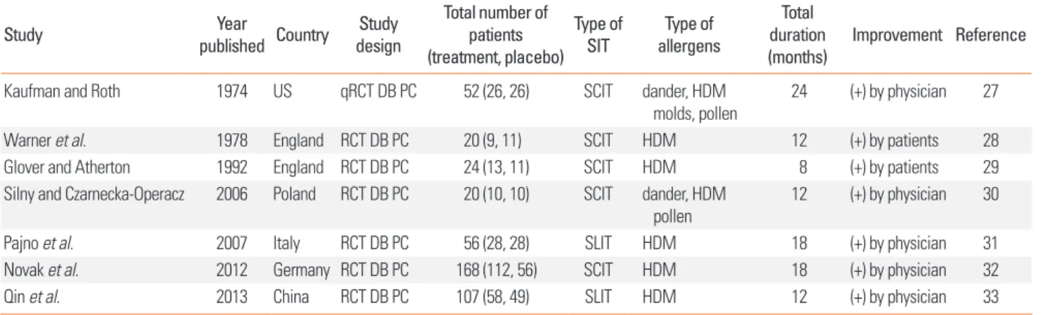
Results from previously performed randomized controlled SIT are summarized in Table 1. First, in Kaufman and Roth’s study in 1974 (United States), quasi-randomized controlled study was performed among total of 52 adult and pediatric AD patients.27 A total of 26 patients completed the SCIT trial for a period of 2 years, and significant clinical improvement was seen in 81% of the treatment group and 40% of the placebo group. Warner et al.28 conducted randomized, double-blind, placebo-controlled study for children with asthma (United Kingdom) and among 20 children who possessed additional atopic features, there was subjective improvement of clinical eczema features as judged by the patients and parents in active treatment group (77.8%) com- pared to minimal improvement in the placebo group (27.3%) af- ter 1 year of treatment. Later, Glover and Atherton performed randomized, double-blind, placebo-controlled trials for HDM SCIT for 24 pediatric AD patients.29 The first study did not reveal
any statistical difference between the treatment and placebo groups. The second study was conducted with patients who un- derwent active treatment in the first study and found greater clinical improvement, suggesting that long-term treatment for at least 1 year is necessary. Persistent efforts of SIT in AD contin- ued, and a double-blind, placebo-controlled trial was conduct- ed, for 20 adult and pediatric AD patients.30 They used W- Atopowe zapalenie skóry (W-AZS), a Polish acronym for atopic dermatitis severity score to assess the extent and severity of skin inflammation index in AD patients concerning pruritus, sleep disturbances and extent and severity of skin inflammation, to evaluate the clinical efficacy. There was a significant decrease in clinical score of W-AZS index after a period of 12 months, sup- porting growing number of evidence for efficacy. A random- ized, double-blind, placebo-controlled trial was performed among a larger population of pediatric patients31 as a sublingual method for 18 months. Scoring atopic dermatitis (SCORAD) showed a dramatic decrease 9 months after the treatment and disease-control medication for treatment of AD was significant- ly reduced in treatment group compared to placebo group. In addition, compared to baseline, visual analogue scale (VAS) showed tendency to decrease only in the treatment group, al- though did not show statistical significance. Another random- ized double-blind placebo-controlled trial by Novak et al.32 was conducted with 168 adult AD patients for 18 months. Even though, the study did not reveal the efficacy in overall AD pa- tients, SIT showed statistical significance of SCORAD reduction in subgroup of severe AD patients with SCORAD >50. Median reduction of total SCORAD of 18% was observed. The best out- come was shown during September to February, due to the use of indoor heating and subsequent high HDM exposure. The ef- ficacy was more pronounced with longer duration. Lastly, most recent randomized control trial carried out by Qin et al.33 ana- lyzed 107 patients undergoing SLIT for 12 months. A total of 84 patients finished the trial, compared to the placebo group
(53.85%), treatment group (77.78%) showed improvement in symptoms. SIT for AD patients are practiced in Korea as well.
But only little clinical studies have been conducted. There was one pilot study of SIT published by Nahm et al.34 Even though 20 AD patients showed significant decreased in SCORAD score with noticeable clinical improvement after 12 months, since it was modified treatment methods combining SIT and hista- mine-immunoglobulin complex treatment, it was difficult to see the sole and exclusive efficacy of SIT. Our institution per- formed retrospective review on patients who underwent at least 3 years of HDM SIT for 217 extrinsic AD patients selected through total IgE and CAP test or skin prick test with hypersen- sitivity to HDM.35 Clinical improvement was judged based on investigator global assessment (IGA) and patients’ subjective assessment of symptoms. In overall, 88.4% of patients showed clinical improvement and among these patients, 63.9% patients showed complete or near-complete remission. Pruritus and loss of sleep was also significantly reduced with 87.2% of pa- tients reporting improvement in pruritus, and 92.7% of patients with only mild or no disturbance of sleep. Hence, although the efficacy of SIT for extrinsic AD patients with positive reactions to HDM was believed to have controversial results for patients in the past, there is a growing trend of thought through many double-blind placebo-controlled trials and meta-analysis, that SIT is indeed an efficient and safe treatment modality for AD patients. While 3 to 6 years of treatment period is generally rec- ommended in literature, there is no set evidence stating long- term efficacy for AD patients receiving SIT for more than 3 years. Yet retrospective review from our institution support the long term efficacy of SIT indicating clinical improvements are most significant when the treatment is continued for a mini- mum of 3 years.
Side-effects and complications
Both local and systemic complications can occur due to SIT.
Table 1. Summary of characteristics and results from randomized controlled trials included in this review
Study Year
published Country Study design
Total number of patients (treatment, placebo)
Type of
SIT Type of
allergens
Total duration
(months) Improvement Reference
Kaufman and Roth 1974 US qRCT DB PC 52 (26, 26) SCIT dander, HDM
molds, pollen
24 (+) by physician 27
Warner et al. 1978 England RCT DB PC 20 (9, 11) SCIT HDM 12 (+) by patients 28
Glover and Atherton 1992 England RCT DB PC 24 (13, 11) SCIT HDM 8 (+) by patients 29
Silny and Czarnecka-Operacz 2006 Poland RCT DB PC 20 (10, 10) SCIT dander, HDM
pollen 12 (+) by physician 30
Pajno et al. 2007 Italy RCT DB PC 56 (28, 28) SLIT HDM 18 (+) by physician 31
Novak et al. 2012 Germany RCT DB PC 168 (112, 56) SCIT HDM 18 (+) by physician 32
Qin et al. 2013 China RCT DB PC 107 (58, 49) SLIT HDM 12 (+) by physician 33
SIT, specific immunotherapy; qRCT, quasi-randomized controlled trial; DB PC, double-blind placebo-controlled; SCIT, subcutaneous immunotherapy; SLIT, sublingual immunotherapy; HDM, house-dust mite; (+), Presence.
Based on a survey of systemic side-effects occurring SCIT in the past 3 years (2008-2011), there were noticeable systemic side- effects in only 0.1% of the total 18.9 million SCIT treatment per- formed, and there was no single case of fatal complications.36 Majority of systemic complications occurred within 30 minutes of injection, and some of the delayed type response were mild symptoms such as a flu-like illness.37
Common local side-effects that can occur in SCIT are urticaria or pruritus, but majority of these reactions persist for less than 24 hours and are rarely regarded as noticeable complications.
Comparing with results from our institution and other RCTs previously performed, mild urticarial eruptions and pruritus occurred in only <1% of patients.38 Furthermore, in one dou- ble-blind placebo-controlled trial, incidence of pruritus lasting for 1-2 days and discomfort, mild exacerbation of atopic lesions, urticaria, headaches or rhinitis were almost similar between the treatment and placebo group,26,27,31 leading to a conclusion that SIT is relatively a safe treatment modality. From RCTs of those who underwent SLIT,30,32 fatigue, headache, localized delayed hyper-responsiveness (>1 hour) occurred in first injection of the treatment, and among these side-effects, localized pruritus was most common. Other noted side effects were facial edema and gastrointestinal discomfort. Yet most of symptoms were mild with spontaneous resolution. There were reports of sud- den worsening of allergic reactions or generalized pruritus in both placebo and control group, but the patients were all man- ageable with a brief symptomatic treatment; no other serious adverse events were reported.39,40
According to data collected from our institution, we witnessed urticaria, localized eruption, pruritus, exacerbation of previous atopic lesions, and relapse of previously known asthma in <1%
of patients. However, the degree of symptoms was very mild, and the symptoms were all controllable with antihistamines.
We believe that it is actually very difficult to accurately deter- mine whether such reactions occur due to SIT or by exposure to other exogenous trigger factors. Nevertheless, from evidenc- es collected thus far, SIT is a very safe treatment modality to in- corporate in a clinic setting.
Biologic effect of allergen SIT in AD Immunologic effect and other serologic effect
There are only few reports on immunological changes ob- served in serum or skin after SIT since most of studies thus far were concentrated on clinical efficacy and safety. Articles elab- orating on changes in serum level of IgE and IgG4 are beginning to appear on surface, but works on variety of cytokines and che- mokines are lacking. Considering a role of allergen as a potent aggravating factor in AD and complicated axes of immunology in AD pathomechanism, it is an important task to find efficacy of inducing immune tolerance through SIT and acquiring data that shows intricate interplay of immunologic changes before and after treatment.
An explanation is needed for serum IgE level changes in re- sponse to SIT in regards to highly activated B cells and deregu- lation of IgE synthesis,41 but there is no clean-cut evidence.
Studies up until now show trend of allergen-specific IgE level gradually decreasing after SIT.31,42-46 For total IgE, there was a general trend for decrease, but statistically, the results were con- flicting with those showing significance31,46 and those that did not.27,29 Serum IgE begins to change relatively at a slow rate with no noticeable drop in its levels; moreover, since there is no evi- dent correlation between clinical improvements after SIT treat- ment, it is hard to explain loss or decrease of response to specif- ic allergen only through changes in IgE.47 To many scholars, the role of IgE as a measurement of clinical sensitivity remains questionable in reality.43 Other works have stressed a significant decrease in Der p-specific IgG4,31,32,45,48,49 and in one pilot study, treatment with SIT led to decrease in markers of AD activity such as IL-16 and thymus and activation regulated chemokine (CCL17) in accordance with clinical improvement.44
Examples of biomarker studies
Although finding a biomarker that can accurately predict treatment response is a necessary task, it is a challenging pro- cess considering multifaceted and intricately woven immuno- logic mechanisms and axes involved in allergic patients. Since the late 1990s, there have been many attempts to find biomark- er candidates. In the early years, most studies concentrated on endothelial cell adhesion molecules,50-54 and works on chemo- kines were published in 2000.55-58 Brief summary on history of biomarker studies are summarized in Table 2. Reviewing stud- ies on biomarker up until now, there have been reports of aller- gen-specific non-IgE antibody increasing through SIT,59 and several studies have shown that serum antibodies can reduce in vitro responses mimicking allergic reactions, such as IgE bind- ing to allergen, IgE-facilitated antigen presentation and baso- phil activation.60-62 In a double-blind placebo controlled study of grass pollen SIT, there was increase in IgG4, IgE blocking fac- tor along with suppression of facilitated allergen binding.63 The authors stressed that not only IgG4, but combined assessment of IgG4 and IgE blocking factor can be done in order to more comprehensively observe functional and clinical efficacy.
Recently, grass pollen SLIT experiment was performed in ex- perimental exposure chamber, and the results showed comple- ment component 1 and the receptor stabilin-1, 2 protein induc- tion from tolerogenic DC also known as regulatory DC has cor- relation with clinical tolerance induced by SIT.64 In addition, the study opened a possibility into readily selecting clinically re- sponsive and unresponsive group through proteins that are easily detected through quantitative polymerase chain reaction in peripheral blood mononuclear cells, and explained relation- ship of short-term efficacy with regulatory immune response.
What exact immunologic mechanisms underlie changes in- duced in SIT is a field of excitement that raises many questions.
There still remains a room to search deeper into discovering biomarkers and choose appropriate candidates, mechanism, treatment response, surrogate end points, and clinical trial for new drug development.65 Hence, it will not be an understate- ment to say that the new era of AD expects a discovery of bio- marker that can assess and standardize treatment response. If biologic marker can show clear-cut correlation with clinical symptoms, it will be an outbreak in the field of science, but con- sidering variegated clinical pictures and associated immuno- logical changes, the quest for a search will not be easily an- swered upon. But through future endeavors in creating more high-quality standardized experiments that reflect clinical im- provement and enable predictions of treatment end-points, we
will be building cornerstone for biomarker discovery.
Further considerations and conclusion
The effectiveness of SIT has been proven through many clini- cal studies recently published and more studies are expected in the future. However, there are still issues that need to be ad- dressed before clinically applying SIT in hospital-settings. Stan- dardized method in selecting candidate patients should be ap- plied for institutions along with objective qualifying criteria.
Also, effective treatment modality for those who are not solely sensitized to HDM (polysensitized patients) raises attention.
There are different routes and schedules for SIT at the moment, and there is rush or ultra-rush protocol besides the well-known Table 2. Prior studies on biomarker candidates of atopic dermatitis
Candidate marker Action Clinical results Reference sE-selectin An adhesion molecule on endothelial cells Reflection of disease severity 50,52-54,66,67 sVCAM-1 An adhesion molecule on endothelial cells Not correlated with disease severity 51,52,54,67
sICAM-1 An adhesion molecule on endothelial cells Not correlated with disease severity 52,54
TARC/CCL17 A chemokine that attracts CCR4+ or CCR8+ cells Reflection of disease severity 55,56,58,68-72
MDC/CCL22 A chemokine that attracts CCR4+ cells Reflection of disease severity 56,57,68,69,72,73
CTACK A chemokine that attracts CCR10+ cells Reflection of disease severity 58
IL-13 An inducer of IgE production Reflection of disease severity 74
IgE Primes the IgE-mediated allergic reaction Reflection of disease severity
No significant result 32,46,68,71,75
29,30,72,76 ECP A basic protein located in the eosinophil primary matrix Reflection of disease severity
No significant result 75,77-80
67 TEC Eosinophils control mechanisms associated with allergy Reflection of disease severity 75,79,81
sIL-2R Expressed by antigen-activated T lymphocytes Reflection of disease severity 79-81
IL-16 A chemokine that attracts CD4+ cells Reflection of disease severity 44,73,75
IL-18 An interferon-γ inducing factor Reflection of disease severity 71,82,83
BDNF A peripheral neurotrophin Reflection of disease severity 84,85
NGF A potent mediator in neuroinflammatory processes Reflection of disease severity Positive staining on AD skin only No significant result
86 87 88 Substance P A neurotransmitter and a neuromodulator Reflection of disease severity
Not correlated with disease severity 86 89 CCL18 A chemokine that attracts both innate and adaptive immune cells Reflection of disease severity
Significantly decreased after Tx 90 72 MEC/CCL28 A chemokine that attracts CCR3+, CCR10+ cells Reflection of disease severity 91,92
PF-4 A platelet chemokine Reflection of disease severity 93,94
Beta-TG A platelet chemokine Reflection of disease severity 93,94
IL-31 Associated with skin-homing CLA-positive T cells Reflection of disease severity 95
CLSP A modulator of calcium-dependent proteins Positive relation in AEAD skin 96
Der p-specific IgG4 A specific IgG molecule for Der p Reflection of disease severity 32,33,45,48,49 Underlyng bar: studies and results of SIT in AD.
sE-selectin, soluble E-selectin; sICAM-1, soluble intercellular adhesion molecule-1; sVCAM-1, soluble vascular adhesion molecule-1; TARC, thymus and activation- regulated chemokine; CCL, C-C motif ligand; CCR, chemokine receptor; MDC, macrophage-derived chemokine; CTACK, cutaneous T cell-attracting chemokine; IL-13, interleukin-13; ECP, eosinophil cationic protein; TEC, total eosinophil count; sIL-2R, soluble IL-2 receptor; BNDF, brain-derived neurotrophic factor; NGF, nerve growth factor; Tx, treatment; MEC, mucosa-associated epithelial chemokine; PF-4, platelet factor 4; beta-TG, beta-thromboglobulin; CLA, cutaneous lymphocyte antigen;
CLSP, calmodulin-like skin protein; AEAD, acute exacerbated.
conventional protocol. In the future, a development for a safe protocol which enables faster immune reaction is promising. If we can perform further studies to see whether early interven- tion allows for blocking progression into allergic march, we will be opening many doors into prevention and treatment of vari- ous allergic diseases. Endeavors in optimizing preparations used and improving treatment response with more refined al- lergoids, potent adjuvants, or recombinant vaccine are also suggested. Lastly, more work should be done in an attempt to discover biomarkers for SIT that will allow clinicians to predict the outcomes or to judge appropriate treatment duration for the patients. Uncovering a new biomarker shall advance the upcoming development and applications of SIT.
ACKNOWLEDGMENTS
This study was supported by a grant from the Korean Health 21 R&D Project, the Ministry of Health & Welfare, Republic of Korea (A111718).
REFERENCES
1. Wüthrich B. Clinical aspects, epidemiology, and prognosis of atop- ic dermatitis. Ann Allergy Asthma Immunol 1999;83:464-70.
2. Shaw TE, Currie GP, Koudelka CW, Simpson EL. Eczema preva- lence in the United States: data from the 2003 National Survey of Children’s Health. J Invest Dermatol 2011;131:67-73.
3. Stensen L, Thomsen SF, Backer V. Change in prevalence of atopic dermatitis between 1986 and 2001 among children. Allergy Asth- ma Proc 2008;29:392-6.
4. Odhiambo JA, Williams HC, Clayton TO, Robertson CF, Asher MI;
ISAAC Phase Three Study Group. Global variations in prevalence of eczema symptoms in children from ISAAC Phase Three. J Aller- gy Clin Immunol 2009;124:1251-8.e23.
5. Perkin MR, Strachan DP, Williams HC, Kennedy CT, Golding J; AL- SPAC Study Team. Natural history of atopic dermatitis and its rela- tionship to serum total immunoglobulin E in a population-based birth cohort study. Pediatr Allergy Immunol 2004;15:221-9.
6. Sandström MH, Faergemann J. Prognosis and prognostic factors in adult patients with atopic dermatitis: a long-term follow-up ques- tionnaire study. Br J Dermatol 2004;150:103-10.
7. Fischer J, Wu Z, Kantyka T, Sperrhacke M, Dimitrieva O, Koblyako- va Y, et al. Characterization of Spink6 in mouse skin: the conserved inhibitor of kallikrein-related peptidases is reduced by barrier inju- ry. J Invest Dermatol 2014;134:1305-12.
8. Hoppe T, Winge MC, Bradley M, Nordenskjöld M, Vahlquist A, Törmä H, et al. Moisturizing treatment of patients with atopic der- matitis and ichthyosis vulgaris improves dry skin, but has a modest effect on gene expression regardless of FLG genotype. J Eur Acad Dermatol Venereol. Forthcoming 2013.
9. Mócsai G, Gáspár K, Nagy G, Irinyi B, Kapitány A, Bíró T, et al. Se- vere skin inflammation and filaggrin mutation similarly alter the skin barrier in patients with atopic dermatitis. Br J Dermatol 2014;
170:617-24.
10. Sprecher E, Leung DY. Atopic dermatitis: scratching through the complexity of barrier dysfunction. J Allergy Clin Immunol 2013;
132:1130-1.
11. Sugiura A, Nomura T, Mizuno A, Imokawa G. Reevaluation of the non-lesional dry skin in atopic dermatitis by acute barrier disrup- tion: an abnormal permeability barrier homeostasis with defective processing to generate ceramide. Arch Dermatol Res 2014;306:427- 40.
12. van Smeden J, Janssens M, Gooris GS, Bouwstra JA. The important role of stratum corneum lipids for the cutaneous barrier function.
Biochim Biophys Acta 2014;1841:295-313.
13. Novak N, Tepel C, Koch S, Brix K, Bieber T, Kraft S. Evidence for a differential expression of the FcepsilonRIgamma chain in dendritic cells of atopic and nonatopic donors. J Clin Invest 2003;111:1047-56.
14. Traidl-Hoffmann C, Mariani V, Hochrein H, Karg K, Wagner H, Ring J, et al. Pollen-associated phytoprostanes inhibit dendritic cell interleukin-12 production and augment T helper type 2 cell polar- ization. J Exp Med 2005;201:627-36.
15. Shreffler WG, Castro RR, Kucuk ZY, Charlop-Powers Z, Grishina G, Yoo S, et al. The major glycoprotein allergen from Arachis hypo- gaea, Ara h 1, is a ligand of dendritic cell-specific ICAM-grabbing nonintegrin and acts as a Th2 adjuvant in vitro. J Immunol 2006;
177:3677-85.
16. Bieber T. Atopic dermatitis. N Engl J Med 2008;358:1483-94.
17. Brown A, Farmer K, MacDonald L, Kalsheker N, Pritchard D, Has- lett C, et al. House dust mite Der p 1 downregulates defenses of the lung by inactivating elastase inhibitors. Am J Respir Cell Mol Biol 2003;29:381-9.
18. Cork MJ, Robinson DA, Vasilopoulos Y, Ferguson A, Moustafa M, MacGowan A, et al. New perspectives on epidermal barrier dys- function in atopic dermatitis: gene-environment interactions. J Al- lergy Clin Immunol 2006;118:3-21.
19. Kawabata A, Kawao N. Physiology and pathophysiology of protein- ase-activated receptors (PARs): PARs in the respiratory system: cel- lular signaling and physiological/pathological roles. J Pharmacol Sci 2005;97:20-4.
20. Cork MJ, Robinson D, Vasilopoulos Y, Ferguson A, Moustafa M, Mac Gowan A, et al. Predisposition to sensitive skin and atopic eczema.
Community Pract 2005;78:440-2.
21. Darsow U, Forer I, Ring J. Allergen-specific immunotherapy in atop- ic eczema. Curr Allergy Asthma Rep 2011;11:277-83.
22. Akkoc T, de Koning PJ, Rückert B, Barlan I, Akdis M, Akdis CA. In- creased activation-induced cell death of high IFN-gamma-produc- ing T(H)1 cells as a mechanism of T(H)2 predominance in atopic diseases. J Allergy Clin Immunol 2008;121:652-8.e1.
23. Akdis CA, Akdis M. Mechanisms of allergen-specific immunother- apy. J Allergy Clin Immunol 2011;127:18-27.
24. Ozdemir C, Kucuksezer UC, Akdis M, Akdis CA. Under the skin or under the tongue: differences and similarities in mechanisms of sublingual and subcutaneous immunotherapy. Immunotherapy 2013;5:1151-8.
25. Frati F, Dell’Albani I, Incorvaia C. Long-term efficacy of allergen im- munotherapy: what do we expect? Immunotherapy 2013;5:131-3.
26. Bae JM, Choi YY, Park CO, Chung KY, Lee KH. Efficacy of allergen- specific immunotherapy for atopic dermatitis: a systematic review and meta-analysis of randomized controlled trials. J Allergy Clin Immunol 2013;132:110-7.
27. Kaufman HS, Roth HL. Hyposensitization with alum precipitated extracts in atopic dermatitis: a placebo-controlled study. Ann Al- lergy 1974;32:321-30.
28. Warner JO, Price JF, Soothill JF, Hey EN. Controlled trial of hypo-
sensitisation to Dermatophagoides pteronyssinus in children with asthma. Lancet 1978;2:912-5.
29. Glover MT, Atherton DJ. A double-blind controlled trial of hypo- sensitization to Dermatophagoides pteronyssinus in children with atopic eczema. Clin Exp Allergy 1992;22:440-6.
30. Silny W, Czarnecka-Operacz M. Specific immunotherapy in the treatment of patients with atopic dermatitis--results of double blind placebo controlled study. Pol Merkur Lekarski 2006;21:558-65.
31. Pajno GB, Caminiti L, Vita D, Barberio G, Salzano G, Lombardo F, et al. Sublingual immunotherapy in mite-sensitized children with atopic dermatitis: a randomized, double-blind, placebo-controlled study. J Allergy Clin Immunol 2007;120:164-70.
32. Novak N, Bieber T, Hoffmann M, Fölster-Holst R, Homey B, Werfel T, et al. Efficacy and safety of subcutaneous allergen-specific im- munotherapy with depigmented polymerized mite extract in atop- ic dermatitis. J Allergy Clin Immunol 2012;130:925-31.e4.
33. Qin YE, Mao JR, Sang YC, Li WX. Clinical efficacy and compliance of sublingual immunotherapy with Dermatophagoides farinae drops in patients with atopic dermatitis. Int J Dermatol 2014;53:
650-5.
34. Nahm DH, Lee ES, Park HJ, Kim HA, Choi GS, Jeon SY. Treatment of atopic dermatitis with a combination of allergen-specific immu- notherapy and a histamine-immunoglobulin complex. Int Arch Allergy Immunol 2008;146:235-40.
35. Lee J, Lee H, Noh S, Bae BG, Park CO, Lee KH. Concurrent Session 02 Dermatitis and Skin Allergy: CS02-5. Efficacy of house dust mite specific immunotherapy in patients with atopic dermatitis. EADC 2nd Eastern Asian Dermatology Congress; 2012 Jun 13-15; Beijing, China. Chinese Society of Dermatology: Beijing; 2012.
36. Epstein TG, Liss GM, Murphy-Berendts K, Bernstein DI. AAAAI and ACAAI surveillance study of subcutaneous immunotherapy, Year 3: what practices modify the risk of systemic reactions? Ann Allergy Asthma Immunol 2013;110:274-8, 278.e1.
37. Epstein TG, Liss GM, Murphy-Berendts K, Bernstein DI. Immedi- ate and delayed-onset systemic reactions after subcutaneous im- munotherapy injections: ACAAI/AAAAI surveillance study of sub- cutaneous immunotherapy: year 2. Ann Allergy Asthma Immunol 2011;107:426-31.e1.
38. Werfel T, Breuer K, Ruéff F, Przybilla B, Worm M, Grewe M, et al.
Usefulness of specific immunotherapy in patients with atopic der- matitis and allergic sensitization to house dust mites: a multi-cen- tre, randomized, dose-response study. Allergy 2006;61:202-5.
39. Canonica GW, Bousquet J, Casale T, Lockey RF, Baena-Cagnani CE, Pawankar R, et al. Sub-lingual immunotherapy: World Allergy Organization Position Paper 2009. Allergy 2009;64 Suppl 91:1-59.
40. Calderón MA, Simons FE, Malling HJ, Lockey RF, Moingeon P, De- moly P. Sublingual allergen immunotherapy: mode of action and its relationship with the safety profile. Allergy 2012;67:302-11.
41. Novak N. Allergen specific immunotherapy for atopic dermatitis.
Curr Opin Allergy Clin Immunol 2007;7:542-6.
42. Akdis CA, Akdis M, Blesken T, Wymann D, Alkan SS, Müller U, et al. Epitope-specific T cell tolerance to phospholipase A2 in bee venom immunotherapy and recovery by IL-2 and IL-15 in vitro. J Clin Invest 1996;98:1676-83.
43. Sulzberger MB. Allergic manifestations in dermatology. N Y State J Med 1936;36:1717-23.
44. Ozdemir C, Kucuksezer UC, Akdis M, Akdis CA. Specific immuno- therapy and turning off the T cell: how does it work? Ann Allergy Asthma Immunol 2011;107:381-92.
45. Bussmann C, Maintz L, Hart J, Allam JP, Vrtala S, Chen KW, et al.
Clinical improvement and immunological changes in atopic der- matitis patients undergoing subcutaneous immunotherapy with a house dust mite allergoid: a pilot study. Clin Exp Allergy 2007;37:
1277-85.
46. Cadario G, Galluccio AG, Pezza M, Appino A, Milani M, Pecora S, et al. Sublingual immunotherapy efficacy in patients with atopic dermatitis and house dust mites sensitivity: a prospective pilot study. Curr Med Res Opin 2007;23:2503-6.
47. Burks AW, Calderon MA, Casale T, Cox L, Demoly P, Jutel M, et al.
Update on allergy immunotherapy: American Academy of Allergy, Asthma & Immunology/European Academy of Allergy and Clini- cal Immunology/PRACTALL consensus report. J Allergy Clin Im- munol 2013;131:1288-96.e3.
48. Soyer OU, Akdis M, Akdis CA. Mechanisms of subcutaneous aller- gen immunotherapy. Immunol Allergy Clin North Am 2011;31:
175-90, vii-viii.
49. Jutel M, Van de Veen W, Agache I, Azkur KA, Akdis M, Akdis CA.
Mechanisms of allergen-specific immunotherapy and novel ways for vaccine development. Allergol Int 2013;62:425-33.
50. Hirai S, Kageshita T, Kimura T, Tsujisaki M, Okajima K, Imai K, et al. Soluble intercellular adhesion molecule-1 and soluble E-selec- tin levels in patients with atopic dermatitis. Br J Dermatol 1996;134:
657-61.
51. Chun WH, Lee HJ, Lee KH. Soluble vascular cell adhesion mole- cule-1 (VCAM-1) in the serum of patients with atopic dermatitis.
Br J Dermatol 1997;136:136.
52. Yamashita N, Kaneko S, Kouro O, Furue M, Yamamoto S, Sakane T.
Soluble E-selectin as a marker of disease activity in atopic dermati- tis. J Allergy Clin Immunol 1997;99:410-6.
53. Laan MP, Koning H, Baert MR, Oranje AP, Buurman WA, Savelkoul HF, et al. Levels of soluble intercellular adhesion molecule-1, solu- ble E-selectin, tumor necrosis factor-alpha, and soluble tumor ne- crosis factor receptor p55 and p75 in atopic children. Allergy 1998;
53:51-8.
54. Wolkerstorfer A, Laan MP, Savelkoul HF, Neijens HJ, Mulder PG, Oudesluys-Murphy AM, et al. Soluble E-selectin, other markers of inflammation and disease severity in children with atopic dermati- tis. Br J Dermatol 1998;138:431-5.
55. Kakinuma T, Nakamura K, Wakugawa M, Mitsui H, Tada Y, Saeki H, et al. Thymus and activation-regulated chemokine in atopic der- matitis: serum thymus and activation-regulated chemokine level is closely related with disease activity. J Allergy Clin Immunol 2001;
107:535-41.
56. Fujisawa T, Fujisawa R, Kato Y, Nakayama T, Morita A, Katsumata H, et al. Presence of high contents of thymus and activation-regulated chemokine in platelets and elevated plasma levels of thymus and activation-regulated chemokine and macrophage-derived chemo- kine in patients with atopic dermatitis. J Allergy Clin Immunol 2002;
110:139-46.
57. Kakinuma T, Nakamura K, Wakugawa M, Mitsui H, Tada Y, Saeki H, et al. Serum macrophage-derived chemokine (MDC) levels are closely related with the disease activity of atopic dermatitis. Clin Exp Immunol 2002;127:270-3.
58. Hijnen D, De Bruin-Weller M, Oosting B, Lebre C, De Jong E, Bruijnzeel-Koomen C, et al. Serum thymus and activation-regulat- ed chemokine (TARC) and cutaneous T cell- attracting chemokine (CTACK) levels in allergic diseases: TARC and CTACK are disease- specific markers for atopic dermatitis. J Allergy Clin Immunol 2004;
113:334-40.
59. van Neerven RJ, Knol EF, Ejrnaes A, Würtzen PA. IgE-mediated al- lergen presentation and blocking antibodies: regulation of T-cell activation in allergy. Int Arch Allergy Immunol 2006;141:119-29.
60. Francis JN, James LK, Paraskevopoulos G, Wong C, Calderon MA, Durham SR, et al. Grass pollen immunotherapy: IL-10 induction and suppression of late responses precedes IgG4 inhibitory anti- body activity. J Allergy Clin Immunol 2008;121:1120-5.e2.
61. James LK, Shamji MH, Walker SM, Wilson DR, Wachholz PA, Fran- cis JN, et al. Long-term tolerance after allergen immunotherapy is accompanied by selective persistence of blocking antibodies. J Al- lergy Clin Immunol 2011;127:509-16.e1-5.
62. Würtzen PA, Lund G, Lund K, Arvidsson M, Rak S, Ipsen H. A dou- ble-blind placebo-controlled birch allergy vaccination study II: cor- relation between inhibition of IgE binding, histamine release and facilitated allergen presentation. Clin Exp Allergy 2008;38:1290-301.
63. Shamji MH, Ljørring C, Francis JN, Calderon MA, Larché M, Kim- ber I, et al. Functional rather than immunoreactive levels of IgG4 correlate closely with clinical response to grass pollen immuno- therapy. Allergy 2012;67:217-26.
64. Zimmer A, Bouley J, Le Mignon M, Pliquet E, Horiot S, Turfkruyer M, et al. A regulatory dendritic cell signature correlates with the clinical efficacy of allergen-specific sublingual immunotherapy. J Allergy Clin Immunol 2012;129:1020-30.
65. Shamji MH, Ljørring C, Würtzen PA. Predictive biomarkers of clin- ical efficacy of allergen-specific immunotherapy: how to proceed.
Immunotherapy 2013;5:203-6.
66. Wolkerstorfer A, Savelkoul HF, de Waard van der Spek FB, Neijens HJ, van Meurs T, Oranje AP. Soluble E-selectin and soluble ICAM-1 levels as markers of the activity of atopic dermatitis in children. Pe- diatr Allergy Immunol 2003;14:302-6.
67. Gutgesell C, Heise S, Seubert A, Stichtenoth DO, Frölich JC, Neu- mann C. Comparison of different activity parameters in atopic der- matitis: correlation with clinical scores. Br J Dermatol 2002;147:914- 9.
68. Jahnz-Rozyk K, Targowski T, Paluchowska E, Owczarek W, Kuchar- czyk A. Serum thymus and activation-regulated chemokine, mac- rophage-derived chemokine and eotaxin as markers of severity of atopic dermatitis. Allergy 2005;60:685-8.
69. Mostafa GA, Tomoum HY, Salem SA, Abd El-Aziz MM, Abou El- Maged DI, El-Sayed El-Far I. Serum concentrations of CCR4 li- gands in relation to clinical severity of atopic dermatitis in Egyptian children. Pediatr Allergy Immunol 2008;19:756-62.
70. Furue M, Matsumoto T, Yamamoto T, Takeuchi S, Esaki H, Chiba T, et al. Correlation between serum thymus and activation-regulated chemokine levels and stratum corneum barrier function in healthy individuals and patients with mild atopic dermatitis. J Dermatol Sci 2012;66:60-3.
71. Kou K, Aihara M, Matsunaga T, Chen H, Taguri M, Morita S, et al.
Association of serum interleukin-18 and other biomarkers with disease severity in adults with atopic dermatitis. Arch Dermatol Res 2012;304:305-12.
72. Kwon YS, Oh SH, Wu WH, Bae BG, Lee HJ, Lee MG, et al. CC che- mokines as potential immunologic markers correlated with clini- cal improvement of atopic dermatitis patients by immunotherapy.
Exp Dermatol 2010;19:246-51.
73. Angelova-Fischer I, Hipler UC, Bauer A, Fluhr JW, Tsankov N, Fischer TW, et al. Significance of interleukin-16, macrophage-de- rived chemokine, eosinophil cationic protein and soluble E-selec-
tin in reflecting disease activity of atopic dermatitis--from laborato- ry parameters to clinical scores. Br J Dermatol 2006;154:1112-7.
74. La Grutta S, Richiusa P, Pizzolanti G, Mattina A, Pajno GB, Citarrel- la R, et al. CD4(+)IL-13(+) cells in peripheral blood well correlates with the severity of atopic dermatitis in children. Allergy 2005;60:
391-5.
75. Wu KG, Li TH, Chen CJ, Cheng HI, Wang TY. Correlations of serum Interleukin-16, total IgE, eosinophil cationic protein and total eo- sinophil counts with disease activity in children with atopic der- matitis. Int J Immunopathol Pharmacol 2011;24:15-23.
76. Gerdes S, Kurrat W, Mrowietz U. Serum mast cell tryptase is not a useful marker for disease severity in psoriasis or atopic dermatitis.
Br J Dermatol 2009;160:736-40.
77. Czech W, Krutmann J, Schöpf E, Kapp A. Serum eosinophil cation- ic protein (ECP) is a sensitive measure for disease activity in atopic dermatitis. Br J Dermatol 1992;126:351-5.
78. Halmerbauer G, Frischer T, Koller DY. Monitoring of disease activi- ty by measurement of inflammatory markers in atopic dermatitis in childhood. Allergy 1997;52:765-9.
79. Kägi MK, Joller-Jemelka H, Wüthrich B. Correlation of eosinophils, eosinophil cationic protein and soluble interleukin-2 receptor with the clinical activity of atopic dermatitis. Dermatology 1992;185:88- 92.
80. Furue M, Sugiyama H, Tsukamoto K, Ohtake N, Tamaki K. Serum soluble IL-2 receptor (sIL-2R) and eosinophil cationic protein (ECP) levels in atopic dermatitis. J Dermatol Sci 1994;7:89-95.
81. Walker C, Kägi MK, Ingold P, Braun P, Blaser K, Bruijnzeel-Koomen CA, et al. Atopic dermatitis: correlation of peripheral blood T cell activation, eosinophilia and serum factors with clinical severity.
Clin Exp Allergy 1993;23:145-53.
82. Trzeciak M, Gleń J, Bandurski T, Sokołowska-Wojdyło M, Wilkows- ka A, Roszkiewicz J. Relationship between serum levels of interleu- kin-18, IgE and disease severity in patients with atopic dermatitis.
Clin Exp Dermatol 2011;36:728-32.
83. Hon KL, Leung TF, Ma KC, Wong CK, Wan H, Lam CW. Serum concentration of IL-18 correlates with disease extent in young chil- dren with atopic dermatitis. Pediatr Dermatol 2004;21:619-22.
84. Raap U, Werfel T, Goltz C, Deneka N, Langer K, Bruder M, et al. Cir- culating levels of brain-derived neurotrophic factor correlate with disease severity in the intrinsic type of atopic dermatitis. Allergy 2006;61:1416-8.
85. Namura K, Hasegawa G, Egawa M, Matsumoto T, Kobayashi R, Yano T, et al. Relationship of serum brain-derived neurotrophic factor level with other markers of disease severity in patients with atopic dermatitis. Clin Immunol 2007;122:181-6.
86. Toyoda M, Nakamura M, Makino T, Hino T, Kagoura M, Morohashi M. Nerve growth factor and substance P are useful plasma markers of disease activity in atopic dermatitis. Br J Dermatol 2002;147:71-9.
87. Oh SH, Bae BG, Park CO, Noh JY, Park IH, Wu WH, et al. Associa- tion of stress with symptoms of atopic dermatitis. Acta Derm Vene- reol 2010;90:582-8.
88. Schulte-Herbrüggen O, Fölster-Holst R, von Elstermann M, Augus- tin M, Hellweg R. Clinical relevance of nerve growth factor serum levels in patients with atopic dermatitis and psoriasis. Int Arch Al- lergy Immunol 2007;144:211-6.
89. Izu K, Tokura Y. The various effects of four H1-antagonists on serum substance P levels in patients with atopic dermatitis. J Dermatol 2005;32:776-81.
90. Park CO, Lee HJ, Lee JH, Wu WH, Chang NS, Hua L, et al. Increased
expression of CC chemokine ligand 18 in extrinsic atopic dermati- tis patients. Exp Dermatol 2008;17:24-9.
91. Ezzat MH, Sallam MA, Shaheen KY. Serum mucosa-associated ep- ithelial chemokine (MEC/CCL28) in atopic dermatitis: a specific marker for severity. Int J Dermatol 2009;48:822-9.
92. Ezzat MH, Shaheen KY. Serum mucosa-associated epithelial che- mokine in atopic dermatitis: a specific marker for severity. Indian J Dermatol 2009;54:229-36.
93. Tamagawa-Mineoka R, Katoh N, Ueda E, Masuda K, Kishimoto S.
Elevated platelet activation in patients with atopic dermatitis and psoriasis: increased plasma levels of beta-thromboglobulin and platelet factor 4. Allergol Int 2008;57:391-6.
94. Kasperska-Zajac A. Recovery of platelet factor 4 (PF-4) and beta- thromboglobulin (beta-TG) plasma concentrations during remis- sion in patients suffering from atopic dermatitis. Platelets 2010;21:
522-4.
95. Ezzat MH, Hasan ZE, Shaheen KY. Serum measurement of interleu- kin-31 (IL-31) in paediatric atopic dermatitis: elevated levels corre- late with severity scoring. J Eur Acad Dermatol Venereol 2011;25:
334-9.
96. Donovan M, Ambach A, Thomas-Collignon A, Prado C, Bernard D, Jammayrac O, et al. Calmodulin-like skin protein level increases in the differentiated epidermal layers in atopic dermatitis. Exp Der- matol 2013;22:836-7.