http://dx.doi.org/10.12671/jkfs.2014.27.2.113
113
Copyright ⓒ 2014 The Korean Fracture Society. All rights reserved.
This is an Open Access article distributed under the terms of the Creative Commons Attribution Non-Commercial License (http://creativecommons.org/licenses/
by-nc/3.0) which permits unrestricted non-commercial use, distribution, and reproduction in any medium, provided the original work is properly cited.
Received October 30, 2013 Revised December 2, 2013 Accepted December 18, 2013
Address reprint requests to: Jae-Sung Yoo, M.D.
Department of Orthopedic Surgery, Dankook University Hospital, 201 Manghyang-ro, Dongnam-gu, Cheonan 330-715, Korea Tel: 82-41-550-6579ㆍFax: 82-41-556-3238
E-mail: [email protected]
Financial support: None. Conflict of interest: None.
중증 외상 환자에서 초기 진단에 실패한 골절
박희곤⋅유재성 ⋅이형석
단국대학교 의과대학 단국대학교병원 정형외과학교실
Missed Fractures in Severely Injured Patients
Hee-Gon Park, M.D., Jae-Sung Yoo, M.D. , Hyung-Suk Yi, M.D.
Department of Orthopedic Surgery, Dankook University Hospital, Dankook University Medical College, Cheonan, Korea
Purpose: The purpose of this study is to analyze anatomic distributions, diagnostic methods, and prognosis of missed fractures in patients with severe injury.
Materials and Methods: A review of single-institutional medical records between January 2001 and May 2012 identified 58 patients with 62 delayed diagnoses of fractures among 4,643 severely injured patients older than 20 years with Injury Severity Scores higher than 16. We evaluated combined injuries, location of fractures, diagnostic methods, and reasons for missed diagnosis at initial exam.
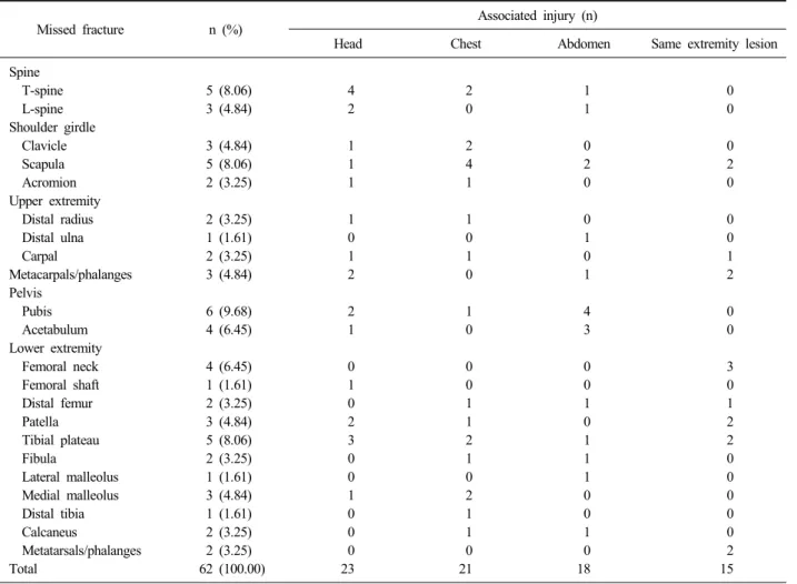
Results: Among 62 missed fractures, there were eight cases of spine fracture, 10 cases of peri-shoulder joint fracture, eight cases of upper extremity fracture, 10 cases of pelvis of acetabulum fracture, and 26 cases of lower extremity fracture.
Head injury was the most common concomitant injury (23 cases). Initially missed fractures were most commonly discovered by official reading by radiologists. The most common reasons for misdiagnosis were the use of improper radiologic study and missed-reading of proper radiologic studies.
Conclusion: In order to prevent misdiagnosis of fractures in patients with severe injury, meticulous physical examination with suspicion of fractures should come first. In addition, obtaining proper radiologic study and thorough evaluation of radio- logic images are important to decreasing the rates of missed fracture diagnoses. In addition, thorough surveillance for ipsi- lateral fractures is important in extremities with identified fractures.
Key Words: Severely injured patient, Fracture, Missed fracture
서 론
사고로 인해 발생하는 중증 외상 환자들의 수는 계속해 서 증가하고 있으며, 생명을 위협하는 손상들은 주로 흉부, 복부 및 골절에서 발생한다.1) 따라서 이러한 손상들을 초 기 진단하기 위해서 본원에서는 흉부, 골반 단순방사선 촬 영 및 focus assessed sonography in trauma (FAST)를 시 행하고 있다. 초기 진단을 위한 노력에도 불구하고 중증 외상 환자에서의 초기 진단 실패는 지속적으로 발생하고 있으며 1.3%-39.0%로 다양하게 보고되고 있다.2-5) 중증 외
Table 1. Fractures Missed in 56 of 4,643 Polytrauma Patients
Missed fracture n (%) Associated injury (n)
Head Chest Abdomen Same extremity lesion
Spine T-spine L-spine Shoulder girdle Clavicle Scapula Acromion Upper extremity Distal radius Distal ulna Carpal
Metacarpals/phalanges Pelvis
Pubis Acetabulum Lower extremity Femoral neck Femoral shaft Distal femur Patella Tibial plateau Fibula
Lateral malleolus Medial malleolus Distal tibia Calcaneus
Metatarsals/phalanges Total
5 (8.06) 3 (4.84)
3 (4.84) 5 (8.06) 2 (3.25)
2 (3.25) 1 (1.61) 2 (3.25) 3 (4.84)
6 (9.68) 4 (6.45)
4 (6.45) 1 (1.61) 2 (3.25) 3 (4.84) 5 (8.06) 2 (3.25) 1 (1.61) 3 (4.84) 1 (1.61) 2 (3.25) 2 (3.25) 62 (100.00)
4 2
1 1 1
1 0 1 2
2 1
0 1 0 2 3 0 0 1 0 0 0 23
2 0
2 4 1
1 0 1 0
1 0
0 0 1 1 2 1 0 2 1 1 0 21
1 1
0 2 0
0 1 0 1
4 3
0 0 1 0 1 1 1 0 0 1 0 18
0 0
0 2 0
0 0 1 2
0 0
3 0 1 2 2 0 0 0 0 0 2 15 The result of sum showed errors related with rounding off.
상과 동반된 골절은 초기 손상에서 간과되기 쉬우며, 지연 진단될 경우 기능적, 미용적인 장애가 남을 수 있으므로 더욱 주의를 요한다.6)
따라서 저자들은 후향적 연구를 통하여 단국대학교병원 에 내원한 중증 외상 환자에서 어떠한 골절들이 초기 진단 에 실패하였는지와, 그 원인과 진단 과정에 대해 살펴보고 자 하였다.
대상 및 방법
본 연구는 Institutional Review board 승인 후, 2001년 1월부터 2012년 5월까지 본원 응급실에 내원한 20세 이상 의 성인에서 Injury Severity Score (ISS) 16 이상의 중증 외상 환자 4,643명 중 골절의 초기 진단에 실패하였던 58 명, 62예의 골절을 대상으로 후향적으로 실시하였다. 모든 경우에서 advanced trauma life support protocols에 따라
초기 처치를 시행하였다. 초기 처치로 기본적인 호흡, 맥박 등의 생체징후 및 신경학적 증상을 조사하였다. 이어서 손 상부위를 알아내기 위해서 면밀한 이학적 검사를 시행하였 고 흉부, 골반, 경추부에 대해 우선적으로 단순방사선 촬영 을 실시하였다. 초기 방사선 검사는 응급실에서 외상전문 의에 의해 판독되었으며 근골격계 방사선과 전문의의 판독 은 입원 후에 시행되었고 모든 영상은 picture archiving and communication system을 이용하여 판독하였다. 정형 외과 전문의의 검진 및 진료는 외상전문의가 필요하다고 판단하여 진료의뢰를 한 경우에 시행되었다. 지연 진단된 골절의 정의는 두개골, 안면골, 늑골을 제외한 모든 골절에 서 입원 12시간 이내에 진단내리거나 의심하지 못하였던 경우로 하였다.7) 골절이 의심되나 혈역학적으로 불안정하 여 초기 입원기간에 더 이상의 방사선학적 검사 시행이 어 려워 향후 검사가 예정된 경우는 지연 진단된 골절에서 제 외하였다.
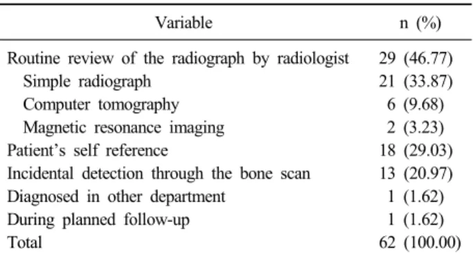
Table 2. How the Diagnosis Was Made
Variable n (%)
Routine review of the radiograph by radiologist Simple radiograph
Computer tomography Magnetic resonance imaging Patient’s self reference
Incidental detection through the bone scan Diagnosed in other department
During planned follow-up Total
29 (46.77) 21 (33.87) 6 (9.68) 2 (3.23) 18 (29.03) 13 (20.97) 1 (1.62) 1 (1.62) 62 (100.00) The result of sum showed errors related with rounding off.
Table 4. Patients with More than One Lesion in the Same Extremity
Lesion diagnosed Lesion missed n
Femoral shaft fracture Femoral shaft fracture Distal clavicle fracture Distal radius fracture Distal radioulnar fracture Bimalleolar fracture Total
Ipsilateral femoral neck fracture Ipsilateral patella fracture Acromion fracture Scaphoid fracture
5th Metacarpal shaft fracture 2nd Metatarsal base fracture
3 2 1 1 1 1 9 Table 3. Reasons for Missing the Diagnosis
Variable n (%)
Radiographic misinterpreted Subtle by still visible fractures Radiographically imperceptible Improper radiographic images Block by splinting devices Poor positioned radiographs Metal artifact
No history taking and physical examination Head injury
Chest injury Abdominal injury
Combined injuries of head, chest, or abdomen Cause of poor general condition of patients No radiograph done
Inadequate physical examination More than one lesion in the same region Total
21 (33.87) 9 (14.52) 12 (19.35) 5 (8.06) 2 (3.23) 2 (3.23) 1 (1.61) 16 (25.81) 7 (11.29) 3 (4.84) 2 (3.23) 4 (6.45) 12 (19.35) 7 (11.29) 5 (8.06) 9 (14.52) 63 (100.00) The result of sum showed errors related with rounding off.
환자의 성별 및 나이, 수상기전, 내원 후 처치, 초기 방사 선 검사, 중환자실 치료, 수술적 치료 등에 대해 조사하였다.
통계적 분석에는 IBM SPSS ver.19.0 (IBM Co., Armonk, NY, USA)을 이용하였다.
결 과
2001년부터 2012년까지 본원 응급실에 내원한 중증 외상 환자 중 골절의 초기 진단에 실패하였던 58명의 평균 연령 은 45.69세(20-86세)였고, 남자 47명(81.03%) 여자 11명 (18.97%)이었다. ISS는 평균 23.78 (16-57)이었고, 58명 중 14명(24.14%)은 내원 당시 기관 삽관이 필요한 상태였으며, 7명(12.07%)은 수축기 혈압이 90 이하로 저혈량성 쇼크가 의심되는 상태였다. 교통사고로 47명이 수상하였으며 그 중 보행자 사고가 12명, 모터싸이클 사고가 21명, 자동차 사고 가 14명이었다. 그 외 낙상 9명, 압착 손상 2명이었다.
총 62예의 초기 진단에 실패한 진단 중 척추 골절이 8 예, 견관절 주위 골절이 10예, 상지 골절이 8예, 골반 및 비구 골절이 10예, 하지 골절이 26예였다(Table 1). 초기 진단에 실패한 골절환자에서 동반 손상은 두부 손상이 23 예로 가장 많았으며, 각 골절과 동반된 손상에 대해 살펴 보면 척추 골절에서는 두부 손상이 75.0% (8예 중 6예), 견관절 주위 골절에서는 흉부 손상이 70.0% (10예 중 7 예), 상지 골절에서는 두부 손상과 동측 상지 손상이 모두 25.0% (8예 중 2예), 골반 및 비구 골절에서는 복부 손상 이 70.0% (10예 중 7예), 하지 골절에서는 동측 하지 손상 이 38.46% (26예 중 10예)로 가장 많았다(Table 1).
지연 진단 방법으로는 방사선의학 전문의의 판독에 의한 경우가 가장 많았고, 환자가 불편감을 호소하여 발견한 경 우, 골주사 검사에 의해 우연히 발견된 경우의 순서였다 (Table 2). 초기 진단에 실패한 원인을 살펴보면 골절선이 명확하지 않거나 판독을 잘못하여 골절을 발견하지 못한
경우가 33.87%로 가장 많았고, 환자의 전신 상태가 불량하 여 문진 및 이학적 검사가 불가능하였던 경우, 같은 부위 에 다른 골절이 동반되어 간과된 경우 순서였다(Table 3).
초기에 전신상태가 불량하여 병력청취나 이학적 검사가 시행되지 않아 진단에 실패하였던 16예에 대해 살펴보면 두부 손상이 7예로 가장 많았고, 흉부 손상이 3예, 복부 손상이 2예였고, 두부 손상, 흉부 손상 및 복부 손상이 동 반된 경우는 4예였다(Table 3).
동측에 다른 골절이 동반되어 간과된 9예에 대해 살펴보 면 대퇴골 간부 골절에서 대퇴골 경부의 비전위 골절이 동 반된 경우가 3예로 가장 많았고, 대퇴골 간부골절과 동반 된 슬개골의 비전위 골절 2예, 쇄골 원위부 골절과 동반된 견갑골 견봉의 비전위 골절이 1예, 요골 원위부 골절과 동 반된 주상골의 비전위 골절 1예, 요척골 원위부 골절과 동 반된 중수지골 골절 1예, 발목 양과골절과 동반된 중족골
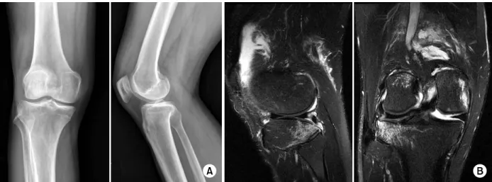
Fig. 1. (A) Antero-posterior and lateral radiographs of the knee in a 42-year-old man show no definite fracture lines. (B) Sagittal and coronal T2-weighted magnetic resonance images show a tibial plateau fracture.
Table 5. Delay from Accident to the Diagnosis of an Injury
Delay time n (%)
12-24 hours 1-7 days
More than 1 week Total
19 (30.65) 32 (51.61) 11 (17.74) 62 (100.00)
기저부 골절 1예였다(Table 4).
지연 진단된 시기를 살펴보면 1일 이내 진단한 경우가 19예(30.65%), 1일 이상 1주 이내에 진단한 경우가 32예 (51.61%), 1주 이상 경과되어 진단된 경우가 11예(17.74%) 로 1일 이상 1주 이내에 진단되었던 경우가 가장 많았다 (Table 5).
1. 증례 1
42세 남자환자로 모터싸이클 운전자 사고로 수상하였으며 경막하 출혈이 있었고, 다발성 늑골 골절과 동반된 혈흉이 있었다. ISS는 27점이었다. Glows coma scale 8점이었고, 내원 당시 기관 삽관이 필요한 상태였다. 내원 3일째 우측 슬관절 주위에 연부조직 종창 및 동통 및 압통이 관찰되어 단순방사선 사진을 촬영하였다. 단순방사선 사진상 골절선이 관찰되지 않아 자기공명영상을 시행하였으며, 이를 통하여 우측 슬관절 경골 고평부 골절을 진단하였다(Fig. 1).
2. 증례 2
34세 남자환자로 자동차 운전자 사고로 수상하였으며,
복강 내 출혈 및 비장 파열이 동반되어 있었다. ISS는 17 점이었다. 내원 당시 좌측 대퇴골 간부 골절을 발견하여 내원 2일째 도수정복술 및 골수강 내 고정술을 시행하였으 나, 술 후 2일째 간과된 대퇴골 근위부 골절이 발견되어 추가적으로 나사못 고정술을 시행하였다(Fig. 2).
고 찰
응급실에서 골절의 초기 진단 실패는 적절한 치료과정을 지연시켜 환자에게 합병증을 야기할 수 있으며,8) 또한 법 적 의료 분쟁의 가장 흔한 이유 중 하나이다.9) 다발성 외 상을 동반한 중증 외상 환자에서의 전위가 심하지 않은 골 절은 정확한 이학적 평가가 어려우며 만족스러운 영상의학 검사를 통해 정확한 진단을 내리기에 어려움이 있다.10) Irving 과 Irving11)은 특히 두부 손상이 동반된 환자에서 초기 진 단에 실패할 가능성이 높다고 보고한 바 있으며, McLaren 등12)은 두부 손상이 동반되었거나 의식이 저하된 경우에 초기 평가가 어려우므로 면밀한 이학적 검사를 통해 골절 을 의심하는 것이 중요하다고 하였다. 본 연구에서도 골절 의 초기 진단에 실패한 예의 동반 손상 중에서 두부 손상 이 37.09%로 가장 흔한 동반 손상이었다.
지금까지 보고된 연구마다 중증 외상 환자 및 다발성 외 상 환자에 대한 기준이 일치되지 않았기 때문에 정확한 비 교는 어렵지만, Guly8)는 응급실에서 초기 진단에 실패한 경우가 0.6%라고 보고한 바 있으며, Juhl 등13)은 초기 진 단에 실패한 골절이 0.5%라고 보고한 바 있다. 본 연구에 서도 9,643명 중 58명(0.6%)에서 초기 진단에 실패하여 유 사한 발생률을 보였다.
단순방사선 사진은 아직까지도 다발성 외상 환자 및 중
Fig. 2. (A) Anter-posterior and lateral radiographs of the femur in a 34 year-old man show a femoral shaft fracture. (B) Antero-posterior and lateral radiographs of the femur after the operation show intramedullary nailing fixation. (C) Antero-posterior radiograph of the hip after the operation shows a neglected femoral neck fracture. (D) Antero-posterior radiograph of the hip after the secondary operation shows multiple screw fixation.
증 외상 환자에서 골절의 진단에 가장 중요한 역할을 하고 있으며 응급실에서 단순방사선 사진의 판독의 오류로 초기 진단에 실패하는 경우가 41%-80%로 가장 흔하게 보고되고
있다.1,14) 또한 Wei 등10)은 판독에서의 오류만큼 중요한 것
이 적절한 영상을 얻는 것이라고 강조하였으며, 석고붕대 를 착용한 채로 촬영한 단순방사선 사진에서 골절 진단에 실패하였던 경우, 단순방사선 촬영 당시 환자의 자세가 적 절하지 않아 정확한 영상을 얻지 못하여 진단에 실패하였 던 경우 등의 예시를 제시하며 적절한 영상을 얻는 것이 정확한 골절 진단을 위하여 필수적인 요소라고 하였다. 본 연구에서도 단순방사선 사진이 부적절하였거나 판독과정에 서 오류를 범하였던 경우가 33.87%로 가장 많았고, 적절하 지 않은 영상으로 진단이 불가능하였던 경우도 8.06%로 관찰되었다.
그러나 Blackmore 등15)과 Herzog 등16)은 척추 골절에서 는 단순방사선 사진의 진단 정확성이 높지 않으며 대신 컴 퓨터단층촬영이나 자기공명영상이 정확한 진단을 필요한
검사라고 하였다. 본 연구에서도 초기 진단에 실패하였던 8예의 척추 골절에서 모두 단순방사선 사진을 통한 진단에 서 실패하였으며, 이후 컴퓨터단층촬영, 자기공명영상, 골 주사 검사 등의 추가적인 검사를 통하여 진단하였다.
Juhl 등13)은 다발성 외상 환자에서 동측의 다른 골절을 초기 진단에서 실패한 11예를 보고한 바 있으며, 골절이 발생하였을 경우 동측에 다른 골절이 있을 가능성이 높으 므로 더욱 주의를 기울여야 한다고 하였다. 본 연구에서도 62예 중 17예에서 동측에 동반 골절이 존재하였으며, 그 중 9예는 전위가 심하지 않아 초기 진단에 실패하였다. 9 예 중에서 대퇴골 간부 골절과 동반된 비전위성 대퇴골 경 부 골절의 초기 진단 실패가 3예로 가장 많았다(Fig. 1).
대퇴 간부 골절 중 대퇴 경부 골절을 동반하는 경우는 약 6%-9%로 보고되고 있으나, 대퇴 경부가 외회전되어 방사 선 사진이 촬영되는 경우, 대퇴 간부의 심한 통증 때문에 고관절부 통증을 호소하지 않는 경우 등의 원인 때문에 대퇴 경부 골절이 간과되기 쉬우며, 초기 진단 실패는 13%-31%
로 보고되고 있다.17,18)
본 연구의 한계점으로는 후향적 연구라는 점, 단일 기관 의 연구로 골절의 초기 진단에 실패하였던 경우의 수가 많 지 않은 점, ISS 16 이상의 중증 환자만을 대상으로 하였 기에 ISS 16 미만의 환자에서 발생한 골절의 초기 진단 실 패의 예는 대상에서 제외된 점, 환자가 사망하였거나 지속 적으로 전신상태가 불량하여 골절 진단을 위한 추가적인 처치가 이루어지지 않은 경우 타 병원으로 전원을 통하여 향후 추가적 평가가 이루어지지 않는 예에서는 간과된 골 절의 진단이 이루어지지 않았다는 점이 있으나, 중증 외상 환자를 대상으로 하여 초기 진단에 실패한 골절의 원인, 진단과정, 위치 등에 대한 분석으로 그 의의가 있을 것으 로 생각된다.
결 론
중증 외상 환자에서 골절의 초기 진단 실패를 방지하기 위해서는 전신 상태가 좋지 않을수록 환자의 증상 호소에 주의를 기울여 골절 및 손상을 의심하는 것이 중요할 것이 다. 또한 응급실 내원 초기에 골절의 정확한 진단을 위해 서는 전문의에 의한 정확한 진단이 필요할 것으로 생각된 다. 또한 적절한 영상 평가를 위해서는 정확한 자세에서 석고붕대에 의한 영상의 간섭 없이 정확한 영상을 얻는 것 이 중요하며, 척추 골절에서는 컴퓨터단층촬영이나 자기공 명영상 등의 추가적인 검사가 진단의 정확성을 높일 수 있 을 것이다. 특히 골절이 발견된 경우 동측 상지 또는 하지 의 다른 골절의 평가에 더욱 주의를 기울여야 할 것이다.
References
1) Richmond J, Egol KA, Koval KJ: Management of or- thopaedic injuries in polytrauma patients. Bull Hosp Jt Dis, 60: 162-167, 2001-2002.
2) Houshian S, Larsen MS, Holm C: Missed injuries in a level I trauma center. J Trauma, 52: 715-719, 2002.
3) Janjua KJ, Sugrue M, Deane SA: Prospective evaluation of early missed injuries and the role of tertiary trauma survey. J Trauma, 44: 1000-1006; discussion 1006-1007, 1998.
4) Kalemoglu M, Demirbas S, Akin ML, et al: Missed in- juries in military patients with major trauma: original
study. Mil Med, 171: 598-602, 2006.
5) Pfeifer R, Pape HC: Missed injuries in trauma patients: a literature review. Patient Saf Surg, 2: 20, 2008.
6) Born CT, Ross SE, Iannacone WM, Schwab CW, DeLong WG: Delayed identification of skeletal injury in multisystem trauma: the 'missed' fracture. J Trauma, 29:
1643-1646, 1989.
7) Laasonen EM, Kivioja A: Delayed diagnosis of ex- tremity injuries in patients with multiple injuries. J Trauma, 31: 257-260, 1991.
8) Guly HR: Missed diagnoses in an accident & emergency department. Injury, 15: 403-406, 1984.
9) Berlin L: Defending the "missed" radiographic diagnosis.
AJR Am J Roentgenol, 176: 317-322, 2001.
10) Wei CJ, Tsai WC, Tiu CM, Wu HT, Chiou HJ, Chang CY: Systematic analysis of missed extremity fractures in emergency radiology. Acta Radiol, 47: 710-717, 2006.
11) Irving MH, Irving PM: Associated injuries in head in- jured patients. J Trauma, 7: 500-511, 1967.
12) McLaren CA, Robertson C, Little K: Missed orthopaedic injuries in the resuscitation room. J R Coll Surg Edinb, 28: 399-401, 1983.
13) Juhl M, Møller-Madsen B, Jensen J: Missed injuries in an orthopaedic department. Injury, 21: 110-112, 1990.
14) Williams SM, Connelly DJ, Wadsworth S, Wilson DJ:
Radiological review of accident and emergency radio- graphs: a 1-year audit. Clin Radiol, 55: 861-865, 2000.
15) Blackmore CC, Mann FA, Wilson AJ: Helical CT in the primary trauma evaluation of the cervical spine: an evidence-based approach. Skeletal Radiol, 29: 632-639, 2000.
16) Herzog C, Ahle H, Mack MG, et al: Traumatic injuries of the pelvis and thoracic and lumbar spine: does thin-slice multidetector-row CT increase diagnostic accu- racy? Eur Radiol, 14: 1751-1760, 2004.
17) Watson JT, Moed BR: Ipsilateral femoral neck and shaft fractures: complications and their treatment. Clin Orthop Relat Res, (399): 78-86, 2002.
18) Wolinsky PR, Johnson KD: Ipsilateral femoral neck and shaft fractures. Clin Orthop Relat Res, (318): 81-90, 1995.
Copyright ⓒ 2014 The Korean Fracture Society. All rights reserved.
This is an Open Access article distributed under the terms of the Creative Commons Attribution Non-Commercial License (http://creativecommons.org/licenses/
by-nc/3.0) which permits unrestricted non-commercial use, distribution, and reproduction in any medium, provided the original work is properly cited.
http://dx.doi.org/10.12671/jkfs.2014.27.2.113
중증 외상 환자에서 초기 진단에 실패한 골절
박희곤⋅유재성 ⋅이형석
단국대학교 의과대학 단국대학교병원 정형외과학교실
목 적: 중증 외상 환자에서 지연 진단된 골절의 종류와 원인, 진단방법 및 처치에 대해 알아보고자 한다.
대상 및 방법: 2001년 1월부터 2012년 5월까지 단국대학교병원 응급실에 내원한 20세 이상의 성인에서 Injury Severity Score 16 이상의 중증 외상 환자 4,643명 중 골절의 초기 진단에 실패하였던 58명, 62예의 골절을 대상으로 하였다. 동반 손상의 종류, 골절의 위치, 진단방법, 초기 진단에 실패하였던 이유에 대해 조사하였다.
결 과: 총 62예의 초기 진단에 실패한 진단 중 척추 골절 8예, 견관절 주위 골절 10예, 상지 골절 8예, 골반 및 비구 골절 10예, 하지 골절 26예였다. 동반 손상은 두부 손상이 23예로 가장 많았으며, 지연 진단 방법으로는 방사선의학 전문의의 판독에 의한 경우가 가장 많았고, 초기 진단에 실패한 원인을 살펴보면 방사선 검사가 적절하지 않거나 골절을 발견하지 못한 경우가 가장 많았다.
결 론: 중증 외상 환자에서 골절의 초기 진단 실패를 방지하기 위해서는 면밀한 이학적 검사를 통해 골절을 의심하는 것이 우선시되어야 할 것이며, 적절한 영상 평가를 위해서는 정확한 영상을 얻는 것이 중요할 것이다. 특히 골절이 발견된 경우 동측 상지 또는 하지의 다른 골절의 평가에 더욱 주의를 기울여야 할 것이다.
색인 단어: 중증 외상 환자, 골절, 지연 진단
접수일 2013. 10. 30 수정일 2013. 12. 2 게재확정 2013. 12. 18 책임저자 유 재 성
천안시 동남구 망향로 201, 단국대학교병원 정형외과
Tel 041-550-6579, Fax 041-556-3238, E-mail [email protected]
119