PGHN
Case Report
A Case of Mesenteric Cyst in a 4-Year-Old Child with Acute Abdominal Pain
Jae Woong Yoon, Du Young Choi, Yeon Kyun Oh, Seung Hyun Lee, Dong Baek Gang*, and Seung Taek Yu
Departments of Pediatrics and *General Surgery, Wonkwang University School of Medicine, Iksan, Korea
Mesenteric cysts are rare intra-abdominal lesions occurring during childhood, which were first described in 1507.
Cases of mesenteric cysts have been continuously reported, but these cases were very small in number. They are often asymptomatic and incidentally found while patients are undergoing work-up or receiving treatment for other conditions such as appendicitis, small-bowel obstruction, or diverticulitis; however, patients may still have lower ab- dominal pain and symptoms that are frequently associated with other abdominal conditions. The symptoms are varia- ble and non-specific, including pain (82%), nausea and vomiting (45%), constipation (27%), and diarrhea (6%). An abdominal mass may be palpable in up to 61% of patients. We are to report the clinical course and literature of a child with mesenteric cysts who complained of acute abdominal pain, distension, and vomiting and were surgically treated after being diagnosed with mesenteric cysts based on radiological examination.
Key Words: Mesenteric cyst, Abdominal pain, Child
Received:October 6, 2016, Revised:November 1, 2016, Accepted:November 5, 2016
Corresponding author: Seung Taek Yu, Department of Pediatrics, Wonkwang University Hospital, Wonkwang University School of Medicine, 895 Muwang-ro, Iksan 54538, Korea. Tel: +82-63-859-1510, Fax: +82-63-853-3670, E-mail: [email protected]
Copyright ⓒ 2017 by The Korean Society of Pediatric Gastroenterology, Hepatology and Nutrition
This is an openaccess article distributed under the terms of the Creative Commons Attribution NonCommercial License (http://creativecommons.org/licenses/by-nc/4.0/) which permits unrestricted noncommercial use, distribution, and reproduction in any medium, provided the original work is properly cited.
INTRODUCTION
Mesenteric cysts are rare intra-abdominal lesions that occur during childhood, which may vary in pre- sentation from an asymptomatic mass to an acute abdomen. They can occur anywhere in the mesen- tery of the gastrointestinal tract from the duodenum to the rectum and may extend from the base of the mesentery to the retroperitoneum [1]. Incidences of pediatric mesenteric cyst are very low (lower than adult cases) [2]. Moreover, the rarity of this con-
dition is one of the causes of incorrect preoperative diagnosis. In this report, we present a case of mesen- teric cyst on the proximal ileum of a patient with ab- dominal pain.
CASE REPORT
A previously healthy 4-year-old boy was trans- ferred to our hospital from a primary clinic com- plaining persistent vomiting, abdominal pain, and an abdominal mass. He didn’t have any other symp-
Fig. 2. Abdominal computed tomography (CT) scan show- ing a large unicystic mass with fine septations (about 12 cm×11 cm). A low atte- nuated intra-abdominal uni- cystic mass was found in an abdominal CT, measured as 12 cm in diameter with thin intracystic septations.
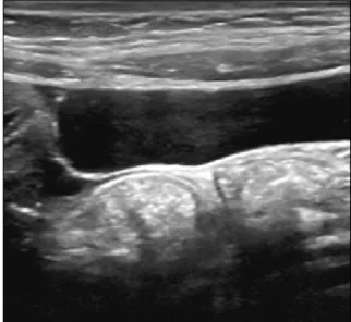
Fig. 1. Ultrasonography scans showing a large unicystic mass that has a well-defined wall with anechoic lesion. On an abdominal ultrasonography, a large cystic lesion was found with a thin wall superior to the bladder, composed of homogenous fluid material inferior to the umbilicus.
toms such as fever, jaundice, melena, or hemate- mesis. There was no familial record of any similar symptoms or history. Vital signs such as blood pres- sure, heart rate, respiratory rate, body temperature were all in normal ranges. However, on physical ex- amination, bowel sounds were decreased, and a 10-cm-sized mass was palpable in the lower abdomen. It was freely movable and apparently fluctuant. There was no tenderness. Blood test re- sults found that hemoglobin level was 12.5 g/dL;
white blood cell was 8,930 /μL with 84.2% neutrophil and 12.0% lymphocyte; platelet count was 301,000 /μL; erythrocyte sedimentation rate was 10 mm/h;
C-reactive protein level was 6.73 mg/L. Liver func- tion tests, basic metabolic panel, amylase and lipase levels, and urinalysis were within normal limits. In an abdominal ultrasonography to rule out in- tussusception, a large cystic lesion was found sur- rounded by a thin wall superior to the bladder, com- posed of homogenous fluid material inferior to the umbilicus; no solid lesion was observed (Fig. 1).
Abdominal computed tomography (CT) revealed a low attenuated intra-abdominal cystic mass meas- ured as 12 cm in diameter with thin intracystic septa- tions (Fig. 2). He was transferred to the general sur- gery department for operation. Intraoperatively, the
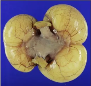
cyst was resected with the attached small bowel. A grossly unicystic mass was found, and the intestinal serosa was intact with a thin cystic wall measuring 9×8.5×4.2 cm, 152 g (Fig. 3). Immunohistochemical markers were negative. Histopathology of the mes-
Fig. 3. Operative photograph showing a giant mesenteric cyst. An unicystic mass was found grossly, and the intact intestinal serosa was found with a thin cystic wall measuring 9×8.5×4.2 cm.
Fig. 4. Histopathology of the mesenteric cystic wall; a single layer of epithelial lining surrounded by fibrous structure (H&E,
×100). A single layer of epithelial lining surrounded by fibrous structure was found at the histopathology of the mesenteric cyst which was a lymphatic mesenteric cyst at the final result.
enteric cystic wall shows a single layer of epithelial lining surrounded by fibrous structure. And the final pathophysiologic result was a lymphatic mesenteric cyst (Fig. 4). He made a good clinical recovery with- out complications after the operation. He was dis- charged home on the fifth post-operative day. There was no recurrence or complications during the fol- low-up period for one year.
DISCUSSION
A mesenteric cyst is defined as any cyst in the mesenterium. It was initially described by the Italian pathologist Antonio Benivieni during the autopsy of an 8-year-old child in 1507 [1,3]. Cases of mesenteric cysts have been reported continuously, but these cas- es were very small in number; thus, the etiology of mesenteric cysts has not been ascertained exactly.
Mesenteric cyst may vary in size from a few centi- meters to over 10 cm [4]. Its location is within the mesentery of the small intestine, most frequently followed by the mesentery of the colon and retro- peritoneum [5]. In a review series of 162 patients, 60% of mesenteric cysts occurred in the small-bowel mesentery, 24% in the large-bowel mesentery, and
14.5% in the retroperitoneum [2,6,7].
Mesenteric cyst is usually asymptomatic, but it can be accompanied by gastrointestinal symptoms which make it misunderstood as appendicitis, small bowel obstruction, or diverticulitis, before operation.
Complications such as rupture, torsion, or intestinal obstruction rarely occur, which cause more severe symptoms [8,9]. According to a large review of the literature by de Perrot et al. [10], pain (82%), nausea and vomiting (45%), constipation (27%), or diar- rhoea (6%) were the presenting symptoms while an abdominal mass was the clinical finding in up to 61%
of the patients. There were also non-specific symp- toms in this case, such as abdominal pain, dis- tension, and vomiting. It is likely that young patients under the age of 10 take less time to show some symptoms compared with patients aged over 10 [6].
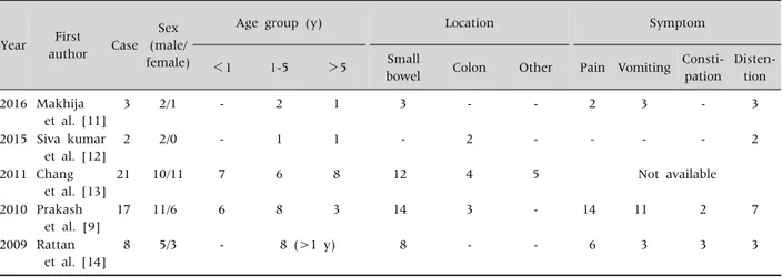
Various clinical characteristics were shown at the re- cent case series reports of mesenteric cysts in pedia- tric patients (Table 1) [9,11-14]. Nearly no case in childhood has been reported in Korea.
Because mesenteric cyst in childhood is occasion- ally accompanied by symptoms like pain, history taking and physical examination should be per- formed precisely, and radiologic findings such as ab- dominal ultrasound (USG) and CT scan are impor-
Table 1.Characteristics of Case Series Reports about Mesenteric Cysts in Childhood
Year First author Case
Sex (male/
female)
Age group (y) Location Symptom
<1 1-5 >5 Small
bowel Colon Other Pain Vomiting Consti- pation
Disten- tion 2016 Makhija
et al. [11]
3 2/1 - 2 1 3 - - 2 3 - 3
2015 Siva kumar et al. [12]
2 2/0 - 1 1 - 2 - - - - 2
2011 Chang et al. [13]
21 10/11 7 6 8 12 4 5 Not available
2010 Prakash et al. [9]
17 11/6 6 8 3 14 3 - 14 11 2 7
2009 Rattan et al. [14]
8 5/3 - 8 (>1 y) 8 - - 6 3 3 3
tant. According to study of Senocak et al. [15], ab- dominal USG by experienced hands is the most reli- able diagnostic tool, and CT scan should be per- formed additionally in a suspected case. USG and CT of the abdomen can distinguish between solid and cystic characteristics of abdominal mass.
Mesenteric cyst has a possibility of secondary complications such as hemorrhage, torsion, ob- struction, or infection, hence complete excision of cyst with or without bowel resection is the procedure of choice to prevent recurrence or malignant trans- formation [6,16]. Once the cyst is removed, it recurs rarely, and the prognosis of the patient is usually excellent. Recurrence is reported to be higher after partial wall excision and drainage than after total excision. There are little reports of malignant cysts at childhood yet. According to Kurtz et al. [6], only 3%
of mesenteric cysts were malignant, all of which were found in adults.
In conclusion, we reviewed the clinical course and literature of a child with mesenteric cysts who com- plained of acute abdominal pain, distension, and vomiting and were surgically treated after being di- agnosed with mesenteric cysts based on radiological examination.
ACKNOWLEDGEMENTS
This work was supported by a research grant funded
by Wonkwang University in 2017.
REFERENCES
1. Dequanter D, Lefebvre JC, Belva P, Takieddine M, Vaneukem P. Mesenteric cysts. A case treated by lapa- roscopy and a review of the literature. Surg Endosc 2002;16:1493.
2. Chung MA, Brandt ML, St-Vil D, Yazbeck S.
Mesenteric cysts in children. J Pediatr Surg 1991;26:
1306-8.
3. Mohanty SK, Bal RK, Maudar KK. Mesenteric cyst--an unusual presentation. J Pediatr Surg 1998;33:792-3.
4. Richard RR. Mesenteric and omental cysts. In: Grosfeld JL, O'Neill JA Jr, Coran AG, Fonkalsrud EW, eds.
Pediatric surgery. 6th ed. Philadelphia: Mosby Elsevier, 2006:1399-406.
5. Vanek VW, Phillips AK. Retroperitoneal, mesenteric, and omental cysts. Arch Surg 1984;119:838-42.
6. Kurtz RJ, Heimann TM, Holt J, Beck AR. Mesenteric and retroperitoneal cysts. Ann Surg 1986;203:109-12.
7. Saviano MS, Fundarò S, Gelmini R, Begossi G, Perrone S, Farinetti A, et al. Mesenteric cystic neoformations:
report of two cases. Surg Today 1999;29:174-7.
8. Marte A, Papparella A, Prezioso M, Cavaiuolo S, Pintozzi L. Mesenteric cyst in 11-year old girl: a techni- cal note. Case report. J Pediatr Surg 2013;1:84-6.
9. Prakash A, Agrawal A, Gupta RK, Sanghvi B, Parelkar S. Early management of mesenteric cyst prevents cata- strophes: a single centre analysis of 17 cases. Afr J Paediatr Surg 2010;7:140-3.
10. de Perrot M, Bründler M, Tötsch M, Mentha G, Morel P. Mesenteric cysts. Toward less confusion? Dig Surg
2000;17:323-8.
11. Makhija D, Shah H, Tiwari C, Jayaswal S, Khedkar K, Waghmare M. Mesenteric cyst(s) presenting as acute intestinal obstruction in children: three cases and liter- ature review. Int J Pediatr Adolesc Med 2016. doi:
10.1016/j.ijpam.2016.04.003.
12. Siva kumar S, Kireeti AS, Mohan TV. Giant mesenteric cysts of large intestine in children: two case reports and review of the literature. J Dent Med Sci 2015;14:93-5.
13. Chang TS, Ricketts R, Abramowsky CR, Cotter BD, Steelman CK, Husain A, et al. Mesenteric cystic mass- es: a series of 21 pediatric cases and review of the
literature. Fetal Pediatr Pathol 2011;30:40-4.
14. Rattan KN, Nair VJ, Pathak M, Kumar S. Pediatric chy- lolymphatic mesenteric cyst-a separate entity from cystic lymphangioma: a case series. J Med Case Rep 2009;3:111.
15. Senocak ME, Gündoğdu H, Büyükpamukçu N, Hiçsönmez A. Mesenteric and omental cysts in children. Analysis of nineteen cases. Turk J Pediatr 1994;36:295-302.
16. Hassan M, Dobrilovic N, Korelitz J. Large gastric mes- enteric cyst: case report and literature review. Am Surg 2005;71:571-3.