ABSTRACT
Objective: Decompressive craniectomy (DC) is one of the treatment modalities in severe traumatic brain injury (TBI), however, there was a lack of evidence for optimal craniectomy size. The authors aimed to investigate optimal DC size and analyze clinical outcome according to craniectomy size.
Methods: We retrospectively reviewed the medical data of 87 patients with a space occupying lesion following TBI who underwent unilateral DC. Craniectomy size was measured by anterior-posterior (AP) diameter and surface estimate (SE). Mortality, clinical outcome, and complications were collected and analyzed according to craniectomy size.
Results: Nineteen patients (21.8%) died and 35 patients (40.2%) had a favorable outcome at last follow-up (a mean duration, 30.3±39.4 months; range, 0.2–132.6 months). Receiver operating curve analyses identified AP diameter more than 12.5 cm (area under the curve [AUC]=0.740; p=0.002) and SE more than 98.0 cm2 (AUC=0.752; p=0.001) as cut-off values for survival, and AP diameter more than 13.4 cm (AUC=0.650; p=0.018) and SE more than 107.3 cm2 (AUC=0.685; p=0.003) for favorable outcome. Large craniectomy resulted in a significantly lower mortality rate and a higher rate of favorable outcome than small craniectomy (p=0.005 and p=0.014, respectively). However, procedure related bleeding occurred more frequently in the large craniectomy group (p=0.044).
Conclusion: Unilateral DC size is associated with clinical outcome of patients with a space occupying lesion following severe TBI. Large craniectomy is needed for survival and favorable outcome.
Keywords: Traumatic brain injury; Decompressive craniectomy; Mortality; Prognosis
INTRODUCTION
Intracranial hypertension, with or without a space occupying lesion, is still an important prognostic factor in severe traumatic brain injury (TBI).1,16,32) Decompressive craniectomy (DC) is a surgical procedure with the purpose of decompression of the intracranial space without reinserting the bone flap as the management of refractory intracranial pressure (ICP).17,28) However, it is still a controversial modality for treatment of severe TBI with
Clinical Article
Received: Dec 15, 2020 Revised: Feb 20, 2021 Accepted: Mar 18, 2021 Address for correspondence:
Jeongjun Lee
Department of Neurosurgery, Dongguk University Ilsan Hospital, 27 Dongguk-ro, Ilsandong-gu, Goyang 10326, Korea.
E-mail: [email protected]
Copyright © 2021 Korean Neurotraumatology Society
This is an Open Access article distributed under the terms of the Creative Commons Attribution Non-Commercial License (https://
creativecommons.org/licenses/by-nc/4.0/) which permits unrestricted non-commercial use, distribution, and reproduction in any medium, provided the original work is properly cited.
ORCID iDs Jinhwan Koo
https://orcid.org/0000-0003-1488-193X Jeongjun Lee
https://orcid.org/0000-0001-6847-1130 Su Hwan Lee
https://orcid.org/0000-0002-9605-6476 Jung Hyeon Moon
https://orcid.org/0000-0003-1582-2288 Seung-Yeob Yang
https://orcid.org/0000-0002-4051-2751 Keun-Tae Cho
https://orcid.org/0000-0002-8372-7250 Conflict of Interest
The authors have no financial conflicts of interest.
Jinhwan Koo , Jeongjun Lee , Su Hwan Lee , Jung Hyeon Moon , Seung-Yeob Yang , and Keun-Tae Cho
Department of Neurosurgery, Dongguk University Ilsan Hospital, Goyang, Korea
Does the Size of Unilateral
Decompressive Craniectomy Impact
Clinical Outcomes in Patients with
Intracranial Mass Effect after Severe
Traumatic Brain Injury?
refractory intracranial hypertension. The Decompressive Craniectomy in Diffuse Traumatic Brain Injury (DECRA) trial found poorer clinical outcomes for patients in the DC group compared with standard care.5) To the contrary, the latest randomized controlled trial (RCT), the Randomized Evaluation of Surgery with Craniectomy for Uncontrollable Elevation of Intracranial Pressure (RESCUEicp) trial, demonstrated that DC contributed to a lower mortality rate and obtained some advantages of functional outcomes compared with medical care.10) Nevertheless, DC is widely used as a treatment modality for intracranial hypertension in severe TBI patients. Jiang et al.15) and Qiu et al.24) demonstrated better outcomes in a large frontotemporoparietal DC (not less than 12×15 cm or 15 cm diameter) group compared with a routine temporoparietal craniectomy (6×8 cm or 8 cm diameter). However, those studies could not show outcomes of a frontotemporoparietal DC between a diameter of 8 cm and 15 cm. Although several studies reported clinical outcomes according to DC size, they had a limitation of small sample size for severe TBI patients.26,27,32) There is also a lack of evidence for optimal size of unilateral DC in severe TBI with space occupying lesion. The goal of this study was to investigate optimal size of unilateral DC and to demonstrate the impact of DC size on mortality and clinical outcomes in severe TBI with space occupying lesions.
MATERIALS AND METHODS
Patient selection
A retrospective analysis was conducted on all patients with severe TBI who underwent emergent DC in Dongguk University Ilsan Hospital between March 2007 and December 2019.
Indications for inclusion in this series were as follows: 1) initial Glasgow Coma Scale (GCS) of 8 or less; 2) a space occupying lesion (subdural hematoma [SDH], cerebral contusion, or epidural hematoma [EDH]) with or without cerebral edema on initial computed tomography (CT) scan; and 3) the midline shift of at least 5 mm or the effacement of basal cisterns.
Patients who underwent bifrontal DC were excluded from the study. Finally, 87 patients treated with unilateral DC were enrolled in our study. This study was conducted according to the principles outlined in the Declaration of Helsinki and was approved by our Institutional Review Board with signed consent being waived (IRB number: 2020-06-015).
Data collection
Medical records and imaging data were reviewed for basal characteristics, radiologic results, clinical outcomes, morbidity, and mortality. Multiple patient variables were retrieved as follows: age, gender, mechanism of injury, initial GCS,33) pupillary light reflexes, Injury Severity Score (ISS), medical parameters (hypertension, diabetes, cerebrovascular disease, coronary heart disease, liver disease, chronic kidney disease, and use of antiplatelet or anticoagulant agents), ICP (72 hours post-operatively), and complications (procedure related bleeding, procedure related infection, and hydrocephalus). Clinical outcomes were evaluated using the Glasgow Outcome Scale (GOS).13) Radiographic data including a Rotterdam CT score,19) types of space occupying hematomas, and craniectomy size were also collected.
Craniectomy size
Craniectomy size was measured on immediately postoperative CT scans using dedicated 3D program (syngo InSpace4D available on a syngo CT Workplace, VA44; Siemens Healthineers, Erlangen, Germany). A measure of craniectomy size was used from the previously published equation of Tanrikulu and Schur.26,32) The size of the DC was calculated as follows: largest anterior-posterior (AP) and largest cranio-caudal (CC) diameter, surface estimate (SE) of
bony decompression (SE=AP/2×CC/2×π).32) The ratio of the flap circumference to the skull hemi-circumference (F-S ratio) was calculated using the previously published equation of Schur (SUPPLEMENTARY FIGURE 1).26) Two experienced neurosurgeons (J Koo and J Lee) who were blinded to pertinent radiologic information independently reviewed all postoperative CT scans. If disagreement between the 2 neurosurgeons occurred, a consensus was established by a third neurosurgeon (KT Cho).
Surgical procedure
All DC was performed unilaterally within 24 hours on the side of the most significant mass effect, which was determined by CT scans. After hair shaving and preparation of the skin, a frontotemporoparietal hemicraniectomy was performed. The dura mater was opened in a C-shaped or a stellate fashion and removal of the hematoma was performed when necessary.
A large duraplasty was performed to allow for maximal expansion of the brain. An ICP monitor was routinely installed in the epidural space, and all procedures were performed by 5 different neurosurgeons.
Reoperation and postoperative management
Brain CT scan was performed immediately following the procedure and on postoperative days 1, 3, and 7. If ICP was above 25 mmHg, an emergent brain CT scan was obtained at the time. Reoperation was performed in any of following cases: 1) procedure-related SDH or EDH with a thickness of 10 mm or more within the field of DC on the CT scan or 2) when ICP was more than 25 mmHg and procedure-related bleeding or unilateral hemispheric swelling was identified on the CT scan. Postoperative medical management included optimized sedation, the normalization of arterial carbon dioxide pressure and electrolytes, use of mannitol, and control of blood pressure while keeping cerebral perfusion pressure over 70 mmHg.
Statistical analyses
Continuous variables were presented as mean±standard deviation and categorical data were expressed as frequencies and percentages. The χ2 or Fisher's exact tests were used to assess categorical variables. Multivariate analysis was conducted using variables with p-values <0.05 in univariate analysis or that were clinically important. Receiver operating characteristic (ROC) curve analysis was used to determine optimal cut-off values for parameters found to be significantly correlated with survival and favorable outcome. The cutoff value was defined as the highest sensitivity and specificity produced by the Youden index. Statistical significance was set at p<0.05, utilizing SPSS (version 22; IBM Corp., Armonk, NY, USA) for all purposes.
RESULTS
Patient characteristics and clinical outcome
The baseline characteristics of patients and clinical outcomes are summarized in
SUPPLEMENTARY TABLE 1. The mean patient age was 61.4±20.0 years (range, 18–98 years) and male patients were 58 (66.7%). Initial mean GCS was 5.0±2.2 (range, 3–8) and mean ISS was 25.1±9.7 (range, 9–57). The mean Rotterdam CT score was 3.6±0.9 (range, 2–6) and 79 (90.8%) patients showed greater than 5 mm of midline shift. The mean post-operative ICP was 20.0±14.7 (range, 3.6–82.0). On postoperative CT scan, the mean AP diameter was measured as 12.9±1.25 cm (range, 8.5–16.0 cm), the CC diameter was 10.0±1.04 cm (range, 7.0–12.6 cm), and the SE was 102.5±18.2 cm2 (range, 51.6–158.9 cm2). The mean F-S ratio was observed as 61.2±7.8% (range, 34.7–77.3%). The mortality rate (GOS 1) was 21.8% and
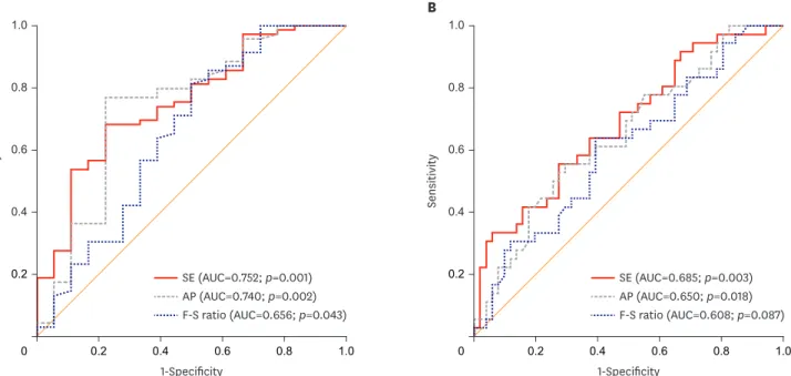
favorable outcome (GOS 4–5) was observed in 40.2% of the patients, with a mean follow- up duration of 30.3±39.4 months (range, 0.2–132.6 months). Univariate analysis indicated that age, initial GCS, bilateral unreactive pupil, Rotterdam CT score, post-operative ICP and craniectomy size were significantly correlated with mortality and favorable outcomes (TABLE 1). In multivariate analysis, post-operative ICP (odds ratio [OR]=1.269; p=0.024) and craniectomy size (SE) (OR=0.791; p=0.034) were identified as significant factors for mortality.
And age (OR=0.914, p=0.001), initial GCS (OR, 1.841; p=0.010), and craniectomy size (SE) (OR=1.048; p=0.042) were statistically significant factors for favorable outcome (TABLE 2).
SUPPLEMENTARY TABLE 2 shows GOS at last follow-up according to type of hemorrhage.
TABLE 1. Univariate analysis for mortality and favorable outcome
Variables Mortality Favorable outcome
Survival (n=69) Death (n=18) p-value Favorable (n=36) Poor (n=51) p-value
Age (years) 59.5±16.3 59.3±20.0 0.048* 49.6±19.1 69.7±15.3 <0.001*
Male 49 (71.0) 9 (50.0) 0.092 24 (66.7) 34 (66.7) 1.000
Underlying disease
Hypertension 27 (39.1) 7 (38.9) 0.985 10 (27.8) 24 (47.1) 0.069
Diabetes 14 (20.3) 5 (27.8) 0.528 5 (13.9) 14 (27.5) 0.132
Coronary heart disease 6 (8.7) 3 (16.3) 0.296 2 (5.6) 7 (13.7) 0.296
Cerebrovascular disease 7 (10.1) 1 (5.6) 0.686 2 (5.6) 6 (11.8) 0.461
Liver disease 7 (10.1) 0 (0.0) 0.216 2 (5.6) 5 (9.8) 0.695
Chronic kidney disease 1 (1.4) 2 (11.1) 0.107 0 (0.0) 3 (5.9) 0.264
Falling 33 (47.8) 6 (33.3) 0.271 13 (36.1) 26 (51.0) 0.170
Initial GCS 5.8±1.9 4.7±1.6 0.033* 6.4±1.8 5.0±1.7 0.001*
Bilateral unreactive pupil 24 (34.8) 12 (66.7) 0.014* 8 (22.2) 28 (54.9) 0.002*
ISS 24.7±9.9 26.5±9.0 0.477 23.5±10.6 26.2±9.0 0.218
Rotterdam CT score 3.4±0.9 4.0±0.8 0.019* 3.3±0.9 3.7±0.9 0.040*
Post-operative ICP (mmHg) 15.9±11.0 35.0±17.0 <0.001* 16.4±11.0 22.5±16.4 0.041*
Craniectomy size
AP 13.2±1.1 12.0±1.5 <0.001* 13.4±1.1 12.7±1.3 0.007*
SE 106.2±16.3 88.2±19.1 <0.001* 109.5±18.0 97.4±17.0 0.002*
F-S ratio 62.4±6.5 56.7±10.7 0.043* 63.2±6.7 59.9±8.4 0.049*
Data are shown as mean±standard deviation or number (%).
GCS: Glasgow Coma Scale, ISS: Injury Severity Score, CT: computed tomography, ICP: intracranial pressure, AP: anterior-posterior; SE: surface estimate, F-S ratio: the ratio of the flap circumference to the skull hemi-circumference.
*p<0.05, statistically significant variables.
TABLE 2. Multivariate analysis for the predictor of mortality and favorable outcome
Variables Mortality Favorable outcome
OR (95% CI) p-value OR (95% CI) p-value
Age (years) 0.992 (0.904–1.088) 0.860 0.914 (0.869–0.963) 0.001*
Hypertension 4.286 (0.100–183.813) 0.448 0.414 (0.082–2.105) 0.414
Diabetes 15.725 (0.244–1,013.317) 0.917 0.312 (0.043–2.279) 0.279
Coronary heart disease 0.019 (0.000–5.524) 0.170 0.696 (0.058–8.414) 0.696 Cerebrovascular disease 0.236 (0.008–7.122) 0.406 5.285 (0.347–80.398) 0.231
Liver disease - - 7.974 (0.757–83.958) 0.084
Chronic kidney disease 0.006 (0.000–133.399) 0.070 - -
Falling 0.127 (0.007–2.393) 0.168 0.630 (0.158–2.504) 0.511
Initial GCS 0.446 (0.159–1.252) 0.125 1.841 (1.156–2.932) 0.010*
Bilateral unreactive pupil 0.060 (0.001–3.145) 0.164 1.397 (0.240–8.129) 0.710
ISS 0.957 (0.814–1.124) 0.590 1.005 (0.931–1.084) 0.907
Rotterdam CT score 6.762 (0.883–51.814) 0.066 0.713 (0.282–1.801) 0.474 Post-operative ICP (mmHg) 1.269 (1.032–1.559) 0.024* 0.986 (0.938–1.036) 0.572 Craniectomy size (SE) 0.791 (0.637–0.982) 0.034* 1.048 (1.002–1.097) 0.042* OR: odds ratio, CI: confidence interval, GCS: Glasgow Coma Scale, ISS: Injury Severity Score, CT: computed tomography, ICP: intracranial pressure, SE: surface estimate.
*p<0.05, statistically significant variables.
Clinical outcomes according to craniectomy size
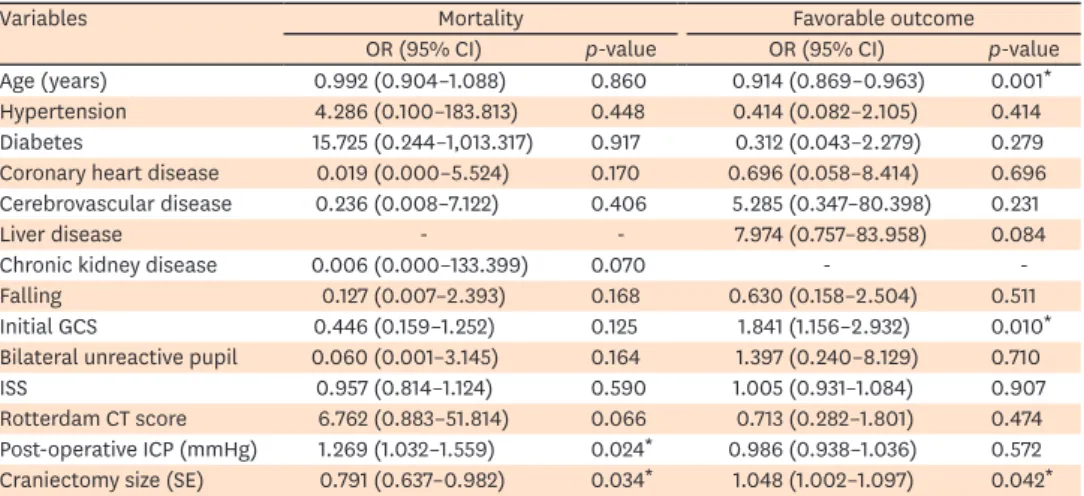
FIGURE 1 shows GOS at last follow up according to 3 parameters of craniectomy size. The mortality rate tended to decrease as the AP diameter, SE, and F-S ratio increased. As the AP diameter increased, the rate of patients who showed favorable outcome increased at last follow-up. The rate of good recovery (GOS 5) had a tendency to increase with increasing AP diameter and SE.
Optimal craniectomy size
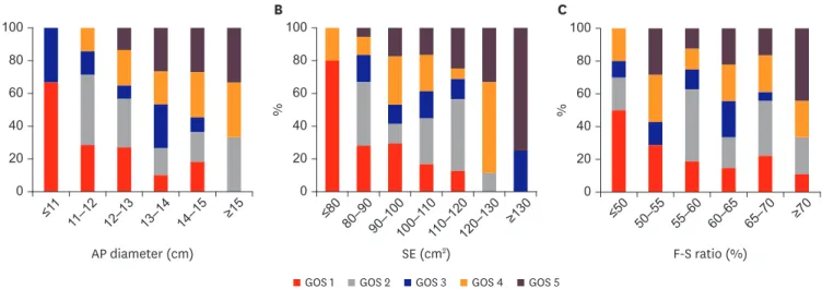
In ROC curve analyses for 3 parameters of craniectomy size, cut-off values for predicting survival and area under the curve (AUC) resulted in the following: AP diameter more than 12.5 cm (AUC=0.740; p=0.002), SE more than 98.1 cm2 (AUC=0.752; p=0.001), and F-S ratio more than 57.7% (AUC=0.656, p=0.043) (FIGURE 2 and TABLE 3). AP diameter more than 13.4 cm and SE>107.3 cm2 were parameters found to significantly predict favorable outcome with AUC of 650 and 0.685, respectively.
Large vs. small craniectomy
The patients were categorized into small and large craniectomy groups based on SE ≤107 cm2 or more than 107 cm2 (TABLE 4). Large craniectomy group showed significantly lower mortality rate compared with small craniectomy group (5.7% vs. 30.8%, respectively;
p=0.005). Favorable outcome occurred in 57.1% of the patients in the large craniectomy group, as compared with 30.8% of those in the small craniectomy group (p=0.014). The mean post-operative ICP was not statistically significant difference between 2 groups, but had borderline significance (p=0.066). Procedure-related bleeding occurred more frequently in the large craniectomy group (p=0.044). Of 29 procedure related bleedings, 27 needed further hematoma removal. There were no significant differences in the rate of procedure-related infection and hydrocephalus.
DISCUSSION
DC is a surgical procedure for refractory intracranial hypertension following severe TBI.17,28) DC has been shown to decrease ICP and increase brain compliance, cerebral blood flow and AP diameter (cm)
20
% 40
100
60 80
0
11–12 13–14
≤11
12–13 14–15 ≥15
50–55 60–65
≤50 55–60 65–70 ≥70
SE (cm2) 20
% 40
100
60 80
0
80–90 100–1
≤80 10
90–100 110–120 ≥130
120–130
F-S ratio (%) 20
% 40
100
60 80
0
GOS 1 GOS 2 GOS 3 GOS 4 GOS 5
A B C
FIGURE 1. Clinical outcome according to parameters of craniectomy Size. Histogram of GOS according to (A) largest AP diameter, (B) SE, and (C) F-S ratio.
GOS, Glasgow Outcome Scale, AP: anterior-posterior, SE: surface estimate, F-S ratio: the ratio of the flap circumference to the skull hemi-circumference.
TABLE 3. Receiver operating characteristic curve analysis results for predictions of clinical outcome
Parameter Cutoff value AUC p-value 95% CI TP TN FP FN Sensitivity (%) Specificity (%) PPV (%) NPV (%) Survival
AP 12.5 0.740 0.002* 0.597–0.883 52 13 6 16 76.8 77.8 89.7 44.8
SE 98.1 0.752 0.001* 0.628–0.876 46 14 5 22 68.1 77.8 90.2 38.9
F-S ratio 57.7 0.656 0.043* 0.496–0.815 55 9 10 13 81.2 50.0 84.6 40.9
Favorable outcome
AP 13.4 0.650 0.018* 0.534–0.766 20 37 15 15 55.6 70.6 57.1 71.2
SE 107.3 0.685 0.003* 0.572–0.798 20 38 14 15 55.6 72.5 58.8 71.7
F-S ratio 62.4 0.608 0.087 0.488–0.728 23 32 20 12 63.9 60.8 53.5 72.7
AUC: area under the curve, CI: confidence interval, TP: true positive, TN: true negative, FP: false positive, FN: false negative, PPV: positive predictive value, NPV:
negative predictive value, AP: anterior-posterior, SE: surface estimate, F-S ratio: the ratio of the flap circumference to the skull hemi-circumference.
*p<0.05, statistically significant variables.
TABLE 4. Clinical outcomes and complications according to SE 107 cm2
Clinical outcomes and complications SE <107 (n=52) SE >107 (n=35) p-value
Mortality 16 (30.8) 2 (5.7) 0.005*
Favorable outcome 16 (30.8) 20 (57.1) 0.014*
Post-operative ICP (mmHg) 22.4±17.1 16.9±10.0 0.066
Procedure related bleeding 13 (25.0) 16 (45.7) 0.044*
EDH 3 7
SDH 4 2
Contusion or ICH 3 2
EDH + SDH 1 4
EDH + contusion or ICH 1 1
SDH + contusion or ICH 1 0
Procedure related infection 2 (3.8) 5 (14.3) 0.112
Hydrocephalus 5 (9.6) 4 (11.4) 1.000
Data are shown as mean±standard deviation or number (%).
ICP: intracranial pressure, EDH: epidural hematoma, SDH: subdural hematoma, ICH: intracranial hemorrhage, SE:
surface estimate.
*p<0.05, statistically significant variables.
1-Specificity 0.2
Sensitivity 0.4 1.0
0.6 0.8
0 0.2 0.4 0.6 0.8 1.0
A
SE (AUC=0.752; p=0.001) AP (AUC=0.740; p=0.002) F-S ratio (AUC=0.656; p=0.043)
1-Specificity 0.2
Sensitivity 0.4 1.0
0.6 0.8
0 0.2 0.4 0.6 0.8 1.0
B
SE (AUC=0.685; p=0.003) AP (AUC=0.650; p=0.018) F-S ratio (AUC=0.608; p=0.087)
FIGURE 2. Receiver operating characteristic curve analysis for survival (A) and favorable outcome (B) SE, largest AP diameter, and F-S ratio predicting survival were used as parameters of craniectomy size.
AUC, area under the curve, SE: surface estimate, AP: anterior-posterior, F-S ratio: the ratio of the flap circumference to the skull hemi-circumference.
oxygen perfusion.11,29,30) However, the clinical effect of DC for patients with severe TBI is still controversial. Two multicenter RCTs, the DECRA and RESCUEicp trials have assessed DC exclusively as management for patients with refractory intracranial hypertension after severe TBI.5,10) In the DECRA trial, patients with DC had worse outcomes compared with standard care at 6 months. Patients with intracranial hematoma were excluded and only bifrontotemporoparietal craniectomy was allowed in the DECRA trial.5) The RESCUEicp trial demonstrated that DC could decrease mortality and improve clinical outcome compared with medical care.10) In the RESCUEicp trial, 109 (63%) patients underwent bifrontal DC and 64 (37%) underwent unilateral DC. Unlike the DECRA trial, patients with intracranial hematoma represented almost 20% in the RESCUEicp trial.10) Different inclusion criteria and strategy of management could lead to various results between the 2 RCTs. And it is difficult to know the exact effect of unilateral DC for severe TBI with space occupying lesion in 2 RCTs.
In current series, we focused on clinical outcome of patients with space occupying lesion who treated with unilateral DC.
In unilateral DC, bone flap size of DC could be an important factor that affects the clinical outcome. In the recent guidelines for the management of severe TBI, a large frontotemporoparietal DC (not less than 12×15 cm or 15 cm diameter) is recommended over a small DC to reduce mortality and improve neurologic outcomes based on 2 RCTs.3) Jiang et al.15) demonstrated that favorable outcome occurred in 96 of 241 (39.8%) patients in the standard trauma craniectomy (12×15 cm), as compared with 70 of 245 (28.6%) patients in the limited craniectomy (6×8 cm). The mortality rate was lower in standard trauma craniectomy compared with limited craniectomy (26.2% vs. 35.1%, respectively;
p<0.05). Qiu et al.24) reported that the mortality rates at 1 month after treatment were 27%
in the frontopareitotemporal craniectomy (15 cm) group and 57% in the temporoparietal craniectomy (8 cm) group (p=0.01). Favorable outcome rates 1 year after TBI were 56.8%
and 32.4%, respectively (p=0.035). These studies reported a clinical outcome comparing 2 extremes of DC size, a frontotemporoparietal DC approximately 15 cm size in diameter and a temporoparietal DC approximately 8cm size in diameter, but they did not show the clinical outcome of frontotemporoparietal DC with a size between 8cm and 15 cm in severe TBI.
Several studies reported clinical outcomes according to DC size that ranged from 7.7 cm to 24 cm in severe TBI.21,25-28,32) Sedney et al.27) reported that increasing DC size had a significant relationship to decreased mortality rate in 20 TBI patients (p=0.032). However, functional outcomes were not significantly related to craniectomy size. Reid at al.25) demonstrated that SE was an independent factor for ICP reduction but not for the neurologic outcome in 58 patients with TBI. Schur et al.26) showed ICP control was better in patients treated with a large DC (F-S ratio >65%) compared with a small DC (F-S ratio <65%) in 30 patients with severe TBI without space occupying and there were no differences in clinical outcomes between the 2 groups. However, these studies had a limitation of a small sample size to demonstrate significant clinical differences according to DC size. Missori et al.21) reported that SE of bone flap was not related to survival in 73 unilateral DCs (surviving 7,643 mm2 vs. deceased 7,372 mm2). However, the overall craniectomy size of this study was too small compared with other studies to result in significant differences between the 2 groups.21)
There are several parameters to measure DC size. In general, AP diameter and SE have been widely used and the F-S ratio was recently introduced by Schur et al.26) In our current series, we measured DC size using these parameters and analyzed the effect of each parameter on clinical outcomes. All parameters significantly influenced the survival rate in severe
TBI. Considering the favorable outcome, AP dimeter and SE were statistically significant parameters, but the F-S ratio was not. The F-S ratio devised by Schur et al.26) is a novel method of measuring the craniectomy size to account for the size of patient's head circumference.
However, the equation of the F-S ratio was devised for DC flap and skull circumference on the axial plane of the CT scan, not accounting for the surface area. The F-S ratio could be measured differently depending on the angle of the axial plane and the shape of the bone flap. Therefore, the F-S ratio seems to have a limitation to show the actual craniectomy size. In the ROC curve analyses, optimal cut-off values of the AP diameter and SE predicting favorable outcome was greater than those predicting survival, which suggests that larger DC may be needed considering the favorable outcome other than survival.
There is a lack of evidence for optimal DC size in severe TBI. As discussed previously, the current guidelines for the management of severe TBI recommended performing DC not less than 12×15 cm or 15 cm in diameter.3) In our current series, we recommend AP diameter greater than 13.4 cm and SE greater than 107.3 cm2 considering favorable outcome. It also seems that better clinical outcome tended to be observed as DC size increased. However, the largest DC case was 16 cm of AP diameter and 158.9 cm2 of SE in current series. Therefore, the current series could not conclude the clinical outcome for DC over 16 cm of AP diameter.
Tanrikulu et al.32) reported the clinical outcome of patients undergoing very large DC with a diameter of 18–24 cm, not included in the current series. Although not statistically significant, they showed that a trend toward better outcome was observed in patients undergoing DC with a diameter of 12–18 cm or SE <180 cm2 than in patients undergoing DC with a diameter of 18 cm or more or SE >180 cm2.32) Furthermore, when they analyzed the patients into 3 groups of 12–15 cm, 15–20 cm, and 20–24 cm according to the size of DC, there was no difference in clinical results between the groups.32) Among DC size between 8–15 cm that was not included in Jiang et al.15) and Qiu et al.,24) patients underwent unilateral DC between 12–15 cm size seemed to result in a good clinical outcome. However, their study included not only patients with TBI, but also patients with cerebral infarction, intracerebral hemorrhage, abscess, and venous sinus thrombosis.32) Sedney et al.27) reported that all patients with DC less than 10cm died. In the current series, 4 (66.7%) patients who were treated with a DC less than 11cm died and 2 (33.3%) showed GOS 3.
We found that age, initial GCS, bilateral unreactive pupil, Rotterdam CT score, and
craniectomy size were significantly correlated with clinical outcome. Several studies reported that age was a strong prognostic indicator after severe TBI.8,14) Jiang et al.14) suggest that decreased capacity of brain for repair, preexisting disease, and increased frequency of systemic complications after TBI might be the causes for poor outcome in older patients. Many studies have demonstrated that initial GCS had a significant inverse correlation with mortality in
TBI.4,6,23) It is well known that there was a strong association between poor outcome and
bilaterally unreactive pupils.12,22) Narayan et al.22) reported that the mortality rate was 61% of patients with a bilaterally unreactive pupil compared with 16% of those with normal pupillary reaction. The rate of favorable outcome was higher in patients with normal pupillary reaction than in those with bilaterally unreactive pupils (76% vs. 30%, respectively).22) Huang et al.9) demonstrated that the Rotterdam CT score is an independent predictor for poor outcomes of patients who underwent DC after TBI in multivariate analysis (p=0.035).
The overall complication rates of DC could range from 13.4% to 53.9%.2,18) The complications of DC may be classified as hemorrhagic, infectious/inflammatory, and disturbances of the cerebrospinal fluid compartment.18) There were few studies for correlation between
procedure-related adverse events and DC size in severe TBI. Jiang et al.15) demonstrated that a large craniectomy group showed significantly lower delayed hematoma and incision cerebrospinal fluid fistula compared with a small craniectomy (10.6% and 2.4% vs. 17.8%
and 7.5%, respectively). In contrast, Qiu et al.24) reported that the incidence of subdural effusion and delayed intracranial hematoma were 10.8% and 21.6% in a large craniectomy vs. 0% and 5.4% in a small craniectomy, respectively (p=0.040 and p=0.041, respectively).
Sedney et al.27) and Schur et al.26) reported that complication rates did not associate with bone flap size. In the current series, rate of procedure related bleeding occurred significantly greater in the large craniectomy group compared with the small craniectomy (45.7% vs. 25%, respectively; p=0.044). Newly developed or expanding hematoma may be caused by the loss of the tamponade effect of high ICP after DC.7,20,31) We suggest that this effect may occur more frequently as increasing DC size in current series. Additionally, the larger surface of the DC itself could be a traumatic factor for hemorrhagic complication. The rate of procedure-related bleeding in the current series was much higher compared with other studies. However, of 29 rebleeding cases in the small craniectomy group, only 2 (6.9%) and 20 (69.0%) patients showed favorable outcomes at last follow-up. In the large craniectomy group, 14 of 16 (87.5%) patients treated with rebleeding showed favorable outcomes. Although procedure-related bleeding occurred more frequently in the large craniectomy group, a favorable clinical outcome could be obtained after reoperation and aggressive medical management.
Our study has several limitations. It was a retrospective, observational, and single- center study. The formula of SE is calculated by measuring the AP and CC diameters on 3 dimensional CT. Therefore, this may be slightly different from the actual surface area. In the current series, only 3 patients underwent DC with a size greater than 15 cm, so clinical outcomes of DC over 15 cm could not be reflected. Further study needs to demonstrate effectiveness and safety of the very large craniectomy.
CONCLUSION
Unilateral DC size is associated with clinical outcomes of patients with space occupying lesions following severe TBI. A large craniectomy could have several advantages for reducing the mortality rate and increasing the rate of favorable outcome. However, procedure-related bleeding could occur more frequently in patients with a large craniectomy. Nevertheless, if reoperation and aggressive medical management following large craniectomy were available, favorable clinical outcomes could be obtained at final follow-up.
SUPPLEMENTARY MATERIALS
SUPPLEMENTARY TABLE 1
Baseline characteristics and clinical outcome Click here to view
SUPPLEMENTARY TABLE 2
Clinical outcome according to type of hemorrhage Click here to view
SUPPLEMENTARY FIGURE 1
Immediate postoperative CT scans using dedicated 3D program: (A) the largest AP diameter and the largest CC diameter, (B) surface estimate of craniectomy was calculated by the equation AP/2×CC/2×π, (C) The ratio of the flap circumference to the skull hemi-circumference was calculated by the equation FC/ShC×100, ShC was calculated using the equation to approximate the perimeter of an ellipse 2π�(𝑎𝑎𝑎𝑎2+ 𝑏𝑏𝑏𝑏2)/2/2 , FC was calculated by ShC×α/180. The
craniectomy size of this figure was following: AP was 13.5 cm, SE 107 cm2, and F-S ratio 59.7%.
Click here to view
REFERENCES
1. Balestreri M, Czosnyka M, Hutchinson P, Steiner LA, Hiler M, Smielewski P, et al. Impact of intracranial pressure and cerebral perfusion pressure on severe disability and mortality after head injury. Neurocrit Care 4:8-13, 2006
PUBMED | CROSSREF
2. Ban SP, Son YJ, Yang HJ, Chung YS, Lee SH, Han DH. Analysis of complications following decompressive craniectomy for traumatic brain injury. J Korean Neurosurg Soc 48:244-250, 2010
PUBMED | CROSSREF
3. Carney N, Totten AM, O'Reilly C, Ullman JS, Hawryluk GW, Bell MJ, et al. Guidelines for the management of severe traumatic brain injury, fourth edition. Neurosurgery 80:6-15, 2017
PUBMED | CROSSREF
4. Colohan AR, Alves WM, Gross CR, Torner JC, Mehta VS, Tandon PN, et al. Head injury mortality in two centers with different emergency medical services and intensive care. J Neurosurg 71:202-207, 1989 PUBMED | CROSSREF
5. Cooper DJ, Rosenfeld JV, Murray L, Arabi YM, Davies AR, D'Urso P, et al. Decompressive craniectomy in diffuse traumatic brain injury. N Engl J Med 364:1493-1502, 2011
PUBMED | CROSSREF
6. Fearnside MR, Cook RJ, McDougall P, McNeil RJ. The Westmead Head Injury Project outcome in severe head injury. A comparative analysis of pre-hospital, clinical and CT variables. Br J Neurosurg 7:267-279, 1993 PUBMED | CROSSREF
7. Flint AC, Manley GT, Gean AD, Hemphill JC 3rd, Rosenthal G. Post-operative expansion of hemorrhagic contusions after unilateral decompressive hemicraniectomy in severe traumatic brain injury. J Neurotrauma 25:503-512, 2008
PUBMED | CROSSREF
8. Hernesniemi J. Outcome following head injuries in the aged. Acta Neurochir (Wien) 49:67-79, 1979 PUBMED | CROSSREF
9. Huang YH, Deng YH, Lee TC, Chen WF. Rotterdam computed tomography score as a prognosticator in head-injured patients undergoing decompressive craniectomy. Neurosurgery 71:80-85, 2012
PUBMED | CROSSREF
10. Hutchinson PJ, Kolias AG, Timofeev IS, Corteen EA, Czosnyka M, Timothy J, et al. Trial of decompressive craniectomy for traumatic intracranial hypertension. N Engl J Med 375:1119-1130, 2016
PUBMED | CROSSREF
11. Jaeger M, Soehle M, Meixensberger J. Effects of decompressive craniectomy on brain tissue oxygen in patients with intracranial hypertension. J Neurol Neurosurg Psychiatry 74:513-515, 2003
PUBMED | CROSSREF
12. Jennett B, Teasdale G, Braakman R, Minderhoud J, Knill-Jones R. Predicting outcome in individual patients after severe head injury. Lancet 1:1031-1034, 1976
PUBMED | CROSSREF
13. Jennett B, Bond M. Assessment of outcome after severe brain damage. Lancet 1:480-484, 1975 PUBMED | CROSSREF
14. Jiang JY, Gao GY, Li WP, Yu MK, Zhu C. Early indicators of prognosis in 846 cases of severe traumatic brain injury. J Neurotrauma 19:869-874, 2002
PUBMED | CROSSREF
15. Jiang JY, Xu W, Li WP, Xu WH, Zhang J, Bao YH, et al. Efficacy of standard trauma craniectomy for refractory intracranial hypertension with severe traumatic brain injury: a multicenter, prospective, randomized controlled study. J Neurotrauma 22:623-628, 2005
PUBMED | CROSSREF
16. Juul N, Morris GF, Marshall SB, Marshall LF. Intracranial hypertension and cerebral perfusion pressure:
influence on neurological deterioration and outcome in severe head injury. The Executive Committee of the International Selfotel Trial. J Neurosurg 92:1-6, 2000
PUBMED | CROSSREF
17. Kolias AG, Kirkpatrick PJ, Hutchinson PJ. Decompressive craniectomy: past, present and future. Nat Rev Neurol 9:405-415, 2013
PUBMED | CROSSREF
18. Kurland DB, Khaladj-Ghom A, Stokum JA, Carusillo B, Karimy JK, Gerzanich V, et al. Complications associated with decompressive craniectomy: a systematic review. Neurocrit Care 23:292-304, 2015 PUBMED | CROSSREF
19. Maas AI, Hukkelhoven CW, Marshall LF, Steyerberg EW. Prediction of outcome in traumatic brain injury with computed tomographic characteristics: a comparison between the computed tomographic classification and combinations of computed tomographic predictors. Neurosurgery 57:1173-1182, 2005 PUBMED | CROSSREF
20. Makino H, Yamaura A. Assessment of outcome following large decompressive craniectomy in management of serious cerebral contusion. A review of 207 cases. Acta Neurochir Suppl (Wien) 28:193-194, 1979 PUBMED
21. Missori P, Morselli C, Domenicucci M, Paolini S, Peschillo S, Scapeccia M, et al. Measurement of bone flap surface area and midline shift to predict overall survival after decompressive craniectomy. World Neurosurg 96:11-14, 2016
PUBMED | CROSSREF
22. Narayan RK, Greenberg RP, Miller JD, Enas GG, Choi SC, Kishore PR, et al. Improved confidence of outcome prediction in severe head injury. A comparative analysis of the clinical examination, multimodality evoked potentials, CT scanning, and intracranial pressure. J Neurosurg 54:751-762, 1981 PUBMED | CROSSREF
23. Phuenpathom N, Choomuang M, Ratanalert S. Outcome and outcome prediction in acute subdural hematoma. Surg Neurol 40:22-25, 1993
PUBMED | CROSSREF
24. Qiu W, Guo C, Shen H, Chen K, Wen L, Huang H, et al. Effects of unilateral decompressive craniectomy on patients with unilateral acute post-traumatic brain swelling after severe traumatic brain injury. Crit Care 13:R185, 2009
PUBMED | CROSSREF
25. Reid P, Say I, Shah S, Tolia S, Musku S, Prestigiacomo C, et al. Effect of bone flap surface area on outcomes in decompressive hemicraniectomy for traumatic brain injury. World Neurosurg 119:e922-e927, 2018 PUBMED | CROSSREF
26. Schur S, Martel P, Marcoux J. Optimal bone flap size for decompressive craniectomy for refractory increased intracranial pressure in traumatic brain injury: taking the patient' s head size into account.
World Neurosurg 137:e430-e436, 2020 PUBMED | CROSSREF
27. Sedney CL, Julien T, Manon J, Wilson A. The effect of craniectomy size on mortality, outcome, and
complications after decompressive craniectomy at a rural trauma center. J Neurosci Rural Pract 5:212-217, 2014 PUBMED | CROSSREF
28. Skoglund TS, Eriksson-Ritzén C, Jensen C, Rydenhag B. Aspects on decompressive craniectomy in patients with traumatic head injuries. J Neurotrauma 23:1502-1509, 2006
PUBMED | CROSSREF
29. Soustiel JF, Sviri GE, Mahamid E, Shik V, Abeshaus S, Zaaroor M. Cerebral blood flow and metabolism following decompressive craniectomy for control of increased intracranial pressure. Neurosurgery 67:65- 72, 2010
PUBMED | CROSSREF
30. Stiefel MF, Heuer GG, Smith MJ, Bloom S, Maloney-Wilensky E, Gracias VH, et al. Cerebral oxygenation following decompressive hemicraniectomy for the treatment of refractory intracranial hypertension. J Neurosurg 101:241-247, 2004
PUBMED | CROSSREF
31. Stiver SI. Complications of decompressive craniectomy for traumatic brain injury. Neurosurg Focus 26:E7, 2009
PUBMED | CROSSREF
32. Tanrikulu L, Oez-Tanrikulu A, Weiss C, Scholz T, Schiefer J, Clusmann H, et al. The bigger, the better?
About the size of decompressive hemicraniectomies. Clin Neurol Neurosurg 135:15-21, 2015 PUBMED | CROSSREF
33. Teasdale G, Jennett B. Assessment of coma and impaired consciousness. A practical scale. Lancet 2:81- 84, 1974
PUBMED | CROSSREF