88 Copyright © 2016 Korean Dementia Association
INTRODUCTION
Human Herpes Virus 6 (HHV6) is known to be commonly associated with encephalitis related to immunosuppression and transplantation, and to bone marrow transplantation (BMT) in particular.1 HHV-6 is known to be common in transplant recipients and lead to several clinical manifestations, such as encephalitis.1-4 HHV 6 encephalitis in patients with BMT is known as latent infection. The first such case was re- ported in 1994.5 HHV6 encephalitis in patients receiving im- munosuppressant drugs following BMT has various clinical manifestations, suggesting the involvement of the medial tem-
poral lobe. These include confusion, seizure, and amnesia, which lead to so-called post-transplant acute limbic encephali- tis.3 Neuroimaging findings in patients with HHV6 encephali- tis correspond to those found in patients with acute limbic en- cephalitis.3,6-8 However, cases of HHV6 encephalitis with hippocampal atrophy and glucose hypometabolism have very rarely been reported.
CASE REPORT
A 41-year-old right-handed woman with acute lymphoblas- tic leukemia (AML) presented with fever and mental change 2 weeks after BMT. She was alert, but had no awareness. She had no any other focal neurologic deficit. The treatment regimen following BMT consisted of 2 mg/kg cyclosporine, which was used to suppress immunity. The patient’s past medical history was not significant, except for the presence of AML. She had
Human Herpes Virus 6 Encephalitis Following Bone Marrow Transplantation with Uncommon Magnetic Resonance
Imaging Findings
Jihye Hwang, Ji Eun Kim, Jee Hoon Roh, Jae-Hong Lee
Department of Neurology, Asan Medical Center, University of Ulsan College of Medicine, Seoul, Korea
Background Human Herpes Virus 6 (HHV6) is commonly associated with encephalitis following bone marrow transplantation. However, hippocampal atrophy and global hypometabolism are rare findings in HHV6 encephalitis.
Case Report A 41-year-old right-handed woman with acute lymphoblastic leukemia presented with fever and mental changes 2 weeks af- ter receiving a sibling bone marrow transplant. The patient’s cerebrospinal fluid (CSF) was positive for HHV-6 deoxyribonucleic acid (DNA), but was negative for other viral DNA. Brain magnetic resonance imaging revealed atrophic changes in bilateral medial temporal lobes. Fol- lowing 4 weeks of ganciclovir therapy, a CSF exam was negative for HHV-6 DNA and the patient’s neurological symptoms partially improved.
However, she was disoriented and had severe retrograde and anterograde amnesia. 18F-fluorodeoxyglucose-positron emission tomography indicated global hypometabolism in the medial temporal lobes and the fronto-parietal cortices.
Conclusions This is a rare and unusual case of hippocampal atrophy in the acute stage of HHV6 encephalitis. Our imaging findings may reflect the chronic indolent course of HHV6 encephalitis.
Key Words herpes, human herpes virus, Human Herpes Virus 6 encephalitis, limbic encephalitis, retrograde amnesia, anterograde amnesia.
Received: July 22, 2016 Revised: September 19, 2016 Accepted: September 19, 2016
Correspondence: Jae-Hong Lee, MD, PhD, Department of Neurology, Asan Medical Center, University of Ulsan College of Medicine, 88 Olympic-ro, 43-gil, Songpa-gu, Seoul 05505, Korea
Tel: +82-2-3010-3446, Fax: +82-2-474-4691, E-mail: [email protected]
cc This is an Open Access article distributed under the terms of the Cre- ative Commons Attribution Non-Commercial License (http://creative- commons.org/licenses/by-nc/3.0) which permits unrestricted non-com- mercial use, distribution, and reproduction in any medium, provided the ori- ginal work is properly cited.
DND
Print ISSN 1738-1495 / On-line ISSN 2384-0757
Dement Neurocogn Disord 2016;15(3):88-91 / http://dx.doi.org/10.12779/dnd.2016.15.3.88
CASE REPORT
www.dnd.or.kr 89
DND
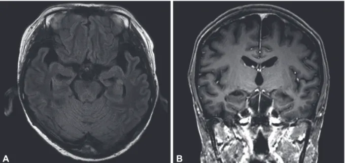
no hypertension, diabetes mellitus, cardiac disease, chronic re- nal disease, or liver disease. She had no family history of a cen- tral nerve system (CNS) disease, hereditary disorder or devel- opmental disorder. She was febrile, with a temperature of up to 39.3°C. Her vital signs were as follows: blood pressure was 115/70 mm Hg, heart rate was elevated up to 134 beats per minute, and respiratory rate was usually 30 breaths per min- ute. A complete blood count revealed anemia and thrombocy- topenia due to AML, and mild leukocytosis. A blood chemis- try study revealed the presence of mild hypocalcemia and elevated liver enzymes. C-reactive protein levels were slightly elevated. A chest X-ray study suggested the presence of inter- stitial pulmonary edema and subsegmental atelectasis and no evidence of pneumonia. Analysis of cerebro-spinal fluid (CSF) indicated 0 red blood cells (RBCs)/mm3, 0 white blood cells (WBCs)/mm3, a protein level of 59.5 mg/dL, and a glucose level of 181 mg/dL. Polymerase chain reaction (PCR) analysis of the CSF was positive for HHV-6 deoxyribonucleic acid (DNA), but was negative for other viral DNA, such as those of herpes simplex virus (HSV)-1, HSV-2, Epstein-Barr virus, cy- tomegalovirus (CMV), and varicella-zoster virus. There was no bacterial or fungal growth in the CSF and the CSF india ink test and the cryptococcal test were negative. Tests for CMV antigenemia and aspergillus antigen were negative in the se- rum. Brain magnetic resonance imaging (MRI) performed 5 days after symptom onset revealed atrophic changes in the bi- lateral medial temporal lobes. There was no abnormal high signal intensity or leptomeningeal enhancement, which sug- gested the presence of meningoencephalitis (Fig. 1). After 2 weeks of therapy with ganciclovir, a second CSF examination indicated 0 RBCs/mm3, 2 WBCs/mm3 (50% monocytes, 20%
lymphocytes, and 1% neutrophils), a protein level of 92.6 mg/
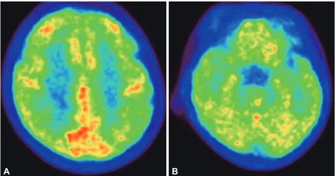
dL, and a glucose level of 118 mg/dL. The CSF was negative for HHV-6 DNA. After 4 weeks of anti-viral therapy, the neuro- logical symptoms were partially improved. However, the pa- tient was disoriented and complained of severe retrograde and anterograde amnesia. Eight weeks after BMT, her Mini-Mental State Examination score was 16; she was incorrect on the time and place orientation, serial 7, recall, interlocking pentagon, and language tests. She was even incorrect when asked if she had graduated from a graduate school. She could not remem- ber the names of her parents, her current address, the name of the high school she graduated from, or recent events a few days old. 18F-fluorodeoxyglucose (FDG) positron emission tomography (PET) performed 8 weeks after BMT indicated global hypometabolism in the bilateral medial temporal lobes and the fronto-parietal cortices (Fig. 2).
DISCUSSION
This is a rare and unusual case with atypical imaging find- ings on brain MRI and FDG-PET in a patient with HHV6 en- cephalitis. An MRI study performed in the acute stage revealed bilateral hippocampal atrophy. An FDG-PET study revealed global hypometabolism in the medial temporal lobes.
HHV6 encephalitis is not rare in immunocompromised pa- tients with hematopoietic stem-cell transplantation.9-11 After HHV6 was first isolated from patients with lymphoprolifera- tive disorders and acquired immunodeficiency syndrome in 1986,12 it came to be known to be commonly associated with encephalitis related to transplantation.5 HHV6 encephalitis in patients with BMT is thought to be due to reactivation of
Fig. 1. Brain MR images in the patient. A: An axial fluid-attenuated inversion recovery image demonstrates an atrophic change in the bilat- eral medial temporal lobes without abnormal high signal intensity. B: A coronal contrast-enhanced T1-weighted image shows an atrophic change in the bilateral medial temporal lobes without leptomeningeal enhancement. MR: magnetic resonance.
B A
Jihye Hwang et al.
Chronic Indolent HHV6 Encephalitis
90 Dement Neurocogn Disord 2016;15(3):88-91
HHV6 virus. HHV6 has been categorized into variants A and B.13 Primary HHV-6B infection is the cause of the common childhood illness roseola infantum,14 latent HHV-6B infection was well known the cause of HHV6 encephalitis in patients with BMT. Numerous such cases have been reported. Clinical- ly, HHV6 encephalitis usually develops 2–6 weeks after BMT.3,10 Like other members of the herpesviridae family, it causes typical limbic encephalitis. Thus, symptoms such as mental change and amnesia are common and may provide di- agnostic clues.3,10,11
CSF PCR is the most definitive and essential test for the di- agnosis of HHV6 encephalitis.6 HHV6 may be detectable in blood, as primary HHV6 infection during infancy results in generally mild, self-limited illnesses, and can be asymptomat- ic.15,16 Therefore, the detection of HHV6 DNA in the CSF by PCR is considered to be substantive evidence of active CNS infection in patients with BMT.6 In this case, the patient showed symptoms of typical limbic encephalitis with fever within 2 weeks of BMT. PCR analysis was positive for HHV6 DNA in the CSF. Serological tests and PCR for other infectious agents were all negative. We were thus able to confirm our diagnosis.
In this case, the brain MRI study revealed chronic atrophic changes in the bilateral medial temporal lobes, rather than ab- normal high signal intensity and leptomeningeal enhance- ment, which would suggest acute meningoencephalitis. Previ- ous studies have shown that patients with HHV6 encephalitis after BMT have typical limbic encephalitis features in brain MRIs. These include high signal intensity in bilateral or unilat- eral medial temporal lobes, including in the hippocampus and
the amygdala in T2/fluid-attenuated inversion recovery/diffu- sion weighted images.3,6,7 Some previous studies have reported HHV6 encephalitis cases where the hippocampal volume was notably reduced after treatment.3,17 In another study, MRI analysis in the late period showed severe atrophic changes in the affected bilateral medial temporal lobes.7 Usually, the atro- phic change of HHV6 encephalitis were presented at the time about 3–4 weeks after symptom onset. In the previous studies, they had described “early volume loss” as the atrophic change after 3 weeks or 26 days after symptom onset.6,7,17 Even consid- ering this, the atrophy of our case was observed at an earlier time. The localization of high signal intensity or atrophy to the medial temporal lobes correlates well with neurologic deficits in patients with HHV6 encepahlitis.18 The clinical association between the medial temporal lobes and the dysfunction of memory formation is well-documented.19-21 The severe retro- grade and anterograde amnesia present in this case also corre- late well with the patient’s medial temporal lobe atrophy. It is unclear whether the medial temporal lobe atrophy was the re- sult of the HHV6 encephalitis, associated finding, or incidental cooccurrence. However, considering the absence of significant previous medical history, her young age, and new symptom of severe retrograde and anterograde amnesia, the atrophic change on the patient’s MRI point to HHV6 encephalitis and not to another preexistent disease. HHV6 encephalitis in im- munocompromised patients is due to the reactivation of latent HHV6 infection. Thus, our imaging findings may be reflecting of the chronic indolent course of HHV6 encephalitis.22 Chron- ic latent HHV6 infection has been suggested to be present in
Fig. 2. 18F-Fludeoxyglucose-PET performed 8 weeks after transplantation shows global hypometabolism in the bilateral medial temporal lobes and the fronto-parietal cortices. A: Hypometabolism in fronto-parietal cortices. B: Hypometabolism in bilateral medial temporal lobes.
PET: positron emission tomography.
A B
www.dnd.or.kr 91
DND
other diseases, such as medial temporal epilepsy.23-25
An analysis of FDG-PET in our patient revealed global hy- pometabolism, in the medial temporal lobes in particular. In previous studies, late-phase FDG-PET findings after anti-viral therapy indicated hypometabolism in areas with intense FDG uptake in the acute stage, such as in the bilateral hippocampi and the amygdala.8,17 Thus, the FDG-PET findings in this case correlate well with previous studies and with the patient’s atro- phy. Other possible considering cause of bilateral medial tem- poral atrophy and global hypometabolism in this case is the effect of immunosuppressive drug. The AML patient in this case received BMT after 6 months after diagnosis, any other medications were not administered to her during the 6 months.
After BMT, cyclosporine and mycofenolate mofetil was ad- ministered as a prophylactic regimen of graft-versus-host dis- ease. But, we thought the 2 weeks of administration was too short period to lead brain atrophy.
This is an unusual case of HHV6 encephalitis with medial temporal lobe atrophy, as seen on brain MRI. Our findings suggest a chronic indolent course for HHV6. HHV6 encepha- litis cases are underestimated in the field of clinical neurology.
With improved efficacy in diverse treatment methods employ- ing transplantations in the hematology and oncology fields, neurologists should consider HHV-6 encephalitis as a differ- ential diagnosis for acute to subacute encephalopathy in pa- tients with transplantation.
Conflicts of Interest
The authors have no financial conflicts of interest.
REFERENCES
1. Yoshikawa T. Human herpesvirus 6 infection in hematopoietic stem cell transplant patients. Br J Haematol 2004;124:421-432.
2. Isaacson E, Glaser CA, Forghani B, Amad Z, Wallace M, Armstrong RW, et al. Evidence of human herpesvirus 6 infection in 4 immuno- competent patients with encephalitis. Clin Infect Dis 2005;40:890- 3. Seeley WW, Marty FM, Holmes TM, Upchurch K, Soiffer RJ, Antin 893.
JH, et al. Post-transplant acute limbic encephalitis: clinical features and relationship to HHV6. Neurology 2007;69:156-165.
4. Vu T, Carrum G, Hutton G, Heslop HE, Brenner MK, Kamble R.
Human herpesvirus-6 encephalitis following allogeneic hematopoiet- ic stem cell transplantation. Bone Marrow Transplant 2007;39:705- 5. Drobyski WR, Knox KK, Majewski D, Carrigan DR. Brief report: 709.
fatal encephalitis due to variant B human herpesvirus-6 infection in a bone marrow-transplant recipient. N Engl J Med 1994;330:1356- 1360.
6. Gorniak RJ, Young GS, Wiese DE, Marty FM, Schwartz RB. MR imaging of human herpesvirus-6-associated encephalitis in 4 patients with anterograde amnesia after allogeneic hematopoietic stem-cell transplantation. AJNR Am J Neuroradiol 2006;27:887-891.
7. Noguchi T, Mihara F, Yoshiura T, Togao O, Atsumi K, Matsuura T, et al. MR imaging of human herpesvirus-6 encephalopathy after hema- topoietic stem cell transplantation in adults. AJNR Am J Neuroradiol 2006;27:2191-2195.
8. Hubele F, Bilger K, Kremer S, Imperiale A, Lioure B, Namer IJ. Se- quential FDG PET and MRI findings in a case of human herpes virus 6 limbic encephalitis. Clin Nucl Med 2012;37:716-717.
9. Ogata M, Fukuda T, Teshima T. Human herpesvirus-6 encephalitis after allogeneic hematopoietic cell transplantation: what we do and do not know. Bone Marrow Transplant 2015;50:1030-1036.
10. Ogata M, Satou T, Kadota J, Saito N, Yoshida T, Okumura H, et al.
Human herpesvirus 6 (HHV-6) reactivation and HHV-6 encephalitis after allogeneic hematopoietic cell transplantation: a multicenter, prospective study. Clin Infect Dis 2013;57:671-681.
11. Ogata M, Kikuchi H, Satou T, Kawano R, Ikewaki J, Kohno K, et al.
Human herpesvirus 6 DNA in plasma after allogeneic stem cell transplantation: incidence and clinical significance. J Infect Dis 2006;193:68-79.
12. Salahuddin SZ, Ablashi DV, Markham PD, Josephs SF, Sturzenegger S, Kaplan M, et al. Isolation of a new virus, HBLV, in patients with lymphoproliferative disorders. Science 1986;234:596-601.
13. Dominguez G, Dambaugh TR, Stamey FR, Dewhurst S, Inoue N, Pellett PE. Human herpesvirus 6B genome sequence: coding content and comparison with human herpesvirus 6A. J Virol 1999;73:8040- 8052.
14. Yamanishi K, Okuno T, Shiraki K, Takahashi M, Kondo T, Asano Y, et al. Identification of human herpesvirus-6 as a causal agent for ex- anthem subitum. Lancet 1988;1:1065-1067.
15. Zerr DM, Meier AS, Selke SS, Frenkel LM, Huang ML, Wald A, et al. A population-based study of primary human herpesvirus 6 infec- tion. N Engl J Med 2005;352:768-776.
16. De Bolle L, Naesens L, De Clercq E. Update on human herpesvirus 6 biology, clinical features, and therapy. Clin Microbiol Rev 2005;18:
217-245.
17. Wainwright MS, Martin PL, Morse RP, Lacaze M, Provenzale JM, Coleman RE, et al. Human herpesvirus 6 limbic encephalitis after stem cell transplantation. Ann Neurol 2001;50:612-619.
18. Provenzale JM, van Landingham K, White LE. Clinical and imaging findings suggesting human herpesvirus 6 encephalitis. Pediatr Neu- rol 2010;42:32-39.
19. Milner B, Klein D. Loss of recent memory after bilateral hippocam- pal lesions: memory and memories-looking back and looking for- ward. J Neurol Neurosurg Psychiatry 2016;87:230.
20. Scoville WB, Milner B. Loss of recent memory after bilateral hippo- campal lesions. 1957. J Neuropsychiatry Clin Neurosci 2000;12:103- 21. Scoville WB, Milner B. Loss of recent memory after bilateral hippo-113.
campal lesions. J Neurol Neurosurg Psychiatry 1957;20:11-21.
22. Yamane A, Mori T, Suzuki S, Mihara A, Yamazaki R, Aisa Y, et al.
Risk factors for developing human herpesvirus 6 (HHV-6) reactiva- tion after allogeneic hematopoietic stem cell transplantation and its association with central nervous system disorders. Biol Blood Mar- row Transplant 2007;13:100-106.
23. Millichap JJ, Millichap JG. Role of HHV-6B Infection in Mesial Temporal Lobe Epilepsy. Pediatr Neurol Briefs 2015;29:40.
24. Kawamura Y, Nakayama A, Kato T, Miura H, Ishihara N, Ihira M, et al. Pathogenic role of human herpesvirus 6B infection in mesial tem- poral lobe epilepsy. J Infect Dis 2015;212:1014-1021.
25. Leibovitch EC, Jacobson S. Human herpesvirus 6 as a viral trigger in mesial temporal lobe epilepsy. J Infect Dis 2015;212:1011-1013.