Received on July 24, 2009. Revised on August 7, 2009. Accepted on August 18, 2009.
*Corresponding Author. Tel: 82-51-890-6434; Fax: 82-51-890-6004; E-mail: [email protected] Keywords: TLR2, T cell co-stimulation, CD4 T cell, CD8 T cell
Expression and Function of TLR2 on CD4 Versus CD8 T Cells
Sun-Mi Lee1, Young-Don Joo2 and Su-Kil Seo1*
Departments of 1Microbiology and Immunology, 2Hemato/Oncology, College of Medicine, Inje University, Busan 614-735, Korea
Background: Toll-like receptors (TLRs) play a fundamental role in innate immunity through their capacity to recognize pathogen-associated molecular patterns. Also, TLRs that are expressed in T cells are reported to function as co-stim- ulatory receptors. However, the functional capacity of TLRs on CD4 T and CD8 T cells has not been directly compared.
Here we compared CD4 and CD8 T cell responses to TLR2 ligand plus TCR-mediated stimulation. Methods: TLR2 ex- pression was analyzed on T cell subsets under naïve and al- loantigen-primed conditions. We analyzed the effects of TLR2 co-stimulation on proliferation and survival of T cell subsets in vitro when stimulated with soluble anti-CD3 in the presence or absence of synthetic ligand Pam3CSK4. Results:
TLR2 expression on CD8 T cells was induced following acti- vation; this expression was much higher than on CD4 T cells.
Thus, the molecule was constitutively expressed on Listeria- specific memory CD8 T cells. Based on these expression lev- els, proliferation and survival were markedly elevated in CD8 T cells in response to the TLR2 co-stimulation by Pam3CSK4
compared with those in CD4 T cells. Conclusion: Our data show that TLR2 co-stimulation is more responsible for pro- liferation and survival of CD8 T cells than for that of CD4 T cells.
[Immune Network 2009;9(4):127-132]
INTRODUCTION
Toll-like receptors (TLRs) are primary sensor molecules that play an integral role in innate immunity via their capacity to recognize pathogen-associated molecular patterns that allow the detection of infection and inflammation (1). TLR stimula- tion of dendritic cells (DCs) and macrophages promotes the production of pro-inflammatory cytokines and the up-regula- tion of MHC and co-stimulatory molecules, which leads to the induction of T cell-mediated adaptive immune responses (2).
Although much of our knowledge of TLR function in the immune system comes from the study of innate immune cells, these molecules also are expressed in T cells. Early studies of TLRs in T cells have been conducted with CD4 T cells.
Naïve human CD4 T cells express TLR2 after activation by TCR stimulation and TLR2 functions as a co-stimulatory receptor. Moreover, TLR2 also participates in the generation and maintenance of CD4 T cell memory (3). TLR3 and TLR9 ligand directly deregulate Bcl-xL in CD4 T cells, thus promot- ing survival (4). CpG DNA-mediated co-stimulation in CD4 T cells proceeds through the MyD88-dependent PI-3 kinase signaling pathway (5). According to a recent study, TLR2 stimulation activates Th1 effector cells without TCR stim- ulation through the enhanced activation of MAPKs. In con- trast, no TLR affects the function of Th2 effector cells (6).
Several studies have reported the co-stimulatory effects of TLR on CD8 T cells. TLR2 engagement on CD8 T cells de- creases the activation threshold for co-stimulatory signals de- livered by APC (7). Quigley et al. showed that direct TLR2- MyD88 signaling in CD8 T cells plays a critical role in clonal expansion and memory formation against vaccinia viral (VV) infection (8). It has been also reported that MyD88-dependent signals are critical for survival of Lymphocytic choriomeningi- tis virus (LCMV)-specific CD8 T cells and sustained accumu- lation for viral clearance (9). Furthermore, TLR2 engagement on cytotoxic T-lymphocytes (CTL) augments antitumor activity against established B16 melanoma tumors (10).
Certain co-stimulatory molecules on activated T cells are known to primarily be involved in either the CD4 or CD8 T cell subset. For example, 4-1BB is preferentially involved in CD8 T cell-mediated immune responses (11). In the pres- ent study, we compared the expression and function of TLR2 on CD4 versus (vs.) CD8 T cells, which have not been di- rectly compared yet. However, we found that TLR2 co-stim-
ulation is more responsible for CD8 T cells than for CD4 T cells.
MATERIALS AND METHODS Mice
Female B6 (H-2b) and Balb/c (H-2d) mice were purchased from Orient Bio Inc. (Seoul, Korea). TLR2-/- (H-2b) mice were provided by S. Akira (Osaka University, Osaka, Japan). All mice were used for the experiments at the age of 8∼10 weeks.
Antibodies and reagents
The following antibodies were purchased from e-Bioscience (San Diego, CA) for flow cytometry: FITC-conjugated an- ti-mouse CD3 (145-2C11), TLR2 (6C2), and H-2b (AF6-88.5);
PE-conjugated anti-mouse CD4 (GK1.5), CD8 (53-6.7), TLR2 (6C2), Bcl-2 (3F11), Bcl- xL (7B2.5), and IFN-γ (XMG1.2);
PE-Cy5-conjugated anti-mouse CD4 (GK1.5) and CD8 (53- 6.7); purified anti-CD16/32 (2.4G2) and purified anti-TLR2 (T2.5). LLO91-99 pentamer was obtained from ProImmune (Oxford, UK). Purified anti-mouse CD3 (145.2C11) was ob- tained from BD Biosciences (San Jose, CA). Pam3CSK4 was purchased from Invivogen (Carlsbad, CA).
Cell preparation, culture, and in vitro proliferation assay
Naïve T cells were isolated from spleen and lymph nodes of B6 using anti-CD90 (Thy1.2) magnetic beads (Miltenyi Biotech, Auburn, CA) after depletion of CD25+ cells. The cells were >97% CD3 T cells with a naïve phenotype. To prepare the CD4 T and CD8 T cells, CD11c+ and CD25+ cells were first depleted using anti-CD11c and -CD25 magnetic beads to remove Treg and lymphoid dendritic cells and then the cells were isolated using anti-CD4 or -CD8 magnetic beads, respectively. The cells were >97% CD3+ CD4+ or CD8+ T cells with a naïve phenotype. 2×10⁵ cells were stimulated with soluble anti-CD3 (0.5 μg/ml) or pulsed with Pam3CSK4
(2 μg/ml) or LPS (2 μg/ml) for 64 h at 37oC in a 5% CO2 environment. Proliferation was measured in triplicate cultures by the incorporation of [3H]thymidine (1 μCi/well, Amers- ham Pharmacia) during the last 12 h of culture. The in- corporation of [3H]thymidine was measured with a β-counter (Wallac, Torrance, CA). For blocking analysis, purified cells were pretreated with anti-TLR2 antibody (2 μg/ml, T2.5) be- fore the stimulation.
In vivo generation of alloantigen activated T cells Responder T cells were purified from the spleen and lymph nodes of B6 (H-2b) mice using the anti-CD90 microbead sepa- ration system (Miltenyi Biotec). Cells (1×10⁷) were sus- pended in PBS and transferred into lethally irradiated (1,000 cGy) Balb/c (H-2d) recipients via tail vein. Recipient spleno- cytes were isolated at 4 days after transplant, and cells were identified as donor T cells with anti-H-2b and -CD4 or -CD8 mAb and analyzed by flow cytometry.
In vivo generation of Listeria-specific memory CD8 T cells
Balb/c mice were infected intravenously (i.v.) with 3000 col- ony-forming units (CFU) of live L. monocytogenes. On day 25, the mice were reinfected with 5000 CFU of live bacteria intraperitoneally (p. i.); 5 days later, LLO91-99-specific CD8 T cells were determined using LLO91-99 pentamer.
Flow cytometry
To measure the expression of TLR2, cells were first incubated with FcR blocker (2.4G2) to block nonspecific antibody bind- ing and then stained with PE-anti-TLR2 and PE-Cy5-anti-CD4 or CD8 and analyzed on a FACSCalibur flow cytometer (BD Biosciences) using the CellQuest software. To measure cell proliferation, cultured cells were treated with BrdU (2 μg, Sigma) for 1 h and washed with PBS. The cells were fixed, permeabilized, treated with DNase I, and stained with FITC-anti-BrdU using a BrdU Flow kit according to the manu- facturer’s instructions. To analyze cell death, cells were stained with FITC-annexin V and 7-AAD. To stain Bcl-2 and Bcl-xL, the cells were fixed, permeabilized, and stained with PE-anti-Bcl-2 or -Bcl-xL mAb. To stain intracellular IFN-γ, cells were fixed, permeabilized using the Cytofix/Cytoperm kit (BD Bioscience) according to the manufacturer’s in- structions, and incubated with PE-anti-IFN-γ mAb.
RESULTS
TLR2 expression is preferentially induced on CD8 T cells vs. CD4 T cells
To assess the expression pattern of TLR2 on CD4 vs. CD8 T cells, we performed flow cytometric analysis on naïve and alloantigen-activated T cells. TLR2 was not expressed on ei- ther naïve CD4 or CD8 T cells before adoptive transfer into allogeneic recipient. Four days after transfer, alloantigen-acti- vated responder T cells induced TLR 2 expression. However,
Figure 1. Expression of TLR2 on CD4 and CD8 T cells. (A) T cells were isolated from spleen and lymph nodes of naïve B6 mice as described in Materials and Methods and stained with PE-Cy5-anti-CD4 or -CD8 and PE-anti-TLR2, and then analyzed by flow cytometry. The cells were adoptively transferred into lethally irradiated (850 cGy) Balb/c recipient. Recipient splenocytes were isolated on day 4 and stained with mAbs against H-2d, TLR2, and CD4 or CD8. The histo- gram for TLR2 expression was gated on H-2b+CD4+ or H-2b+CD8+ cells. (B) Balb/c mice were infected with L. monocytogenes and reinfected on day 25 p. i. with 5000 CFU. Splenocytes were isolated on day 5, stained with FITC-anti-CD8 and PE-LLO91-99 pentamer, and analyzed by flow cytometry.
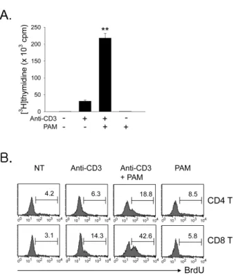
Figure 2. TLR2 ligand co-stimulates T cell proliferation in the total T, CD4, and CD8 T cells. Total T cells were isolated as described in Materials and Methods and incubated with anti-CD3s in the presence or absence of TLR2 ligand Pam3CSK4 (PAM) for 64 h. (A) The T cell proliferation was determined via [3H]thymidine incorporation. Values are means and standard deviations of data from three independent experiments. **p<0.001, anti-CD3 vs. anti-CD3+PAM. (B) Cultured cells were treated with BrdU (2 μg) for the last 1 h and stained with PE-anti-CD4 or -CD8 mAb. The cells were then stained for inc- orporated BrdU and analyzed by flow cytometry. The histogram for BrdU incorporation was gated on CD4+ or CD8+ cells.
the expression levels were much higher on CD8 T cells (45.7±4.8%) than on CD4 T cells (5.4±3.2%) (Fig. 1A).
Furthermore, Listeria-specific memory CD8 T cells constitu- tively expressed TLR2 (Fig. 1B). These data indicate that TLR2 is preferentially expressed on CD8 T cells following activa- tion.
TLR2 co-stimulation dominantly enhances CD8 T cell expansion more than CD4 T cell expansion To test the effect of TLR2 co-stimulation on the proliferation of total T cells or CD4 vs. CD8 T cells, T cells were isolated as described in Materials and Methods and incubated with soluble anti-CD3 (anti-CD3s) in the presence or absence of TLR2 ligand Pam3CSK4. First, we observed that the co-stim- ulation of total T cells with Pam3CSK4 led to a seven-fold en-
hancement of anti-CD3-induced proliferation (Fig. 2A). We next evaluated the ratio of CD4 vs. CD8 T cells in the pro- liferative capacity of the total T cell population that was en- hanced by TLR2 co-stimulation, and performed the BrdU in- corporation assay. Fig. 2B shows that there were more CD8 T cells than CD4 T cells in the increased proliferative capacity of total T cells. To further confirm the direct effect of TLR2 signaling on T cell subsets, we isolated highly purified pop- ulations of naïve CD4 or CD8 T cells after the depletion of CD11c+ and CD25+ cells to remove contaminating lymphoid DCs and natural Treg, respectively (>97% purity). T cell sub- sets were then incubated with anti-CD3s in the presence or absence of Pam3CSK4. The co-stimulation of CD8 T cells with Pam3CSK4 led to a 11-fold enhancement of anti-CD3-induced proliferation. However, the CD4 cell proliferation was in-
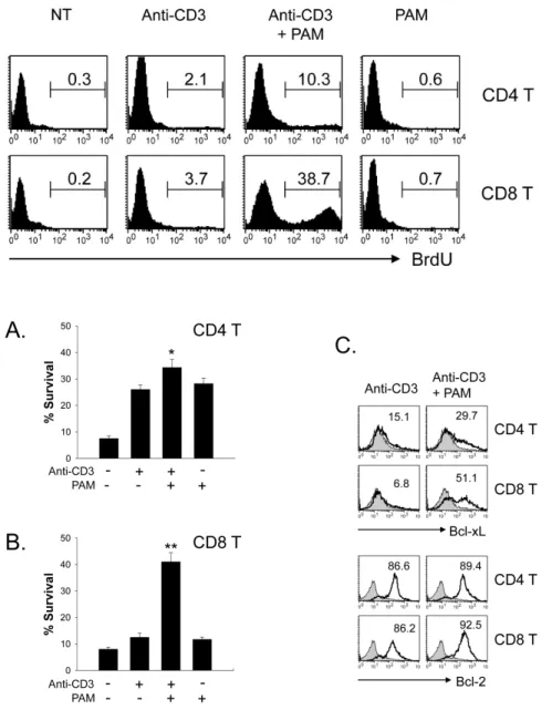
Figure 3. Effect of TLR2 co-stimulation on proliferation of CD4 T cell vs. CD8 T cells.
CD4 and CD8 T cells were isolated as described in Materials and Methods and incubated with anti-CD3s in the presence or absence of TLR2 ligand PAM for 64 h. The cultured cells were treated with BrdU (2 μg) for the last 1 h, stained for incorporated BrdU, and analyzed by flow cytometry.
Figure 4. Effect of TLR2 co-stimulation on the survival of CD4 vs. CD8 T cells. Isolated CD4 (A) and CD8 T cells (B) were incubated with anti-CD3s in the presence or absence of TLR2 ligand PAM for 64 h. The cells were harvested, stained with FITC-Annexin-V and 7-AAD, and then analyzed by flow cytome- try. %Survival was determined on annexin V-7-AAD-cells. Values are means and stan- dard deviations of data from three indep- endent experiments. *p<0.05, **p<0.001, anti-CD3 vs. anti-CD3+PAM. (C) Cells were harvested and stained for intracellular Bcl-xL or Bcl-2.
creased five-fold (Fig. 3). Taken together, these results in- dicate that TLR2 co-stimulation is preferentially involved in CD8 T cell expansion rather than CD4 T cell expansion.
TLR2 co-stimulation elevates CD8 T cell survival more strongly than CD4 T cell survival
To further assess the effect of TLR2 co-stimulation on the sur- vival of CD4 vs. CD8 T cells, isolated naïve CD4 and CD8 T cells were stimulated with anti-CD3s in the presence or ab- sence of Pam3CSK4. Survival was then detected by annexin V plus 7-AAD staining at 64 h following activation. Pam3CSK4
increased the activated CD8 T cell survival from 12% to 40%
(Fig. 4B). However, the CD4 T cell survival was increased from 25% to 35% by TLR2 co-stimulation (Fig. 4A). Members of the Bcl family are reported to be key mediators of acti- vated T cell survival following TLR2 co-stimulation (4). There- fore, we compared the levels of these molecules following TLR2 ligand treatment of CD4 or CD8 T cells. We observed more significant increases in Bcl-xL protein in Pam3CSK4- treated CD8 T cells than in CD4 T cells. However, Bcl-2 pro- tein levels were similar in CD4 and CD8 T cells (Fig. 4C).
These data indicate that TLR2 co-stimulation is preferentially
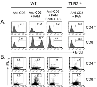
Figure 5. Specificity of TLR2 co-stimulation. CD4 and CD8 T cells were isolated from normal or TLR2-/- mice and incubated with anti-CD3s in the presence or absence of TLR2 ligand PAM for 64 h.
Other cells were pretreated with purified anti-TLR2 mAb (2 μg/ml) before the stimulation. (A) Cultured cells were treated with BrdU (2 μg) for the last 1 h, stained for incorporated BrdU, and then analyzed by flow cytometry. (B) Cultured cells were treated with brefeldin A for the last 4 h and stained for intracellular IFN-γ.
involved in CD8 T cell survival versus that in CD4 T cells.
Thus, it is associated with specific Bcl-xL up-regulation.
Specificity of TLR2 co-stimulation
To exclude the possibility that the effects caused by Pam3CSK4 were non-specific, we tested the assay using an- ti-TLR2 monoclonal antibody, which was added to the culture before the stimulation. As shown in Fig. 5, the enhanced pro- liferation with Pam3CSK4 was completely reversed in both CD4 and CD8 T cells by anti-TLR2 treatment as was IFN-γ production. In addition, T cells purified from TLR2-/- mice ex- hibited no response to Pam3CSK4 in terms of either the pro- liferation (Fig. 5A) or the IFN-γ production (Fig. 5B), in- dicating that Pam3CSK4 acts through TLR2-dependent signal- ing pathways.
DISCUSSION
TLR in T cells can function as a co-stimulatory molecule for both CD4 and CD8 T cell activation (12). In this study, we have confirmed that the TLR2 ligand Pam3CSK4 provides a di- rect potent co-stimulatory effect on TCR-mediated T cell
proliferation. However, we found that TLR2 co-stimulation was biased toward CD8 T cells rather than CD4 T cells. For instance, the addition of Pam3CSK4 increased the anti-CD3- mediated proliferation of total T cells by 7-fold (Fig. 2A). In this increased proliferative capacity, CD8 T cells were found in a higher proliferative ratio than CD4 T cells (Fig. 2B), which was confirmed on an isolated subset of T cells (Fig.
3). We also observed that TLR2 co-stimulation promoted the survival of CD8 T cells more than that of CD4 T cells (Fig.
4A, B). This was caused not by Bcl-2 but by increased Bcl- xL (Fig. 4C). In fact, the different sensitivity to TLR2 co-stim- ulation is probably related to the expression levels on CD4 versus CD8 T cells. The surface expression was more highly induced following activation on CD8 T cells compared with that on CD4 T cells (Fig. 1A). Taken together, these results indicate that CD8 T cells preferentially respond to TLR2 co-stimulation.
CD8 T cells are critical for prevention of acute and chronic viral infections (13) as well as for tumor eradication (14). In recent studies, the physiological significance of TLR2 on CD8 T cell-mediated effector immune responses has been repor- ted. Quigley et al. reported that TLR2-/- and MyD88-/- CD8 T cells had severely diminished clonal expansion in response to vaccinia viral (VV) infection, which involved the TLR2 co-stimulation on VV-specific CD8 T cells (8). The study also reported that long-lived memory CD8 T cells could not devel- op in the absence of direct TLR2-MyD88 signaling. We also observed that TLR2 is constitutively expressed in Listeria-spe- cific memory CD8 T cells (Fig. 1B). Indeed, rapid Listeria- specific memory CD8 T cell formation is affected by primary infection (15). It may be related to TLR2 expression that is induced on Listeria-specific CD8 T cells during the primary infection time. The TLR2 expression may affect rapid ex- pansion of the memory CD8 T cells during the secondary in- fection period.
Our data also indicated that TLR2 co-stimulation decreased the threshold for antigen-specific signaling through TCR. We stimulated T cells with soluble anti-CD3 to provide weak TCR-mediated activation. Although, under these conditions, TLR2 signaling effectively elicited the expansion and IFN-γ production of CD8 T cells (Fig. 5), it can be speculated that TLR2 signaling affects autoreactive CD8 T cell responses.
Autoreactive T cells recognize autoantigens, which are basi- cally presented by immature DCs that give feeble TCR signal- ing, resulting in ignorance or anergy (16). Under pathogen infection conditions, TLR2 signaling enhances the direct path-
way of autoreactive T cell activation by co-stimulation as well as the indirect pathway by induction of DC maturation. A number of animal models for autoimmune disease probably involve TLR signaling in their pathogenesis (17,18). Our data indicate that promoting the expansion and the effector func- tion of CD8 T cells by TLR2 signaling was completely re- versed by the anti-TLR2 mAb, T2.5 (Fig. 5), the therapeutic activity of which has been reported in the sepsis model (19).
Therefore, T2.5 might be become a valuable therapeutic agent for CD8 T cell-mediated pathological conditions in the presence of TLR ligand.
Although it has been recently suggested that TLR2 could be particular in its ability to co-stimulate CD4 and CD8 T cells, in this present study, we find that its dominant effect appears to be the regulation of CD8 T cell activation. These observations suggest a potential therapeutic role for this mol- ecule in the management of cancer and chronic infectious dis- eases as well as autoimmune diseases.
ACKNOWLEDGEMENTS
This work was supported by the Korea Research Foundation Grant funded by the Korean Government (MOEHRD, Basic Research Promotion Fund) (KRF-2006-003-E00098 to SKS), and by the Korea Science and Engineering Foundation (KOSEF) grant funded by the Korean Government (MEST) (No. R01-2008-000-20238-0 to YDJ).
CONFLICTS OF INTEREST
The authors have no financial conflict of interest.
REFERENCES
1. Takeda K, Kaisho T, Akira S: Toll-like receptors. Annu Rev Immunol 21;335-376, 2003
2. Medzhitov R: Toll-like receptors and innate immunity. Nat Rev Immunol 1;135-145, 2001
3. Komai-Koma M, Jones L, Ogg GS, Xu D, Liew FY: TLR2 is expressed on activated T cells as a costimulatory recep- tor. Proc Natl Acad Sci USA 101;3029-3034, 2004 4. Gelman AE, Zhang J, Choi Y, Turka LA: Toll-like receptor
ligands directly promote activated CD4+ T cell survival. J Immunol 172;6065-6073, 2004
5. Gelman AE, LaRosa DF, Zhang J, Walsh PT, Choi Y, Sunyer JO, Turka LA: The adaptor molecule MyD88 activates PI-3
kinase signaling in CD4+ T cells and enables CpG oligo- deoxynucleotide-mediated costimulation. Immunity 25;783- 793, 2006
6. Imanishi T, Hara H, Suzuki S, Suzuki N, Akira S, Saito T:
Cutting edge: TLR2 directly triggers Th1 effector functions.
J Immunol 178;6715-6719, 2007
7. Cottalorda A, Verschelde C, Marçais A, Tomkowiak M, Musette P, Uematsu S, Akira S, Marvel J, Bonnefoy-Berard N: TLR2 engagement on CD8 T cells lowers the threshold for optimal antigen-induced T cell activation 36;1684-1693, 2006
8. Quigley M, Martinez J, Huang X, Yang Y: A critical role for direct TLR2-MyD88 signaling in CD8 T-cell clonal ex- pansion and memory formation following vaccinia viral infection. Blood 113;2256-2264, 2009
9. Rahman AH, Cui W, Larosa DF, Taylor DK, Zhang J, Goldstein DR, Wherry EJ, Kaech SM, Turka LA: MyD88 plays a critical T cell-intrinsic role in supporting CD8 T cell expansion during acute lymphocytic choriomeningitis virus infection. J Immunol 181;3804-3810, 2008
10. Asprodites N, Zheng L, Geng D, Velasco-Gonzalez C, Sanchez-Perez L, Davila E: Engagement of Toll-like re- ceptor-2 on cytotoxic T-lymphocytes occurs in vivo and augments antitumor activity. FASEB J 22;3628-3637, 2008 11. Shuford WW, Klussman K, Tritchler DD, Loo DT, Chalupny
J, Siadak AW, Brown TJ, Emswiler J, Raecho H, Larsen CP, Pearson TC, Ledbetter JA, Aruffo A, Mittler RS: 4-1BB cos- timulatory signals preferentially induce CD8+ T cell pro- liferation and lead to the amplification in vivo of cytotoxic T cell responses. J Exp Med 186;47-55, 1997
12. Marsland BJ, Kopf M: Toll-like receptors: paving the path to T cell-driven autoimmunity? Curr Opin Immunol 19;611- 614, 2007
13. Wong P, Pamer EG: CD8 T cell responses to infectious pathogens. Annu Rev Immunol 21;29-70, 2003
14. Dougan M, Dranoff G: Immune therapy for cancer. Annu Rev Immunol 27;83-117, 2009
15. Wong P, Lara-Tejero M, Ploss A, Leiner I, Pamer EG: Rapid development of T cell memory. J Immunol 172;7239-7245, 2004
16. Steinman RM, Hawiger D, Nussenzweig MC: Tolerogenic dendritic cells. Annu Rev Immunol 21;685-711, 2003 17. Kerfoot SM, Long EM, Hickey MJ, Andonegui G, Lapointe
BM, Zanardo RC, Bonder C, James WG, Robbins SM, Kubes P: TLR4 contributes to disease-inducing mechanisms result- ing in central nervous system autoimmune disease. J Immu- nol 173;7070-7077, 2004
18. Lee EK, Kang SM, Paik DJ, Kim JM, Youn J: Essential roles of toll-like receptor 4 signaling in arthritis induced by type II collagen antibody and LPS. Int Immunol 17;325-333, 2005 19. Meng G, Rutz M, Schiemann M, Metzger J, Grabiec A,
Schwandner R, Luppa PB, Ebel F, Busch DH, Bauer S, Wagner H, Kirschning CJ: Antagonistic antibody prevents toll-like receptor 2-driven lethal shock-like syndromes. J Clin Invest 113:1473-1481, 2004