Endocrinol Metab 2015;30:488-493
http://dx.doi.org/10.3803/EnM.2015.30.4.488 pISSN 2093-596X · eISSN 2093-5978
Original Article
Subclinical Atherosclerosis in Patients with Cushing Syndrome: Evaluation with Carotid Intima-Media Thickness and Ankle-Brachial Index
Luigi Petramala1, D’Elia Lorenzo1, Gino Iannucci1, Antonio Concistré1, Laura Zinnamosca1, Cristiano Marinelli1, Giuseppe De Vincentis2, Antonio Ciardi3, Giorgio De Toma3, Claudio Letizia1
1Department of Internal Medicine and Medical Specialties, 2Section Nuclear Medicine, Department of Radiology, 3Department of Surgery, “P. Valdoni”, University of Rome “Sapienza”, Rome, Italy
Background: Cushing syndrome (CS) has been described as a killing disease due its cardiovascular complications. In fact, chronic cortisol excess leads to a constellation of complications, including hypertension, hyperglycemia, adiposity, and thrombo- embolism. The main vascular alteration associated with CS is atherosclerosis.
Methods: Aim of this study was to analyze carotid intima-media thickness (cIMT) and ankle-brachial index (ABI), two surrogate markers of subclinical atherosclerosis in a consecutive series of CS patients, compared to patients with essential hypertension (EH) and health subjects (HS).
Results: Patients with CS showed a significant increase (P<0.05) of cIMT (0.89±0.17 mm) compared to EH (0.81±0.16 mm) and HS (0.75±0.4 mm), with a high prevalence of plaque (23%; P<0.03). Moreover, CS patients showed a mean ABI values (1.07±0.02) significantly lower respect to HS (1.12±0.11; P<0.05), and a higher percentage (20%) of pathological values of ABI (≤0.9; P<0.03).
Conclusion: In conclusion, we confirmed and extended the data of cIMT in CS, and showed that the ABI represent another sur- rogate marker of subclinical atherosclerosis in this disease.
Keywords: Cushing syndrome; Subclinical atherosclerosis; Carotid intima-media thickness; Ankle brachial index
INTRODUCTION
Cushing syndrome (CS) represents the patter of symptoms and signs caused by prolonged exposure of cortisol excess [1-3].
CS is relatively rare, most common in adults between the ages of 20 and 50, affecting approximately 2 to 3 million people in each year [1]. The CS is secondary to an adrenocorticotrophin (ACTH)-secreting pituitary tumor (ACTH-dependent CS) in
around 70%, a cortisol-secreting adrenal lesion (ACTH-inde- pendent CS) in 15% to 20%, and an ACTH-secreting extrapitu- itary tumor (ectopic CS) in 10% to 15% of the cases [4-6].
Excess cortisol levels determine adverse clinical features in- cluding central adiposity, pigmented striae, muscle weakness, and mood disturbance. CS is associated with hypertension, im- paired glucose tolerance and diabetes, and an increase of car- diovascular disease [6]. The estimated standardized all-cause
Received: 20 February 2015, Revised: 27 March 2015, Accepted: 18 June 2015 Corresponding author: Claudio Letizia
Department of Internal Medicine and Medical Specialties, University of Rome
“Sapienza”, Policlinico Umberto I, Viale del Policlinico 155, Rome 00165, Italy Tel: +39-6-49970944, Fax: +39-6-49970944, E-mail: [email protected]
Copyright © 2015 Korean Endocrine Society
This is an Open Access article distributed under the terms of the Creative Com- mons Attribution Non-Commercial License (http://creativecommons.org/
licenses/by-nc/3.0/) which permits unrestricted non-commercial use, distribu- tion, and reproduction in any medium, provided the original work is properly cited.
pISSN 2093-596X · eISSN 2093-5978
mortality ratio in patients with active CS is 2 to 4 times higher than the general population [7]. In particular, cardiovascular complications associated with CS include coronary artery dis- ease, stroke and congestive heart failure, which significantly increase the mortality rate [3,5,8]. In particular, the main vascu- lar alteration associated with CS is arterial atherosclerosis [9].
Increased carotid intima-media thickness (cIMT) has been proposed as signal of subclinical atherosclerosis [10]. More- over, ankle-brachial index (ABI) has been reported another surrogate marker of carotid or coronary atherosclerosis and predictor of features ischemic events [11].
The aim of this study was to analyze cIMT and ABI, two surrogate markers of atherosclerosis, in a consecutive series of CS patients and compare them with essential hypertension (EH) patients and health subjects (HS).
METHODS
The study was carried out under clinical conditions, and we en- rolled 30 consecutive patients with CS: 20 women and 10 men of mean age 54.9±11.8 (SD). Twelve patients had ACTH-de- pendent CS and 18 had ACTH- independent CS. The CS diag- nosis was based on the clinical signs and symptoms (central re-
distribution of fat, hypertension, osteoporosis, muscle weak- ness), hormonal data and imaging tests (pituitary magnetic res- onance imaging, adrenal computed tomography, adrenal scinti- scan). Endocrine tests for CS showed lack diurnal rhythm of serum cortisol levels (>5 μg/dL at midnight), no suppression of serum cortisol (>1.8 μg/dL) levels after a low-dose (1 mg) dexamethasone suppression test, and increased 24-hour urinary free cortisol excretion. Measurement of plasma ACTH differ- entiated ACTH dependent CS (ACTH ≥10 pg/mL) from ACTH-independent CS (<10 pg/mL). In these patients, possi- ble coexistence of hyperaldosteronism was excluded from the study population by screening test using aldosterone (ng/dL) to renin (ng/mL/hr) ratio less than 30.
Thirty-five age-matched EH patients (49.2±11.3 years) and 30 HS (46.3±19.3 years) served as controls (Table 1). The di- agnosis of EH was made by exclusion of the common forms of secondary hypertension (primary aldosteronism, pheochromo- cytoma, renoparenchymal disease, and renovascular stenosis).
Exclusion criteria for CS patients and controls included: car- diovascular disease defined as history of previous stroke or es- tablished cardiovascular disease or symptomatic peripheral or carotid artery disease, impaired kidney formation (glomerular filtration rate ≤60 mL/min/1.73 m2) and chronic inflammation.
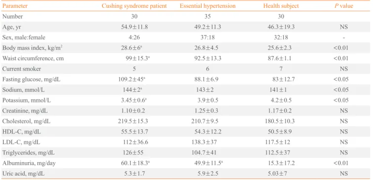
Table 1. Anthropometric and Laboratory Data in All Groups Study
Parameter Cushing syndrome patient Essential hypertension Health subject P value
Number 30 35 30
Age, yr 54.9±11.8 49.2±11.3 46.3±19.3 NS
Sex, male:female 4:26 37:18 32:18 -
Body mass index, kg/m2 28.6±6a 26.8±4.5 25.6±2.3 <0.01
Waist circumference, cm 99±15.3a 92.5±13.3 87.6±1.1 <0.01
Current smoker 5 6 7 NS
Fasting glucose, mg/dL 109.2±45a 88.1±6.9 83±12.7 <0.05
Sodium, mmol/L 144±2a 143±2 141±1 <0.05
Potassium, mmol/L 3.45±0.6a 3.9±0.5 4.2±0.5 <0.05
Creatinine, mg/dL 1.10±0.2 1.25±0.3 1.17±0.2 NS
Cholesterol, mg/dL 219.5±15.3 210.7±9.5 180.5±10.3 NS
HDL-C, mg/dL 55.5±13.7 54.3±12.2 50.5±8.9 NS
LDL-C, mg/dL 112±36.6 138.3±37 117.5±12 NS
Triglycerides, mg/dL 126±55 104.7±41 112.5±37 NS
Albuminuria, mg/day 60.1±18.3a 49.9±11.5a 15.3±17.2 <0.01
Uric acid, mg/dL 5.3±1.7 5.9±2.5 5.03±7 NS
Values are expressed as mean±SD.
NS, not significant; HDL-C, high density lipoprotein cholesterol; LDL-C, low density lipoprotein cholesterol.
aP<0.01 vs. health subject.
Arterial hypertension was defined as systolic blood pressure (SBP) >140 mm Hg and/or diastolic blood pressure >90 mm Hg, often 3 averaged blood pressure (BP) measurements or as receiving antihypertensive treatment. All subject underwent 24-hour ambulatory blood pressure monitoring.
Informed consent was obtained from all subjects and the study was performed in accordance with the Declaration of Helsinki.
Measurement of carotid intima-media thickness
A Hewlett-Packard Sonor 5500 Ultrasound system (Hewlett- Packard, Andover, MA, USA), equipped with a 3.11 MHz real- time B-mode scanner was used for the evaluation. Imaging of the right common carotid artery (CCA) was performed with the subjects turning their head 45° to the left. The high-resolution images were analyzed to calculate cIMT, defined of thickness of the vascular intima-media complex obtained in five consec- utive regions of the wall of the CCA, every 4 to 5 mm begin- ning close to the bifurcation. The value attributed to each sub- ject was the average value among the cIMT measurement, five from the left and five from the right carotid artery. Intra- and interobserved variabilities for cIMT were 4.6±0.4 and 5.2±
0.3, respectively.
Mean common carotid diameter was defined as the line identifying the media-adventitia interface in the near to the far wall calculated automatically by averaging measurements at 0.1 intervals of 1 cm.
Ankle-brachial index
For all subjects we measured ABI after a 5 minutes rest in the supine position. The ABI was determined using automated os- cillometric measurement BOSO-ABI system neo (Bosch+
Sohn GmbH U. Co. KG, Jungingen, Germany), that allows si- multaneous arm-leg BP measurements. This validated device can determine ABI accurately and significantly faster than with the traditional method. Moreover, it is much less influenced by the observed. All the measurements were performed in each subject included in the study by only personal specifically trained for this purpose.
Statistical analysis
Statistical analysis was performed by using SigmaStat program (Jandel Corp., Las Vegas, NV, USA). Data were expressed as mean±standard deviation for numeric data and frequency (per- centage) for categorical data. Differences between data were evaluated by Student t test for paired data or Wilcoxon test for impaired data. P<0.05 was considered statistically significant.
RESULTS
Table 1 shows the anthropometric and laboratory data of all sub- jects enrolled in the study. CS patients showed highest body mass index (28.6±6 kg/m2) and waist circumference (100±15.3 cm) compared to EH patients (26.8±4.5 kg/m2 and 92.5±13.3 cm, respectively; P<0.01) and HS (25.6±2.3 kg/m2 and 87.6±
11 cm, respectively; P<0.01).
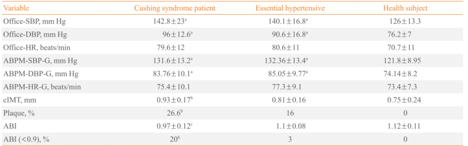
Table 2. Blood Pressure, cIMT, and ABI in All Groups Study
Variable Cushing syndrome patient Essential hypertensive Health subject
Office-SBP, mm Hg 142.8±23a 140.1±16.8a 126±13.3
Office-DBP, mm Hg 96±12.6a 90.6±16.8a 76.2±7
Office-HR, beats/min 79.6±12 80.6±11 70.7±11
ABPM-SBP-G, mm Hg 131.6±13.2a 132.36±13.4a 121.8±8.95
ABPM-DBP-G, mm Hg 83.76±10.1a 85.05±9.77a 74.14±8.2
ABPM-HR-G, beats/min 75.4±10.1 77.3±9.1 73.4±7.3
cIMT, mm 0.93±0.17b 0.81±0.16 0.75±0.24
Plaque, % 26.6b 16 0
ABI 0.97±0.12c 1.1±0.08 1.12±0.11
ABI (<0.9), % 20b 3 0
Values are expressed as mean±SD.
cIMT, carotid intima-media thickness; ABI, ankle-brachial index; SBP, systolic blood pressure; DBP, diastolic blood pressure; HR, heart rate; ABPM, ambulatory blood pressure monitoring; G, global (24 hours).
aP<0.01 vs. health subject; bP<0.03 vs. essential hypertensive and health subject; cP<0.05 vs. health subject.
No statistically significant differences (P>0.05) of clinical BP values were found in all hypertensive groups (CS and EH), whereas significantly higher were in these groups compared to HS (P<0.01). Moreover, Table 1 shows the biochemical pa- rameters revealed in all groups. In particular, patients with CS showed higher fasting blood glucose (109.2±25 mg/dL) re- spect to EH patients (88.1±6.9 mg/dL) and HS (83±12.7 mg/
dL; P<0.05, respectively). Finally, CS and EH patients showed increased levels of albuminuria (60.1±18.3 and 49.9±11.5 mg/day, respectively) compared to HS (15.3±17.2 mg/day;
P<0.01, respectively).
Echocolor-Doppler imaging of CCA showed a significant in- crease (P<0.05) of cIMT in patients with CS (0.89±0.17 mm) compared to EH patients (0.81±0.16 mm) and HS (0.75±0.24 mm). Moreover, we found a higher prevalence of plaque in CS patients (26.6%) compared to in EH patients (16%). None plaque was revealed in HS (Table 2).
Mean ABI measured by automatic method in CS and EH pa- tients were 1.07±0.02 and 1.1±0.08 respectively. A significant difference (P<0.05) was found for ABI between CS patients (0.97±0.12) and HS (1.12±0.11). In particular in CS patients we found a higher percentage (20%) of pathological value for ABI (<0.9) compared to EH and HS (P<0.03) (Table 2).
DISCUSSION
CS, a clinical condition that refers to the manifestation induced by chronic cortisol excess is associated with increased cardio- vascular morbidity, and vascular events are one of the major causes of death in untreated patients [3,5,7,8]. The main vascu- lar alteration associated with CS is arterial atherosclerosis [9].
Carotid ultrasound which is assured both cIMT and carotid plaque is useful in detecting the degree of subclinical athero- sclerosis. In fact, cIMT and carotid plaque is considered a sur- rogate marker of subclinical atherosclerosis and it is able to predict both coronary and cerebrovascular events [10].
The results of the present study show that cIMT was in- creased in patients with CS compared to EH patients and HS.
The increase of cIMT in CS confirmed and reinforced the con- cept that the glucocorticoids may alter the structure of wall ar- teries, predisposing at the atherosclerosis. In fact previous stud- ies reported that arterial wall damage in CS patients is more performed than in EH patients, in part, secondary to excessive cortisol production, thereby resulting in thickening the intima- media layer of carotid artery [12]. Moreover, CS patients have higher degree of early atherosclerosis, and the appropriate treat-
ment not only corrects the high BP values and metabolic disor- ders, but also reverses vascular change in these patients [13].
Several experimental studies have shown that glucocorti- coids excess causes direct cardiovascular effects, such as in- creased renin-angiotensin system, sympathetic nervous system and endothelin system as well as decreased nitric oxide (NO) synthesis and kallikrein-kinin system [13]. Moreover, patients with CS are known to have an irreversible arterial stiffness (de- creased vascular compliance), and several humoral markers of endothelial dysfunction (such as endothelin, homocysteine, vascular endothelial factor, adrenomedulin, and cell adhesion molecules) are believed to be responsible for vascular endothe- lial and smooth muscle proliferation as well as fibrosis around vessels [14-17]. Collectively, it has been suggested that gluco- corticoids excess in CS plays an important role in the develop- ment of endothelial dysfunction that is consistent ad initial event in the development of atherosclerosis plaques.
Another important factor is the dysregulation of the glucose metabolism that we found in our patients with CS. In fact, ob- servational studies in persons with hyperglycemia have shown that glucose concentrations were associated with cIMT [18-21].
Finally, in our study another important result found is the mean ABI measured by automated method. The ABI, which is the ratio of SBP at the ankle to that in the arm, is used to detect peripheral obstructive arterial disease and cerebrovascular dis- ease, and has attracted considerable clinical and scientific inter- est [22]. However, the ABI is also an indicator of generalized atherosclerosis and low ABI has been related to an increased incidence of cardiovascular mortality [23-27]. This increased relative risk has been shown to be independent of baseline car- diovascular and risk factors suggesting that the ABI may have independent role in predicting cardiovascular events.
Initially this method described by Carter [28], was only de- termined with the use of vascular Doppler. More recently, stud- ies have demonstrated the efficacy of using automatic oscillo- metric sphygmomanometers for determination of this index, because it is simples, low cost, and easy to use [29]. In particu- lar, has been reported that the automated oscillometric mea- surement of ABI is a reliable and useful alternative to conven- tional eco-Doppler determination in the general population [30], and some authors [30-32] have reported that only one BP measurement is sufficient to perform the ABI determination as no additional advantages have been shown with a second or a third determination.
An abnormal ABI value is defined as ≤0.90 and values
>1.40 indicate a no compressible artery [25]. Recently, several
investigators have reported that ABI value of 0.91 to 0.99 should be considered borderline and that is associated with an increasing risk of cardiovascular disease [33,34]. Moreover, in comparison with subjects with normal ABI, subjects with al- tered indices are at approximately four times greater risk of de- veloping cardiovascular disease [35]. In our study, the results analysis of the ABI revealed differences among the groups. In particular, arterial wall alterations were found in the CS pa- tients (ABI <0.9 in 20% of subjects), and we hypothesized that the chronic high glucocorticoids levels associated to high blood values and metabolic disorders significantly accelerate the de- velopment of atherosclerosis, leading a decrease in ABI.
CONFLICTS OF INTEREST
No potential conflict of interest relevant to this article was re- ported.
REFERENCES
1. Nieman LK, Biller BM, Findling JW, Newell-Price J, Sav- age MO, Stewart PM, et al. The diagnosis of Cushing’s syndrome: an Endocrine Society Clinical Practice Guide- line. J Clin Endocrinol Metab 2008;93:1526-40.
2. Bertagna X, Guignat L, Groussin L, Bertherat J. Cushing’s disease. Best Pract Res Clin Endocrinol Metab 2009;23:607- 23.
3. Arnaldi G, Mancini T, Polenta B, Boscaro M. Cardiovascu- lar risk in Cushing’s syndrome. Pituitary 2004;7:253-6.
4. Pivonello R, De Martino MC, De Leo M, Lombardi G, Co- lao A. Cushing’s Syndrome. Endocrinol Metab Clin North Am 2008;37:135-49.
5. Arnaldi G, Angeli A, Atkinson AB, Bertagna X, Cavagnini F, Chrousos GP, et al. Diagnosis and complications of Cushing’s syndrome: a consensus statement. J Clin Endo- crinol Metab 2003;88:5593-602.
6. Fardet L, Petersen I, Nazareth I. Risk of cardiovascular events in people prescribed glucocorticoids with iatrogenic Cushing’s syndrome: cohort study. BMJ 2012;345:e4928.
7. Colao A, Pivonello R, Spiezia S, Faggiano A, Ferone D, Filippella M, et al. Persistence of increased cardiovascular risk in patients with Cushing’s disease after five years of successful cure. J Clin Endocrinol Metab 1999;84:2664-72.
8. Etxabe J, Vazquez JA. Morbidity and mortality in Cush- ing’s disease: an epidemiological approach. Clin Endocri- nol (Oxf) 1994;40:479-84.
9. De Leo M, Pivonello R, Auriemma RS, Cozzolino A, Vitale P, Simeoli C, et al. Cardiovascular disease in Cushing’s syn- drome: heart versus vasculature. Neuroendocrinology 2010;92 Suppl 1:50-4.
10. Stein JH, Korcarz CE, Hurst RT, Lonn E, Kendall CB, Mohler ER, et al. Use of carotid ultrasound to identify sub- clinical vascular disease and evaluate cardiovascular dis- ease risk: a consensus statement from the American Society of Echocardiography Carotid Intima-Media Thickness Task Force. Endorsed by the Society for Vascular Medicine. J Am Soc Echocardiogr 2008;21:93-111.
11. Busch MA, Lutz K, Rohl JE, Neuner B, Masuhr F. Low ankle- brachial index predicts cardiovascular risk after acute ischemic stroke or transient ischemic attack. Stroke 2009;40:3700-5.
12. Albiger N, Testa RM, Almoto B, Ferrari M, Bilora F, Petro- belli F, et al. Patients with Cushing’s syndrome have in- creased intimal media thickness at different vascular levels:
comparison with a population matched for similar cardio- vascular risk factors. Horm Metab Res 2006;38:405-10.
13. Isidori AM, Graziadio C, Paragliola RM, Cozzolino A, Am- brogio AG, Colao A, et al. The hypertension of Cushing’s syndrome: controversies in the pathophysiology and focus on cardiovascular complications. J Hypertens 2015;33:44- 60.
14. Ermetici F, Malavazos AE, Corbetta S, Eller-Vainicher C, Cannavo S, Corsi MM, et al. Soluble adhesion molecules levels in patients with Cushing’s syndrome before and after cure. J Endocrinol Invest 2008;31:389-92.
15. Kristo C, Ueland T, Godang K, Aukrust P, Bollerslev J.
Biochemical markers for cardiovascular risk following treatment in endogenous Cushing’s syndrome. J Endocrinol Invest 2008;31:400-5.
16. Letizia C, Di Iorio R, De Toma G, Marinoni E, Cerci S, Celi M, et al. Circulating adrenomedullin is increased in patients with corticotropin-dependent Cushing’s syndrome due to pituitary adenoma. Metabolism 2000;49:760-3.
17. Terzolo M, Allasino B, Bosio S, Brusa E, Daffara F, Ventu- ra M, et al. Hyperhomocysteinemia in patients with Cush- ing’s syndrome. J Clin Endocrinol Metab 2004;89:3745-51.
18. Succurro E, Marini MA, Arturi F, Grembiale A, Lugara M, Andreozzi F, et al. Elevated one-hour post-load plasma glu- cose levels identifies subjects with normal glucose tolerance but early carotid atherosclerosis. Atherosclerosis 2009;
207:245-9.
19. Marini MA, Succurro E, Castaldo E, Cufone S, Arturi F, Sciacqua A, et al. Cardiometabolic risk profiles and carotid
atherosclerosis in individuals with prediabetes identified by fasting glucose, postchallenge glucose, and hemoglobin A1c criteria. Diabetes Care 2012;35:1144-9.
20. Emerging Risk Factors Collaboration, Sarwar N, Gao P, Seshasai SR, Gobin R, Kaptoge S, et al. Diabetes mellitus, fasting blood glucose concentration, and risk of vascular disease: a collaborative meta-analysis of 102 prospective studies. Lancet 2010;375:2215-22.
21. Gast KB, Smit JW, den Heijer M, Middeldorp S, Rippe RC, le Cessie S, et al. Abdominal adiposity largely explains as- sociations between insulin resistance, hyperglycemia and subclinical atherosclerosis: the NEO study. Atherosclerosis 2013;229:423-9.
22. Doobay AV, Anand SS. Sensitivity and specificity of the ankle-brachial index to predict future cardiovascular out- comes: a systematic review. Arterioscler Thromb Vasc Biol 2005;25:1463-9.
23. Diehm C, Allenberg JR, Pittrow D, Mahn M, Tepohl G, Haberl RL, et al. Mortality and vascular morbidity in older adults with asymptomatic versus symptomatic peripheral artery disease. Circulation 2009;120:2053-61.
24. Abbott RD, Petrovitch H, Rodriguez BL, Yano K, Schatz IJ, Popper JS, et al. Ankle/brachial blood pressure in men
>70 years of age and the risk of coronary heart disease. Am J Cardiol 2000;86:280-4.
25. Aboyans V, Criqui MH, Abraham P, Allison MA, Creager MA, Diehm C, et al. Measurement and interpretation of the ankle-brachial index: a scientific statement from the Ameri- can Heart Association. Circulation 2012;126:2890-909.
26. Newman AB. Peripheral arterial disease: insights from popu- lation studies of older adults. J Am Geriatr Soc 2000;48:1157- 62.
27. Resnick HE, Lindsay RS, McDermott MM, Devereux RB, Jones KL, Fabsitz RR, et al. Relationship of high and low ankle brachial index to all-cause and cardiovascular disease mortality: the Strong Heart Study. Circulation 2004;109:733-
9.
28. Carter SA. Indirect systolic pressures and pulse waves in art erial occlusive diseases of the lower extremities. Circu- lation 1968;37:624-37.
29. Takahashi I, Furukawa K, Ohishi W, Takahashi T, Matsu- moto M, Fujiwara S. Comparison between oscillometric- and Doppler-ABI in elderly individuals. Vasc Health Risk Manag 2013;9:89-94.
30. Llisterri Caro JL, Rodriguez Roca GC, Alonso Moreno FJ, Lou Arnal S, Divison Garrote JA, Santos Rodriguez JA, et al. Blood pressure control in Spanish hypertensive patients in Primary Health Care Centres. PRESCAP 2002 Study.
Med Clin (Barc) 2004;122:165-71.
31. Kollias A, Xilomenos A, Protogerou A, Dimakakos E, Ster- giou GS. Automated determination of the ankle-brachial in- dex using an oscillometric blood pressure monitor: valida- tion vs. Doppler measurement and cardiovascular risk fac- tor profile. Hypertens Res 2011;34:825-30.
32. Real de Asua D, Puchades R, Garcia-Polo I, Suarez C. In- fluence of multiple blood pressure measurements on the es- timation of the ankle-brachial index and the consequent di- agnosis of peripheral artery disease. Blood Press Monit 2012;17:73-5.
33. Ankle Brachial Index Collaboration, Fowkes FG, Murray GD, Butcher I, Heald CL, Lee RJ, et al. Ankle brachial in- dex combined with Framingham risk score to predict car- diovascular events and mortality: a meta-analysis. JAMA 2008;300:197-208.
34. Ovbiagele B. Association of ankle-brachial index level with stroke. J Neurol Sci 2009;276:14-7.
35. Criqui MH, McClelland RL, McDermott MM, Allison MA, Blumenthal RS, Aboyans V, et al. The ankle-brachial index and incident cardiovascular events in the MESA (Multi-Ethnic Study of Atherosclerosis). J Am Coll Cardiol 2010;56:1506- 12.