Introduction
The maxillary sinus varies in its extension. It is essential to understand the anatomic relationship between the maxil- lary sinus floor and the root of the maxillary molar for planning preoperative treatments for maxillary posterior teeth.1
The close relationship of the maxillary sinus and the roots of the maxillary molars can lead to accidental oroan- tral communication.2,3The topographical relationship of the roots of the posterior maxillary teeth and the maxillary
sinus floor is an important determinant in the prognosis of orthodontic tooth movement.4 Sinusitis can result from the spread of a periapical or periodontal infection to the sinus or iatrogenic perforation of the sinus floor.3,5
It has been reported that the anatomical relationship be- tween the root and the cortical plates might influence the spread of odontogenic infection originating in the maxil- lary molars.6The thickness of the bone between the root and the alveolar cortical plate might be important in pre- dicting the spread of infection as well as in treatment plan- ning.
The purposes of this study were to investigate the rela- tionship between the roots of the maxillary molars and the maxillary sinus using cone beam computed tomography (CBCT) and to measure the distances between the roots of the maxillary molars and sinus floor and the thickness of the bone between the root and the alveolar cortical plate.
Assessment of the relationship between the maxillary molars and adjacent structures using cone beam computed tomography
Yun-Hoa Jung, Bong-Hae Cho
Department of Oral and Maxillofacial Radiology, College of Dentistry, Pusan National University, Yangsan, Korea ABSTRACT
Purpose: This study investigated the relationship between the roots of the maxillary molars and the maxillary sinus using cone beam computed tomography (CBCT), and measured the distances between the roots of the maxillary molars and the sinus floor as well as the thickness of the bone between the root and the alveolar cortical plate.
Materials and Methods: The study sample consisted of 83 patients with normally erupted bilateral maxillary first and second molars. A total of 332 maxillary molars were examined using CBCT images. The vertical relationship of each root with the maxillary sinus was classified into four types on CBCT cross-sectional images. The distance between the sinus floor and root and the bone thickness between the root and alveolar cortical plate were measured.
Results: In the buccal roots of the maxillary molars, a root protruding into the sinus occurred most frequently. A root projecting laterally along the sinus cavity was most common in the palatal roots of the maxillary first molars.
The mesiobuccal roots of the maxillary second molar were closest to the sinus. The mesiobuccal roots of the first molars were closest to the cortical plate.
Conclusion: The relationship between the roots of the maxillary molars and the sinus differed between the buccal and palatal roots. A root protruding into the sinus occurred more frequent in the buccal roots of the maxillary molars.
The mesiobuccal root of the maxillary second molar was closest to the maxillary sinus floor and farthest from the alveolar cortical plate. (Imaging Sci Dent 2012; 42 : 219-24)
KEY WORDS: Molar; Maxillary Sinus; Bone and Bones; Cone-Beam Computed Tomography
*This study was supported by Clinical Research Grant, Pusan National University Dental Hospital (2012).
Received April 20, 2012; Revised June 3, 2012; Accepted July 23, 2012 Correspondence to : Prof. Bong-Hae Cho
Department of Oral and Maxillofacial Radiology, Pusan National University Dental Hospital, Beomeo-ri, Mulgeum-eup, Yangsan-si, Gyeongsangnam-do 626-787, Korea Tel) 82-55-360-5261, Fax) 82-55-360-5029, E-mail) [email protected]
Copyright ⓒ 2012 by Korean Academy of Oral and Maxillofacial Radiology
This is an Open Access article distributed under the terms of the Creative Commons Attribution Non-Commercial License (http://creativecommons.org/licenses/by-nc/3.0) which permits unrestricted non-commercial use, distribution, and reproduction in any medium, provided the original work is properly cited.
Imaging Science in Dentistry∙pISSN 2233-7822 eISSN 2233-7830
Materials and Methods
The study samples were selected randomly from the pati- ents who had visited Pusan National University Hospital between 2010 and 2011 and had normally erupted bilateral maxillary first and second molars, as shown on the panora- mic radiographs. The selected patients underwent CBCT examination due to pathology such as cysts, tumors, im- pacted third molars, or temporomandibular joint disorders.
Subjects with pathology in the maxillary posterior teeth were excluded from the study. A total of 332 maxillary molars in 83 patients were examined using CBCT images.
The sample of patients comprised 33 males and 50 females with a mean age of 28.8 years, ranging from 20 to 53 years.
CBCT images were acquired with a CBCT scanner (PaX- Zenith 3D, Vatech Co., Hwaseong, Korea). Scanning para- meters were 110 kVp, 24 seconds, 5.7 mA, a voxel size of 0.2 mm or 0.3 mm, and a field of view of 16 cm×14 cm or 24 cm×19 cm. The CBCT volume data were reconstruct- ed using CBCT software (Ez3D2009, Vatech Co., Hwa- seong, Korea). CBCT images were evaluated to assess the roots of the maxillary molars, maxillary sinus, and
cortical plate.
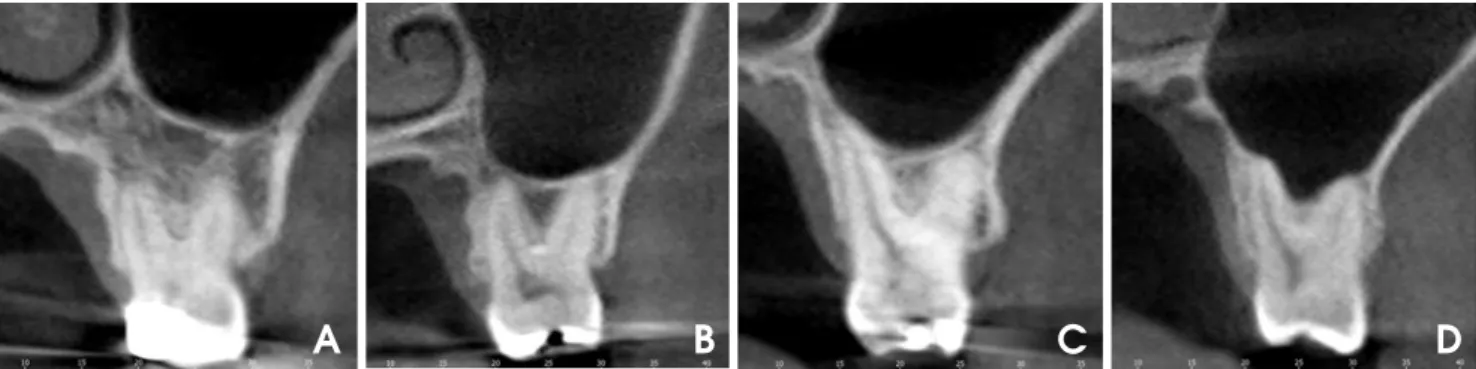
The vertical relationship between each root of the molar and the sinus floor was classified into four types based on the CBCT cross-sectional images: Type 0, the root was not in contact with the cortical borders of the sinus; Type 1, the root was in contact with the cortical borders of the sinus; Type 2, the root was projecting laterally on the sinus cavity, but its apex was outside the sinus borders; and Type 3, the root apex was projecting into the sinus cavity (Fig.
1).7In Types 2 and 3, the horizontal relationship between the roots of the teeth and the sinus floor was classified into three types: Type B, the lowest point of the sinus floor was located on the buccal side; Type BP, the lowest point of the sinus floor was located between the buccal and palatal roots; Type P, the lowest point of the sinus floor was located on the palatal side of the palatal root (Fig. 2).7In Type 0 and Type 3, the distance between the apices of the molars and the sinus floor was measured using CBCT cross-sec- tional images. The measurements were taken from the root apex to the cortical inferior wall of the sinus along the longitudinal axis of the root. The apices extending below the sinus floor were assigned positive values, whereas
A B C D
Fig. 1.CBCT images show 4 types of vertical relationships between the root of the maxillary molars and the sinus floor. A. Type 0, the root is separate from the sinus floor. B. Type 1, the root is in contact with the sinus floor. C. Type 2, the root is projecting laterally along the sinus cavity, but is outside the sinus borders. D. Type 3, the root is projecting into the sinus cavity.
B P
Type B Type BP Type P
Fig. 2.The illustrations show 3 types of the horizontal relationships between the root of the maxillary molars and the sinus floor. Type B, the lowest point of the maxillary sinus floor is located on the buccal side. Type BP, the lowest point of the sinus floor is located between the buccal and palatal roots. Type P, the lowest point of the sinus floor is located on the palatal side of the palatal root.
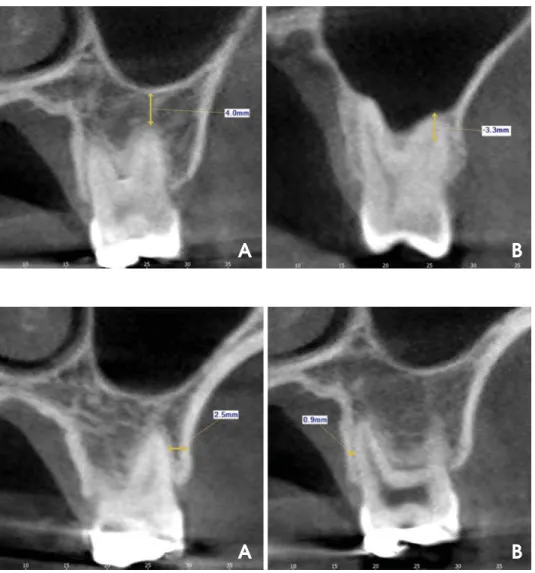
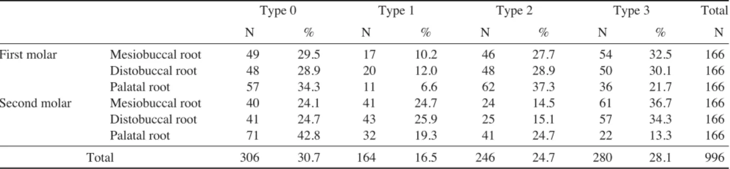
those above the sinus floor were assigned negative values (Fig. 3). The minimum distance from the root below bifur- cation to the appropriate cortical plate was measured in the CBCT cross-sectional images. For buccal roots, the distance to the buccal cortical plate was obtained, and the distance to the palatal cortical plate was obtained for the palatal roots (Fig. 4).
Intra-observer variation was determined by repeating all of the measurements at an interval of four weeks. No statistical differences were found using a paired t-test between the replicate measurements. The averages of two measurements were then computed. A paired t-test was used to compare the measurements of the left and right sides. The results from the right and left molars were aver- aged for each tooth type because no statistically significant differences were found between the right and left side mea- surements. A descriptive analysis of the data was present- ed as frequency, mean, and standard deviation (SD). A one- way analysis of variance (ANOVA) test was used to assess
the differences of bone thickness covering the roots among four vertical relationships between the root and the sinus.
A p-value under 0.01 was considered statistically signifi- cant. All of the statistical analyses were carried out with statistical software (PASW Statistics 18.0, SPSS Inc., Chi- cago, IL, USA).
Results
In the classification of the each root in relationship to the sinus floor, Type 3 was more frequent in the buccal roots, including the mesiobuccal and distobuccal roots, of the maxillary molars. Type 2 was most common in the palatal roots of the maxillary first molar (M1), whereas Type 0 was most frequently observed in the palatal roots of the maxillary second molar (M2) (Table 1). The vertical relationship was classified in each molar, and Type 3 was shown to be most frequent in the molars with more than one root (Table 2). In the horizontal relationship between
A B
Fig. 3.The distance between the root apex and the sinus floor is mea- sured using CBCT cross-sectional images. Apices extending below the floor of the sinus are assigned positive values (A), whereas those above the sinus floor are assigned negative values (B).
A B
Fig. 4.The minimum bone thick- ness between the root and the alve- olar cortical plate is measured using CBCT cross-sectional images. For the buccal roots, the distance to the buccal cortical plate is obtained (A), and for the palatal roots, the distance to the palatal cortical plate is mea- sured (B).
the roots of molars and the sinus floor, Type BP was most frequent in molars and Type B was more frequent in M2 than M1 (Table 3).
The mean distance between the sinus floor and the root apex was the longest for the palatal roots of M2 and the shortest for the mesiobuccal roots of M2 (Table 4).
The mesiobuccal roots of M1 were closest to the corti- cal plate, and the mesiobuccal roots of M2 were farthest from the cortical plate. The thickness of the bone covering the roots showed a statistically significant difference among the four vertical relationships, except in the buccal roots of M2 (p⁄0.01). The bone covering the roots was thinnest in Type 2 (p⁄0.01, Table 5).
Discussion
We examined the relationship between the root of the maxillary molar and the maxillary sinus floor based on
CBCT images. The most frequent relationship in previous studies was that the sinus floor did not contact the roots of the molars.1,8,9 Meanwhile, apical protrusion into the maxillary sinus (Type 3) of one or more roots of the mol- ars was most frequent in our study although the roots being separate from the sinus (Type 0) was most frequent in each root of the molars.
The relationship between the root of the molars and the sinus floor showed a difference between the buccal and palatal roots. The root protruding into the maxillary sinus (Type 3) was most frequent in the buccal roots of the mol- ars. The root projecting laterally along the sinus (Type 2) was most frequent in the palatal root of M1, and the root separate from the sinus floor (Type 0) was most frequent in the palatal root of M2.
Most studies have revealed that the buccal roots of M2 are closely related to the floor of the maxillary sinus.1,8,10-12 Eberhardt et al8 and Georgescu et al12 reported that the mesiobuccal roots of M2 were closest to the sinus floor, and Kilic et al1reported that the distobuccal root of M2 was closest to the sinus floor. Our results showed that the distance between the sinus floor and the root of the molar was shortest for the mesiobuccal roots of M2, for which Type 3 was most frequent and longest for the palatal roots of M2, for which Type 0 was most frequent.
Previous CBCT examinations have revealed a correla- tion between mucosal thickening in the maxillary sinus
Table 1.The vertical relationship between each root of the maxillary molars and the sinus floor
Type 0 Type 1 Type 2 Type 3 Total
N % N % N % N % N
First molar Mesiobuccal root 49 29.5 17 10.2 46 27.7 54 32.5 166
Distobuccal root 48 28.9 20 12.0 48 28.9 50 30.1 166
Palatal root 57 34.3 11 6.6 62 37.3 36 21.7 166
Second molar Mesiobuccal root 40 24.1 41 24.7 24 14.5 61 36.7 166
Distobuccal root 41 24.7 43 25.9 25 15.1 57 34.3 166
Palatal root 71 42.8 32 19.3 41 24.7 22 13.3 166
Total 306 30.7 164 16.5 246 24.7 280 28.1 996
Table 2.The vertical relationship between the maxillary molars and the sinus floor
Type 0* Type 1† Type 2‡ Type 3§ Total
No. % No. % No. % No. % No.
First molar 44 26.5 21 12.7 47 28.3 54 32.5 166
Second molar 39 23.5 42 25.3 24 14.5 61 36.7 166
Total 83 25.0 63 19.0 71 21.4 115 34.6 332
*Maxillary molars in which all roots were Type 0, †Maxillary molars in which more than one root was Type 1, ‡Maxillary molars in which more than one root was Type 2, §Maxillary molars in which more than one root was Type 3
N, number
Table 3. The horizontal relationship between the roots of the maxillary molars and the sinus floor
Type B Type BP Type P Total
N % N % N % N
First molar 3 3.0 98 97.0 0 0.0 101
Second molar 23 27.1 62 72.9 0 0.0 85
Total 26 14.0 160 86.0 0 0.0 186
and decayed posterior maxillary teeth or periodontitis.13 The prevalence and severity of maxillary sinus mucosal thickening have been positively associated with the degree of apical periodontitis.14Bacteria and toxins in apical le- sions may infiltrate the maxillary sinuses via direct diffu- sion through porous maxillary bone or through blood and lymph vessels, causing thickening of sinus mucosa.15We think that more research is needed to determine whether there is a difference in the frequency of odontogenic max- illary sinusitis according to the vertical relationship between the maxillary sinus and molar root.
Ariji et al reported that 80% of patients with buccal cor- tical change had buccal roots that were close to the buccal cortical plate.6Our study showed that the bone thickness covering the roots was thinnest on the mesiobuccal roots of M1 and thickest on the mesiobuccal roots of M2. The thickness of the bone covering the roots varied depending on the vertical relationship between the sinus floor and the root of the molars. It was thinnest in Type 2 and was thickest in Type 3, except in the buccal roots of M2. This might be because Type B was more frequent in M2.
In conclusion, the relationship of the roots of the maxil- lary molars and the sinus floor differed between the buccal and palatal roots. A root protruding into the maxillary sinus was more frequent in the buccal roots of the maxillary molars. The mesiobuccal root of the maxillary second molar was closest to the maxillary sinus floor. The thick-
ness of the bone buccal to the root was markedly thinner in the maxillary first molar than in the maxillary second molar.
References
1. Kilic C, Kamburoglu K, Yuksel SP, Ozen T. An assessment of the relationship between the maxillary sinus floor and the maxillary posterior teeth root tips using dental cone-beam com- puterized tomography. Eur J Dent 2010; 4 : 462-7.
2. Watzek G, Bernhart T, Ulm C. Complications of sinus per- forations and their management in endodontics. Dent Clin North Am 1997; 41 : 563-83.
3. Hauman CH, Chandler NP, Tong DC. Endodontic implications of the maxillary sinus: a review. Int Endod J 2002; 35 : 127- 41.
4. Fuhrmann R, Bücker A, Diedrich P. Radiological assessment of artificial bone defects in the floor of the maxillary sinus.
Dentomaxillofac Radiol 1997; 26 : 112-6.
5. Engström H, Chamberlain D, Kiger R, Egelberg J. Radiographic evaluation of the effect of initial periodontal therapy on thick- ness of the maxillary sinus mucosa. J Periodontol 1988; 59 : 604-8.
6. Ariji Y, Obayashi N, Goto M, Izumi M, Naitoh M, Kurita K, et al. Roots of the maxillary first and second molars in horizon- tal relation to alveolar cortical plates and maxillary sinus: com- puted tomography assessment for infection spread. Clin Oral Investig 2006; 10 : 35-41.
7. Jung YH, Cho BH. Comparison of panoramic radiography and cone beam computed tomography for assessing the relation- ship between the maxillary sinus floor and maxillary molars.
Table 4.Distance from the root apex of the maxillary molars to the sinus floor in Type 0 and Type 3 (unit: mm)
Type 0 Type 3 Total
First molar Mesiobuccal root 2.72±1.98 -3.42±1.50 -0.31±2.79
Distobuccal root 2.38±1.81 -3.59±1.41 -0.39±2.63
Palatal root 3.11±2.13 -4.94±1.59 0.00±3.28
Second molar Mesiobuccal root 2.12±1.62 -3.40±1.87 -0.74±2.59
Distobuccal root 2.40±1.74 -3.16±1.76 -0.49±2.53
Palatal root 2.95±2.10 -2.65±1.60 0.91±2.46
Total 2.67±1.96 -3.53±1.74 -0.17±2.77
Table 5.Thickness of the bone between the roots of the maxillary molars and their respective cortical plate (unit: mm)
Type 0 Type 1 Type 2 Type 3 Total
First molar Mesiobuccal root* 1.04±0.77 1.49±0.83 0.78±0.79 1.72±1.05 1.23±0.96
Distobuccal root* 1.62±1.08 2.21±1.10 1.39±0.94 2.57±1.18 1.91±1.18
Palatal root* 1.16±0.62 1.65±0.54 1.09±0.57 1.66±0.61 1.28±0.64
Second molar Mesiobuccal root 3.59±0.90 3.50±0.91 3.58±1.27 3.12±0.81 3.39±0.95
Distobuccal root 2.90±0.98 3.15±1.05 2.80±1.70 3.18±0.95 3.05±1.12
Palatal root* 1.40±0.99 1.42±0.54 1.05±0.87 1.91±0.83 1.38±0.90
Total* 1.82±1.26 2.51±1.23 1.50±1.30 2.48±1.13 2.04±1.30
* p⁄0.01, one-way ANOVA test
Korean J Oral Maxillofac Radiol 2009; 39 : 69-73.
8. Eberhardt JA, Torabinejad M, Christiansen EL. A computed tomographic study of the distances between the maxillary sinus floor and the apices of the maxillary posterior teeth. Oral Surg Oral Med Oral Pathol 1992; 73 : 345-6.
9. Sharan A, Madjar D. Correlation between maxillary sinus floor topography and related root position of posterior teeth using panoramic and cross-sectional computed tomography imaging. Oral Surg Oral Med Oral Pathol Oral Radiol Endod 2006; 102 : 375-81.
10. Yoon HR, Park CS. A radiologic study of the relationship of the maxillary sinus floor and apex of the maxillary molar. J Korean Acad Oral Maxillofac Radiol 1998; 28 : 111-26.
11. Kwak HH, Park HD, Yoon HR, Kang MK, Koh KS, Kim HJ.
Topographic anatomy of the inferior wall of the maxillary sinus in Koreans. Int J Oral Maxillofac Surg 2004; 33 : 382-8.
12. Georgescu CE, Rusu MC, Sandulescu M, Enache AM, Didi- lescu AC. Quantitative and qualitative bone analysis in the maxillary lateral region. Surg Radiol Anat 2012; 34 : 551-8.
13. Brüllmann DD, Schmidtmann I, Hornstein S, Schulze RK. Cor- relation of cone beam computed tomography (CBCT) findings in the maxillary sinus with dental diagnoses: a retrospective cross-sectional study. Clin Oral Investig 2012; 16 : 1023-9.
14. Lu Y, Liu Z, Zhang L, Zhou X, Zheng Q, Duan X, et al. Asso- ciations between maxillary sinus mucosal thickening and api- cal periodontitis using cone-beam computed tomography scann- ing: a retrospective study. J Endod 2012; 38 : 1069-74.
15. Phothikhun S, Suphanantachat S, Chuenchompoonut V, Nisa- pakultorn K. Cone-beam computed tomographic evidence of the association between periodontal bone loss and mucosal thickening of the maxillary sinus. J Periodontol 2012; 83 : 557- 64.