The treatment of malocclusion after open reduction of maxillofacial fracture: a report of three cases
Sung-Suk Lee, Su-Gwan Kim, Seong-Yong Moon, Ji-Su Oh, Jae-Seek You Department of Oral and Maxillofacial Surgery, School of Dentistry, Chosun University, Gwangju, Korea
Abstract(J Korean Assoc Oral Maxillofac Surg 2014;40:91-95)
The posttraumatic complications of jaw fractures related to jaw function and facial deformity include nonunion, malunion, malocclusion, temporo- mandibular joint dysfunction and facial asymmetry. This report presents cases referred to our department for revision of malunion and malocclusion following inadequate reduction of jaw fractures. Three patients with posttraumatic malocclusions caused by malunion were treated with a LeFort I osteotomy in one case and re-fracture in two cases. All of the patients exhibited stable results without further complications (e.g., malunion or maloc- clusion). Accurate preoperative diagnosis and proper anatomical reduction of the fracture segments are essential to preventing post-surgical malunion and malocclusion.
Key words: Fractures, Malunited, Malocclusion
[paper submitted 2013. 12. 31 / revised 2014. 3. 17 / accepted 2014. 3. 31]
ment. The former condition is caused by a bone defect or displacement and overlap of the bone segment, whereas the latter condition is caused by decreased vascular supply, a nu- tritional disorder, inflammation and inhibiting factors of bone healing4.
It is necessary to consider the location and complexity of the fracture, the existence of teeth on the fracture line and in- fection presenting in an open fracture. The patient’s poor oral hygiene may cause infection, which would require additional surgery5. Additional surgery causes the patient increased pain, recovery time and costs for hospitalization, as well as interruption of daily life. In treating mandibular fractures, it is necessary to make the utmost effort to minimize complica- tions6.
The purpose of this study was to introduce cases in which revision surgery was necessitated by malunion occurring af- ter reduction of a mandibular fracture and to discuss relevant complications.
II. Cases Report
We followed the Helsinki Declaration throughout this study. We obtained approval from the Chosun University Dental Hospital Clinical Trial Center Institutional Review
I. Introduction
The mandibular bone sustains fracture most frequently be- cause it is located in the center of the face at a location prone to external injury1. The goals of treatment are to ensure rapid healing through accurate reduction and fixation and to mini- mize disability and complications. Many techniques have been introduced over the years, including maxillomandibular fixation, which uses intraosseous wire, lag screw fixation, ex- ternal rigid fixation and, recently, internal rigid fixation, such as stable internal fixation using a metal plate2. These methods may cause such complications as infection, malunion, de- layed union, nonunion, hypoesthesia, malocclusion and facial deformity3. Inappropriate union is attributable to the mechan- ical environment, which entails wide gap between segments and movement of segments, and a poor biological environ-
Su-Gwan Kim
Department of Oral and Maxillofacial Surgery, School of Dentistry, Chosun University, 309, Pilmun-daero, Dong-gu, Gwangju 501-759, Korea
TEL: +82-62-220-3815 FAX: +82-62-228-7316 E-mail: [email protected]
This is an open-access article distributed under the terms of the Creative Commons Attribution Non-Commercial License (http://creativecommons.org/licenses/by-nc/3.0/), which permits unrestricted non-commercial use, distribution, and reproduction in any medium, provided the original work is properly cited.
CC
Copyright Ⓒ 2014 The Korean Association of Oral and Maxillofacial Surgeons. All rights reserved.
completed, the patient reported good occlusion on both sides.
The patient had hypoesthesia of the lower lip and discomfort during mouth opening.
2. Case 2
A 22-year-old man was diagnosed with a nasoethmoidal complex fracture, a LeFort II fracture and a subcondylar fracture of the right mandible that were sustained in a mo- torcycle accident. The patient was hospitalized in the plastic surgery department and open reduction was performed on the nasoethmoidal complex and maxilla using an absorbable plate. No treatment was performed for the subcondylar frac- ture. Five weeks later, the patient visited this department with the primary complaint of malocclusion and was diagnosed with malunion of the maxilla.(Fig. 5. A) The absorbable Board (CDMDIRB-1325-146).
1. Case 1
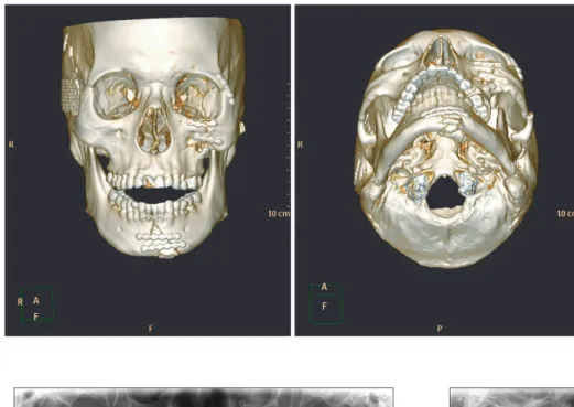
A 50-year-old man was admitted to the emergency room of this hospital after he fell at his worksite from a height of 2 m and sustained an injury to the maxillofacial area. Based on the clinical and radiological examination results, the patient was diagnosed with fractures in both lateral orbital walls and in the left lower and interior orbital walls, a zygo- matico-maxillary complex fracture, a right temporal bone fracture and a mandibular symphysis fracture, as shown in the initial picture.(Fig. 1) In Department of Otolaryngology, open reduction was performed for internal fixation using a metal plate in the area of the left orbit and at the site of the mandibular symphysis; in this surgical approach, the tem- poral bone was used to preserve the right facial nerve. Two months later, the patient visited this hospital with the primary complaint of malocclusion and was transferred to our de- partment. During examination, spacing caused by malunion was found between the central incisors on both sides of the mandibular symphysis and malocclusion was found on both sides.(Figs. 2, 3) Revision surgery to adjust the occlusion was performed under general anesthesia. The existing metal plate was removed, and a chisel was used to re-fracture the mandibular symphysis site. After the occlusion was adjusted to the original position as much as possible, a miniplate was used for the re-fixation, and an arch bar was used to perform the maxillomandibular fixation.(Fig. 4) After the surgery was
Fig. 1. Three-dimensional computed tomography of the initial examination.
Sung-Suk Lee et al: The treatment of malocclusion after open reduction of maxillofacial fracture: a report of three cases. J Korean Assoc Oral Maxillofac Surg 2014
Fig. 2. Panoramic view two months after surgery in Department of Otolaryngology.
Sung-Suk Lee et al: The treatment of malocclusion after open reduction of maxillofacial fracture: a report of three cases. J Korean Assoc Oral Maxillofac Surg 2014
on the subcondyle and parasymphysis before a miniplate was used for fixation.(Fig. 7. B)
III. Discussion
The goal of treatment for mandibular fracture is accurate reduction of the bone segments to recover the pre-traumatic occlusion and to restore normal masticatory function, pro- nunciation, shape and sensation7. It is necessary to perform rigid fixation for the formation of the callus because integra- tion occurs only under mechanically stable conditions, and plates were removed under general anesthesia, and a LeFort
I osteotomy was performed for maxillomandibular fixation to ensure proper occlusion and a miniplate was used for re- fixation. The patient healed without particular discomfort or complications.(Fig. 5. B)
3. Case 3
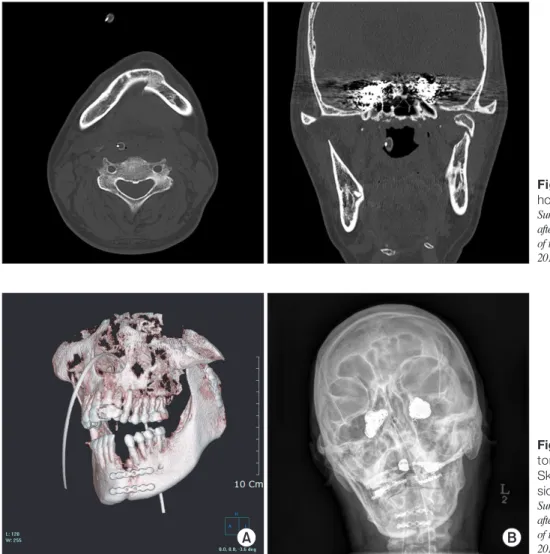
The third patient was a 38-year-old man who visited this hospital after a traffic accident 10 months previously. The examination showed a fracture on the left parasymphysis and a subcondylar fracture.(Fig. 6) After re-fracture of the sub- condylar and parasymphysis sites under general anesthesia, a miniplate was used for fixation with a block bone graft.(Fig.
7. A) The patent was lethargic from the traffic accident, and it was therefore difficult to perform a follow-up examina- tion. The patient visited the hospital for revision surgery six months later. The examination results demonstrated maloc- clusion attributable to malunion. Re-fracture was performed
Fig. 3. Computed tomography two months after surgery in Department of Otolaryngology.
Sung-Suk Lee et al: The treatment of malocclusion after open reduction of maxillofacial fracture: a report of three cases. J Korean Assoc Oral Maxillofac Surg 2014
Fig. 4. Panoramic view after revision surgery.
Sung-Suk Lee et al: The treatment of malocclusion after open reduction of maxillofacial fracture: a report of three cases. J Korean Assoc Oral Maxillofac Surg 2014
Fig. 5. A. Panoramic view after surgery in Department of Plastic Surgery. B. Panoramic view after revision surgery.
Sung-Suk Lee et al: The treatment of malocclusion after open reduction of maxillofacial fracture: a report of three cases. J Korean Assoc Oral Maxillofac Surg 2014
does not take place in that time11. Malunion occurs due to inappropriate reduction, agitation of the bone segment, non- compliance on the part of patient or improper internal fixa- tion12.
The mandible is prone to complications that occur after jaw fracture reduction due to the following reasons. The mandible is the only bone in the facial region that moves and it has less bone support than the facial bones; therefore, a fracture of the mandible generally results in greater degrees of instability.
Because of the muscle attached to the mandible, mandible displacement may occur even after reduction and fixation.
The mandible is located in the lower part of the oral cavity, which increases the possibility of infection. The mandible has lower blood circulation than the maxilla, which contributes to the occurrence of inappropriate integration4.
Physician misdiagnosis is another factor that can cause complications after mandible surgery and is attributable to inexperience or inadequate radiological examination equip- ment. Mandibular fracture is accompanied by edema and hemorrhage, which cause difficulty in precise diagnosis. The mechanical stability is also indispensable to vascularization
of the primary callus. Mechanical stability causes internal growth of the capillary process and removes fibrocartilage, which lead to osteon formation. Distortion and stress prevent such processes8.
Many techniques have been studied and developed to treat mandibular fracture, and it is necessary to consider potential complications for all techniques. Alpert et al.9 introduced four types of complications that were classified as follows:
1) complications that appear after proper treatment is con- ducted; 2) complications that are attributable to inappropriate treatment; 3) complications that are due to failure of surgical treatment; and 4) complications that occur because no treat- ment is conducted.
Mathog and Boies10 reported that the most significant fac- tors causing malunion in mandibular fracture included inap- propriate movement, incomplete reduction, infection, poor blood circulation, and metabolic change. The integration of normal bone typically occurs in 4-8 weeks, depending on the age of the patient, and malunion occurs if bone integration
Fig. 6. Computed tomography at the hospital upon initial examination.
Sung-Suk Lee et al: The treatment of malocclusion after open reduction of maxillofacial fracture: a report of three cases. J Korean Assoc Oral Maxillofac Surg 2014
Fig. 7. A. Three-dimensional computed tomography after the first surgery. B.
Skull posteroanterior view after the revi- sion surgery.
Sung-Suk Lee et al: The treatment of malocclusion after open reduction of maxillofacial fracture: a report of three cases. J Korean Assoc Oral Maxillofac Surg 2014
References
1. Killey HC, Banks P. Killey's fractures of the mandible. 3rd ed.
Bristol: Wright-PSG; 1983.
2. Moraes RB, Landes CA, Luz JG. Fixation of mandibular fractures with plates or miniplates: prospective study. Minerva Stomatol 2010;59:159-66.
3. Mathog RH, Toma V, Clayman L, Wolf S. Nonunion of the man- dible: an analysis of contributing factors. J Oral Maxillofac Surg 2000;58:746-52.
4. Li Z, Zhang W, Li ZB, Li JR. Abnormal union of mandibular frac- tures: a review of 84 cases. J Oral Maxillofac Surg 2006;64:1225- 31.
5. Taher AA. Osteomyelitis of the mandible in Tehran, Iran. Analysis of 88 cases. Oral Surg Oral Med Oral Pathol 1993;76:28-31.
6. Assael LA. Evaluation of rigid internal fixation of mandible frac- tures performed in the teaching laboratory. J Oral Maxillofac Surg 1993;51:1315-9.
7. Punjabi AP, Thaller SR. Late complications of mandibular frac- tures. Oper Tech Plast Reconstr Surg 1998;5:266-74.
8. Schwimmer A. Mandibular pseudoarthrosis and non-unions. In:
Greenberg AM, ed. Craniomaxillofacial fractures: principles of in- ternal fixation using the AO/ASIF technique. New York: Springer- Verlag; 1993:77-84.
9. Alpert B, Engelstad M, Kushner GM. Invited review: small versus large plate fixation of mandibular fractures. J Craniomaxillofac Trauma 1999;5:33-9.
10. Mathog RH, Boies LR Jr. Nonunion of the mandible. Laryngo- scope 1976;86:908-20.
11. Mathog RH. Nonunion of the mandible. Otolaryngol Clin North Am 1983;16:533-47.
12. Ellis E 3rd. Complications of rigid internal fixation for mandibular fractures. J Craniomaxillofac Trauma 1996;2:32-9.
occurrence of multiple fractures could result in a physician erroneously overlooking some bone fracture sites4.
For surgical management of such complications, a thor- ough and corrective examination prior to surgery is neces- sary. The pre-traumatic occlusion must be verified, and an osteotomy to perform internal fixation is required. It might be necessary to perform a new osteotomy on sites other than the malunion site for the recovery of occlusion7.
In this study, malunion after mandibular reduction led to malocclusion, and revision surgery provided satisfactory results. A precise pre-operative examination and appropriate surgical procedure followed by proper post-operative man- agement are necessary to prevent post-surgical complications of jaw fracture fixation. Additional studies are warranted to investigate the surgical and non-surgical management of such complications.
Conflict of Interest
No potential conflict of interest relevant to this article was reported.