대한치주과학회지 : Vol. 37, No. 2(Suppl.), 2007
Establishment of B-1 cell-derived polyreactive monoclonal antibodies and expression of costimulators by B-cell to antigenic stimulation
Ju-Youn Lee1, Jeom-Il Choi1, Jo-Young Suh2*
1. Department of Periodontology, School of Dentistry, Pusan National University 2. Department of Periodontology, School of Dentistry, Kyungpook National University
I. INTRODUCTION
The innate immune system is comprised of the cells and mechanisms that defend the host from infection by other organisms in a non-specific manner. It is mainly directed to recognition of invariant molecules of in- fectious agents. Most of them are essential for pathogen survival, and are conserved and shared by groups of pathogens, such as lip- opolysaccharide (LPS) present in gram-neg- ative bacteria. It is also essential for the ac- tivation of the adaptive immune response, capable of coping with a high mutation rate and an antigen (Ag) heterogeneity of in- fectious agents, and of generating a long- lasting immune memory1).
It is generally thought that the B cell com-
partment is a representative of the adaptive branch of immune defense2). However, be- cause of their anatomical location, B-1 and marginal zone (MZ) B cells are the first cell populations to encounter Ags3-5). These two B cell subsets have evolved to provide a first line of defense against pathogens. With their additional properties to produce factors that can directly mediate microbial destruc- tion and to express Toll-like receptors, B cells provide an important link between the innate and adaptive branches of the immune system6).1)
B-1 cells represent a specialized subset of B cells that are distinct from the majority of recirculating conventional B cells (termed B-2) in an individual.
They are distinguished by tissue dis-
* This study was supported by 2006 Pusan National University Hospital Clinical Research Grant.
* Correspondence: Jo-Young Suh, Department of Periodontology, School of Dentistry, Kyungpook National University, 188-1, Samdeok 2Ga, Jung-Gu, Daegu, 702-412, Korea (E-mail: jysuh@knu.ac.kr)
tribution, cell surface phenotype, B cell re- ceptor signal generation, capacity for self-re- newal and perhaps most importantly, their role in immune defense7-11). B-1 cells, al- though constituting only a minor fraction of B cells in spleen and lymph nodes, represent the main B-cell population in the peritoneal and pleural cavities. B-1 cells only comprise a small percentage of all B cells, but they are the primary source of the serum Abs, known as Natural Antibodies (NAbs)8,12,13). The majority of NAbs have a low affinity and are polyreactive, with the ability to rec- ognize autoantigens as well as Ags from bacterial and parasitic sources. B-1 cells de- velop de novo prior to weaning and persist thereafter as a self-replenishing population and produce IgM autoantibodies, including rheumatoid factor, Abs to single-strand DNA, and Abs to bacterial Ags such as LPS or phosphatidyl choline14-17).
The exact role of B-1 cells during an im- mune response is still under investigation.
Hezenberg's group18) has provided the evi- dence for a mechanism of B-1 cells (NAbs).
They have reported that the natural IgM Abs against viruses are present prior to infection and do not increase. Thus, B-1 cells do not respond to pathogens with an increase of IgM Abs; the increase of the antivirus IgM comes entirely from the B-2 cells.
The demonstration that B-1 cells can bind, process and present many different Ags to T cells, argues that under ordinary circum- stances B-1 cells do not stimulate T cells but do so in the absence of the important
costimulators B7-1 and B7-2. Thus, B-1 cells have all the properties required for in- ducing and maintaining immunological toler- ance and are particularly suited to pick-up the myriad of endogenous Ags to which the host might make an autoimmune response19). Thus, it is tempting to speculate, that where- as high affinity monoreactive Ag-binding B cells provide a defense against foreign in- vaders, low affinity polyreactive Ag-binding B-1 cells may provide a defense against au- toimmune self-destruction by inducing and maintaining immunological tolerance20).
Presentation of Ags to T lymphocytes by B-1 cells is difficult to evaluate at the hu- man level because of problems in readily obtaining normal B-1 cells and Ag-specific T cells from HLA-compatible subjects. To circumvent this problem, use of inbred mice may be suggested. The purpose of this study was to characterize B-1 cell-derived innate immunity and examine the immunoregulatory function of B cells. In the present study, we have established hybridoma producing B-1 cell-derived polyreactive monoclonal Abs.
By using this hybridoma, we have charac- terized B-1 cell-derived monoclonal Ab re- sponses to a panel of exogenous and endog- enous Ags. Expression of costimulators by splenic and peritoneum-derived B cells to antigenic stimulation was also evaluated.
II. MATERIALS AND METHODS
1. Reagents
Cell culture media (Dulbecco's modified Eagle's medium, DMEM) and fetal bovine serum (FBS) were obtained from GibscoBRL (Grand Island, NY, USA). Reagents pur- chased from Sigma (St. Louis, Missouri, USA) were cell freezing medium DMSO (serum free); hypoxanthin, aminopterin, and thymidine (HAT); hypoxanthine and thymi- dine (HT) medium supplements; complete and incomplete Freund's adjuvant, bovine se- rum albumin (BSA), ethylene diamine tetra- acetic acid (EDTA). Polyethylene glycol (PEG) 4000 was purchased from Merck (Darmstadt, Germany).
2. Antigens and Antibodies
We used LPS, Porphyromonas gingivalis hsp60 (Pghsp), PC-KLH (PC coupled to keyhole limpet hemocyanin) as exogenous Ags and Mammalian heat shock protein (Mahsp), Deoxyribonucleic acid (DNA) from calf thymus as endogenous Ags. LPS (Sigma) from Escherichia coli 0111:B4, Mahsp [Recombinant Mouse Hsp60 Protein (Stressgen, Victoria, BC, Canada)], DNA from calf thy- mus (Sigma) and PC-KLH (Biosearch Technologies, Inc., Novata, CA, USA) were used in this experiment. Recombinant Pghsp, was purified from Escherichia coli trans- formed with P. gingivalis GroEL21).
Peroxidase-labeled affinity purified anti- bodies to mouse IgM (μ) produced in goats were purchased from KPL (Gaithersburg MD, USA). Abs used for immunofluorescence were obtained from PharMingen (San Diego,
CA, USA): B220 (CD45R; RA3-6B2-Per cp and PE), MHC class II (2G9 and M5/
114.15.2, -FITC), B7-1 (CD80, 16-10A1,-FITC) and B7-2 (CD86, GL1, -FITC)
3. Medium
DMEM containing 10~20% (v/v) FBS was supplemented with 0.2M glutamine and 10 mg/ml Gentamycin. The standard medium was used for the growing of myeloma and hybridoma cells.
4. Mice
C57BL/6 mice and RAG1 knock-out (KO) mice on the C57BL/6 background, originally obtained from Jackson Laboratories (Bar Harbor, ME, USA), were bred and reared in the Pusan National University Hospital Medical Research Institute and were used at 4~6 weeks of age.
5. Myeloma cell line
The HAT-sensitive Balb/c mouse myeloma cell line SP2/0-Ag14 (ATCC #CRL 1581) was used in fusion experiments.
6. Adoptive transfer of peritoneal B-1 cells into RAG1 knock-out mice
C57BL/6 mice were sacrificed by cervical dislocation and thoroughly cleaned with 70%
ethyl alcohol. A small incision was made in- to the abdomen and the peritoneal cavity
(PerC) was rinsed several times through the opening with freshly prepared cold sterile phosphate-buffered saline (PBS) containing 1% FBS.
The cells collected were devoid of eryth- rocytes by incubating the cells with lysing buffer at 4℃ for 5 min, resuspended in PBS buffer (0.5% BSA, 2mM EDTA) and centri- fuged three times at 1500 rpm for 5 min.
The peritoneal washing was passed through a 30㎛ nylon mesh (Miltenyi Biotec GmbH, Bergisch Gladbach, Germany) to obtain a homogeneous cell population.
The cells were suspended with PBS buffer and a CD90 Microbead (Miltenyi) and in- cubated for 15 min at 4℃. PerC B-1 cells were enriched from peritoneal lavage by re- moving the adherent accessory cells and magnetic bead depletion of T cells. The fi- nal B-1 cells were injected into the peri- toneal cavity of RAG1 KO mice.
7. Establishment of hybridoma producing B-1 cell-derived polyreactive monoclonal antibodies
One week before fusion, the myeloma cell line was expanded into a complete DMEM-10/
HEPES/pyruvate.
RAG1 KO mice were sacrificed by cer- vical dislocation 10 weeks after the adoptive transfer. After the surgical skin preparation, the spleen was exteriorized through a 1-cm left subcostal incision and isolated. Spleen cells that were collected from the mice were
fused with the myeloma cell line using PEG at a 1:1 ratio. After fusion, cells were re- suspended in standard DMEM medium sup- plemented with 20% FBS and 1% (v/v) HAT medium supplement (DMEM-20/HAT media) to exclude non-fused cells. Next, they were distributed in 96-well culture plates at an approximate density of 2.5 × 10⁶ cells/well. The plates were incubated at 37℃ in a CO2 incubator containing 5%
CO2 in the air. Selective growth of the hy- brid cells was carried out for fourteen days.
Finally, the medium was replaced with the standard medium supplemented with 1%
(v/v) HT medium supplement to neutralize the HAT medium.
For initial screening, the supernatant was examined by enzyme-linked immunosorbent assay (ELISA) for the production of Abs against a panel of exogenous and endoge- nous Ags used in the present experiment.
Positive hybridomas were subcloned by lim- iting dilution (0.3 cell/well). After additional screening for monoclonality of cells, the hy- bridoma was selected for production of Abs by ELISA. The hybridoma cells were injected (5 × 10⁷ cells/mice) in nude mice and the peritoneal ascites were collected for purifica- tion of IgM monoclonal Ab using column chromatography (ImmunoPure®, PIERCE, USA).
8. Measurement of monoclonal
antibody titers to exogenous and
endogenous antigens by ELISA
Microtiter plates were coated with 5 differ-ent Ags [LPS, Pghsp, PC-KLH, Mammalian heat shock protein (Mahsp), thymic DNA, 10㎍/ml] in coating buffer and incubated overnight at 4℃. LPS, Pghsp, and PC-KLH are exogenous Ags, while Mahsp and DNA are endogenous Ags. The plates were wash- ed and an aliquot of supernatant serially di- luted was added; following this, they were incubated at room temperature. After wash- ing the plates, peroxidase-conjugated mouse anti-goat IgM was added. After 2 hours of incubation, the plates were washed and 50μ L of substrate solution per well was added.
The reaction was stopped by adding 50μL of stop solution per well. Absorbance was read at 450nm in the ELISA reader (Sunrise®, XFLUOR4, version 4.51).
9. Immunization of Animals
To characterize the immunoregulatory func- tion of peritoneal and splenic B cell to exogenous and endogenous Ags, 8 C57BL/6 mice were initially immunized subcuta- neously with 5μg of each Ags suspended in complete Freund's adjuvant, followed by two subsequent injections of Ags suspended in incomplete Freund's adjuvant at 1-week intervals. The Ags used for immunization in- cluded LPS (LPS group), Porphyromonas gingivalis heat shock protein (Pghsp group), Mammalian hsp (Mahsp group), and thymic DNA (DNA group). Two C57BL/6 mice were included in the control group and im- munized with complete and incomplete ad- juvant only.
10. Flow cytometric analysis
At 1 week after final immunization, mice were sacrificed and their spleen cells and peritoneal cells were collected, respectively, by the method described above. All proce- dures were carried out according to the manufacturer's recommendation. Briefly, the cells were washed twice in PBS, counted, and resuspended in Fluorescence Activated Cell Sorting (FACS) buffer (1% BSA in PBS containing 0.01% sodium azide). For phenotypic analysis, cells (0.2-1×10⁶ cells/
stain) were initially incubated with CD16/CD32 (Fc block) for 20 min at 4℃. Subsequently, cells were incubated with an optimal amount of primary Abs (-FITC, MHC Class, B7-1, B7-2) followed by appropriate secondary Ab (-PE, B220). All incubation were performed on ice for 20 min and were followed by three washes with FACS buffer. Flow cyto- metric analysis was performed using the Cytomics FC 500 system with CXP Software (Beckman Coulter Inc.).
III. RESULTS
1. Monoclonal antibody
production against exogenous and endogenous antigens
Hybridoma producing B-1 cell-derived monoclonal Ab were established. Initially, six wells out of 66 wells demonstrated the homogeneous cell clusters. After limiting di- lution, 2 wells demonstrated monoclonality
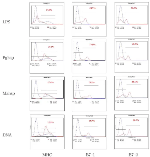
Figure 2. Overlay plot of flow cytometric analysis of splenic B cells. MHC expression was enhanced in the test group and B7-1 and B7-2 was slightly enhanced in the test group. blue line: control group, red line: test group.
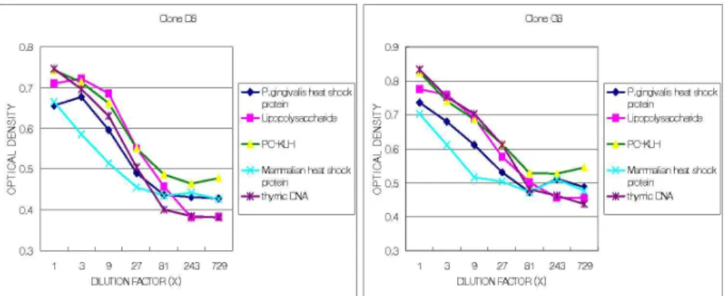
Figure 1. Dose-dependent antibody responses (expressed as optical density) of monoclonal antibodies derived from the two clones against a panel of antigens. Absorbance was read at 450 nm in the ELISA reader. They show the dose-dependent pattern and polyreactivity of monoclonal antibody responses.
Figure 3. Overlay plot of flow cytometric analysis of peritoneal B cells. MHC expression was enhanced in the test group and the expression of B7-1 and B7-2 was pronounced in the test group. blue line: control group, red line: test group.
of cell clusters (clone D8 and clone G8, re- spectively). The monoclonal Ab produced from peritoneal ascites were tested for the Ab response by ELISA. Figure 1 shows the dose-dependent pattern of Ab responses (expressed as an optical density) of the monoclonal Ab obtained from the two clones. The two clones demonstrated a very similar pattern in terms of polyreactive re- activity to the multiple Ags tested.
2. Expression of costimulators by B-cells to antigenic stimulation
One week after final immunization, mice in the 4 test groups (Pghsp, LPS, Mahsp, thymic DNA) and the control group were sacrificed.
MHC expression was enhanced similarly in Splenic and PerC B cells. With regards to B7-1 and B7-2, the two factors were ex- pressed higher in both the splenic and PerC B cells when compared with the control
group, while the expression was more pro- nounced in PerC B cells than in splenic B cells (Figure 2, 3). Thus, the type of Ag used in this experiment did not significantly influence the results but the origin of B cells definitely influenced the results.
IV. DISCUSSION
In the early 1980s, many authors demon- strated that some monoclonal Abs had the capacity to bind to a variety of different Ags22-26). By immortalizing human peripheral B lymphocytes with the Epstein-Barr Virus, Notkins20) found that 10-15% of the cells made broadly reactive Abs which are now referred to as polyreactive Abs (Natural Abs), in addition the majority of polyreactive Abs are of the IgM isotype. Consistent with a major role for B-1 cells in natural IgM Ab production, a number of natural IgM Ab specificities have been identified in the B-1 repertoire. These include specificities for phosphorylcholine27), phosphatidyl chol- ine12,28,29), thymocytes30), LPS31), and the in- fluenza virus32).
To evaluate the exact role of B-1 cells without T-cell help, we have used RAG1 KO mice devoid of mature T- and B-cells, reconstituted with B-1 cells enriched from C57B/6 mice. We were able to show that B-1 cell derived monoclonal Ab was poly- reactive in nature; thus, these Abs are bound to a variety of different Ags in a dose-satu- rable fashion (Figure 1). Notkins20) has found that several typical monoclonal poly-
reactive Abs react to different degrees with a variety of self and foreign Ags. In con- trast to polyreactive Abs, monoreactive Abs bind only to the immunizing Ag. Casali and Schettino33) have shown that a major pro- portion of NAbs are polyreactive, displaying different affinities for different Ags. They have also shown that the binding of most natural polyreactive Abs to a given Ag can be cross-inhibited by a variety of different Ags in a dose saturable fashion. The precise mechanism underlying polyreactivity still re- mains unclear. One possibility is that the confirmational flexibility34,35) at the Ag-bind- ing site of polyreactive Abs is greater than that of monoreactive Abs, thereby allowing a greater number of Ags to bind. Another possibility is a diverse array of binding site structures at the combining site36).
The affinity of monoclonal Ab for most Ags is very low37-39). Because of their weak interactions with self-Ag, B-1 cell derived monoclonal Abs (NAbs) have often been ne- glected as the so-called background serum Abs without significant relevance. However, Kazatchkine and Kaveri40) have demonstrated that NAbs are essential components of ther- apeutic molecules in the form of intravenous Immunoglobulin (IVIg). IVIg has been found to be beneficial in a variety of autoimmune and inflammatory diseases. The biological role of B-1 cells during an immune response is still under investigation. Hezenberg' group18) has demonstrated that NAbs against the influenza virus are important in terms of protection against infection. Ochsenbein et
al.41) have reached the same conclusion from a study that used the lymphocytic choriome- ningitis virus infection as a model.
Therefore, several studies have clearly sug- gested that NAbs are essential for the in- duction of a primary immune response that protects against bacterial and viral pathogens. We demonstrated polyreactivity of B-1 cell-derived monoclonal Ab to various exogenous and endogenous Ags. Therefore, we may assume thar B-1 cell-derived mono- clonal Ab charge the primary defense line to infection in the fetal stage and infancy, prior to induction of the adaptive immune re- sponse with high affinity Ab.
To investigate the expression of cos- timulators of PerC and splenic B cells to several antigenic challenges, we have immu- nized the mice with 2 exogenous Ags (LPS, Pghsp) and 2 endogenous Ags (Mahsp, DNA). The results indicated that MHC ex- pression was enhanced similarly in splenic and PerC B cells. With regards to B7-1 and B7-2, the two factors were expressed higher in both the PerC and splenic B cells when compared with the control group, while the expression was more pronounced in PerC B cells than in splenic B cells. But the ex- pression of MHC, B7-1 and B7-2 were very similar irrespective of the kind of Ags.
Thus, we can assume that the type of Ags does not significantly influence the results but the origin of B cells definitely influen- ces the results. These results coincide with Rothestein and Kolber's study42). They have demonstrated that PerC B cells and splenic
B cells respond differently to phorbol esters.
These results have supported the possibility that residence in the peritoneal cavity influ- ences the different responses of a B cell. It is surprising that specificity for a self-epit- ope does not affect all B cells uniformly43). One possible interpretation of these results is that differentiation to signaling-incompetent B-1 occurs only in specific sites such as the peritoneum. This seems inconsistent with the findings of Liou et al.44), who found that peritoneal B-1 and splenic B-1 cells from Ig transgenic mice were similarly resistant to experimental tolerance induction. These re- sults suggest that B-1 cells were phenotypi- cally and functionally identical regardless of locale.
Wang et al.45) have shown that despite their Ag-presenting ability, B-1 cells from normal mice failed to trigger the pro- liferation of Ag specific T cells. Analysis of the B7-1 and B7-2 showed that these mole- cules were not expressed on B-1 cells from normal mice. Chen et al.46) have revealed that polyreactive B-1 cells expressed high levels of MHC, but little of no B7-1 and B7-2. These findings argue that the lack of costimulators on B-1 cells is the most likely explanation for their failure to stimulate Ag-specific T cells. This suggests that B-1 cells may contribute to the induction and/or maintenance of immunological tolerance. The issue of the expression of costimulators has not fostered a consensus of opinions. Our results indicate that although B cells are similar in character to the cell surface phe-
notype, the role of B cells in immune re- sponse is different according to its origin.
The relationship between infectious disease and B-1 cells has been investigated by sev- eral authors. Interleukin-10 (IL-10), an an- ti-inflammatory Th2 cytokine, is known to down-regulate the functions of anti- gen-presenting cells and Th1 development.
Sasaki47) has demonstrated that IL-10 defi- ciency is associated with a higher suscepti- bility to alveolar bone loss. B-1 cells both produce and utilize IL-10 as an autocrine growth factor48). Therefore, we may hypoth- esis that B-1 cells have a preventive role in alveolar bone loss due to periodontitis. On the other hand, renal ischemia reperfusion injury (IRI) is a leading cause of acute renal failure in both allografts and native kidneys.
B-1 cells also play a critical role in renal IRI49).
B-1 cells had two-sided effects for in- flammatory disease and their exact role was not clearly determined. Therefore, to identify the role of B-1 cells, further studies con- cerning clinical disease were carried out.
However, within the context of our experi- ment, we concluded that B-1 cell derived monoclonal Abs are polyreactive to a variety of Ags in a dose-saturable pattern. In addi- tion, innate immune response elicited by PerC or splenic B cells is a complex proc- ess in nature. It requires the expression of different multiple costimulators by these an- tigen presenting cells in response to different Ags. Based on our findings, it is suggested that PerC and splenic B cells share distinc-
tive characteristics in their modes of im- munoregulatory function.
V. CONCLUSION
We have utilized hybridoma producing B-1 cell-derived monoclonal antibodies to charac- terize B-1 cell-derived monoclonal antibody responses to the exogenous and endogenous antigens. Expression of costimulators (MHC Class, B7-1, 7-2) of peritoneum-derived and splenic B cells was also evaluated by the flow cytometric analysis (FACS). After the limiting dilution, we established two clones demonstrating similarity in their manner of polyreactivity and in terms of their dose-sat- urable response to the multiple antigens tested. PerC and splenic B cells revealed a distinctive pattern in their mode of express- ing costimulators when challenged by exoge- nous or endogenous antigens.
VI. REFERENCES
1. Janeway Jr CA, Medzhitov R. Innate im- mune recognition. Annu Rev Immunol 2002;
20:197-216.
2. Rajewsky K. Clonal selection and learning in the antibody system. Nature 1996;381:
751-758.
3. Tanguay DA, Colarusso TP, Pavlovic S, et al. Early induction of cyclin D2 expression in phorbol ester-responsive B-1 lymphocytes.
J Exp Med 1999;189:1685-1690.
4. Oliver AM, Martin F, Kearney JF.
IgMhighCD21high lymphocytes enriched in the splenic marginal zone generate effector cells more rapidly than the bulk of fol-
licular B cells. J Immunol 1999;162:7198- 7207.
5. Martin F, Oliver AM, Kearney JF.
Marginal zone and B1B cells unite in the early response against T-independent blood- borne particulate antigens. Immunity 2001;
14:617-629.
6. Li Y, Li H, Weigert M. Autoreactive B cells in the marginal zone that express du- al receptors. J Exp Med 2002;195:181-188.
7. Hayakawa KR, Hardy RR, Parks DR, Hezenberg LA. The “Ly-1 B” cell sub- population in normal immunodefective, and autoimmune mice. J Exp Med 1983;157:
202-218.
8. Kantor AB, Hezenberg LA. Origin of mur- ine B cell lineages. Annu Rev Immunol 1993;11:501-538.
9. Hardy RR, Carmack CE, Li YS, Hayakawa K. Distinctive development ori- gins and specificities of murine CD5+ B cells. Immunol Rev 1994;137:91-118.
10. Martin F, Kearney JF. B-cell subsets and the mature preimmune repertoire: marginal zone and B1 B cells as part of a “natural immune memory” Immunol Rev 2000;175:
70-79.
11. Kohler H, Bayry J, Nicoletti A, Kaveri SV. Natural autoantibodies as tools to pre- dict the outcome of immune response?
Scandinavian J Immunol 2003;58:285-289.
12. Hayakawa K, Hardy RR, Honda M, Hezenberg LA, Steinberg AD. Ly-1 B cells: functionally distinct lymphocytes that secrete IgM autoantibodies. Proc Natl Acad Sci USA 1984;81:2494-2498.
13. Stall AM, Wells SM, Lam KP. B-1 cells:
unique origins and functions. Semin Immunol 1996;8:45-59.
14. Casali P, Burastero SE, Nakamura M, Inghirami G, Notkins AL. Human lympho- cytes making rheumatoid factor and anti-
body to ssDNA belong to Leu-1+B-cell subset. Science 1987;236:77-81.
15. Hardy RR, Hayakawa K, Shimizu M, Yamasaki K, Kishimoto T. Rheumatoid factor secretion from human Leu-1 B cells.
Science 1987;236:81-83.
16. Nakamura M, Burastero SE, Notkins AL, Casali P. Human monoclonal rheumatoid factor-like antibodies from CD5(leu-1)+ B cells are polyreactive. J Immunol 1988;
140:4180-4186.
17. Casali P, Notkins AL. CD5+ B lympho- cytes, polyreactive antibodies and the hu- man B-cells repertoire. Immunol Today 1989;10:364-368.
18. Nawata Y, Stall A, Hezenberg LA, Eugui E, Allison A. Surface immunoglobulin li- gands and cytokines differentially affect proliferation and antibody production by human CD5+ and CD5-B lymphocytes.
Intern Immunol 1990;2:603-614.
19. Chumley MJ, Dal Porto JM, Kawaguchi S, et al. VH11Vκ9 B cell antigen receptor drives generation of CD5+ B cells both in vivo and vitro. J Immunol 2000;164:4586- 4593.
20. Notkins AL. Polyreactive antibodies and polyreactive antigen-binding B(PAB) cells.
Curr Top Microbiol Immunol 2000;252:
241-249.
21. Meada H, Miyamoto M, Hongyo H, et al.
Heat shock protein 60(GroEL) from Pophyromonas gingivalis. Molecular clon- ing and sequence analysis of its gene and purification of the recombinant protein.
FEMS Microbiol Lett 1994;119:129-136.
22. Dighiero GP, Lymberi JC, Mazie S, et al.
Murine hybridomas secreting natural mono- clonal antibodies reacting with self antigens. J Immunol 1983;131:2267-2272.
23. Haspel MV, Onodera T, Prabhakar BS, et al. Multiple organ-reactive monoclonal
autoantibodies. Nature 1983;304:73-76.
24. Satoh J, Prabhakar BS, Haspel MV, Ginsberg-Fellner F, Notkins AL. Human monoclonal autoantibodies at react with multiple endocrine organs. N Engle J Med 1983;309:217-220.
25. Prabhakar BS, Saegusa J, Onodera T, Notkins AL. Lymphocytes capable of mak- ing monoclonal antibodies that react with multiple organs are a common feature of the normal B cell repertoire. J Immunol 1984;133:2815-2817.
26. Ternynck T, Avramcas S. Murine natural monoclonal antibodies. A study of their polyspecificities and their affinities.
Immunol Rev 1986;94:99-112.
27. Masmoudi H, Mota-Santos T, Huetz F, Coutinho A, Cazenave PA. All T15 Id-positive antibodies (but not the majority of VHT15+ antibodies) are produced by peritoneal CD5+ B lymphocytes. Int Immunol 1990:2;515-520.
28. Pennell CA, Mercolino TJ, Grdina TA, et al. Biased immunoglobulin variable region gene expression by Ly-1 B cells due to clonal selection. Eur J Immunol 1989;19 :1289-1295.
29. Hardy RR, Carmack CE, Shinton SA, Riblet RJ, Hayakawa K. A single VH gene is utilized predominantly in an- ti-BrMRBC hybridomas derived from puri- fied Ly-1 B cells. Definition of the VH11 family. J Immunol 1989;142:3643-3651.
30. Hayakawa K, Carmack CE, Hyman R, Hardy RR. Natural autoantibodies to thy- mocytes: origin, VH genes, fine specific- ities, and the role of Thy-1 glycoprotein. J Exp Med 1990;172:869-878.
31. Su SD, Ward MM, Apicella MA, Ward RE. The primary B cell response to the O/core region of bacterial lip- opolysaccharide is restricted to the Ly-1
lineage. J Immunol 1991;146:327-331.
32. Baumgarth N, Herman OC, Jager GC, Brown L, Hezenberg LA. Innate and ac- quired humoral immunities to influenza vi- rus are mediated by distinct arms of the immune system. Proc Natl Acad Sci USA 1999;96:2250-2255.
33. Casali P, Schettino EW. Structure and function of natural antibodies. Curr Top Microbiol Immunol 1996;210:167-179.
34. Kramer A, Keitel T, Winkler K, et al.
Molecular basis for the binding pro- miscuity of an anti-p24(HIV-1) monoclonal antibody. Cell 1997;91:799-809.
35. Wedemayer GJ, Patten PA, Wang LH, Schultz PG, Stevens RC. Structural in- sights into the evolution of an antibody combining site. Science 1997;276:1665-1669.
36. Ramstand PA, Guddat LW, Edmundson AB, Raison RL. Diverse binding site struc- tures revealed in homology models of pol- yreactive immunoglobulins. J Commut Aided Mol Des 1997;11:453-461.
37. Nakamura M, Burastero SE, Ucki Y, et al.
Probing and normal and autoimmune B cell repertoire with Epstein-Barr virus.
Frequency of B cells producing mono- reactive high affinity autoantibodies in pa- tients with Hasimoto's disease and sys- temic lupus erythematous. J Immunol 1988;141:4165-4172.
38. Burastero SE, Casali P, Wikder RL, Notkins AL. Monoreactive high affinity and polyreactive low affinity rheumatoid factors and produced by CD5+ B cells from patients with rheumatoid arthritis. J Exp Med 1988;168:1979-1992.
39. Casali P, Notkins AL. Probing the human B-cell repertoire with EBV: polyreactive antibodies and CD5+ B lymphocytes. Annu Rev Immunol 1989;7:513-535.
40. Kazatchkine MD, Kaveri SV. Immunomodula-
tion of autoimmune and inflammatory dis- eases with intravenous immune globulin. N Engl J Med 2001;345:747-755.
41. Ochsenbein AF, Fehr T, Lutz C, et al.
Control of early viral and bacterial dis- tribution and disease by natural antibodies.
Science 1999;286:2156-2159.
42. Rothstein TL, Kolber DL. Peritoneal B cells respond to phorbol ester in the ab- sence of co-mitogen. J Immunol 1988;140:
2880-2885.
43. Chumley MJ, Dal Porto JM, Cambier JC.
The unique antigen receptor signaling phe- notype of B-cells is influenced by locale but induced by antigen. J Immunol 2002;
169:1735-1743.
44. Liou LB, Colosia A, Corley RB, Clarke SH, Scott DW. Differential susceptibility to tolerance induction in vitro of splenic B cells from several transgenic mouse lines.
Role of B1 cells. J Immunol 1995;154:
6262-6274.
45. Wang Z, Chen ZJ, Wheeler J, Shen S, Louis A. Characterization of murine poly- reactive antigen-binding B cells: pre- sentation of antigens to T cells. Eur J Immunol 2001;31:1106-1114.
46. Chen ZJ, Shimizu F, Wheeler J, Notkins AL. Polyreactive antigen-binding B cells in the peripheral circulation are IgD+ and B7-. Eur J Immunol 1996;26:2916-2923.
47. Sasaki H, Okamatsu Y, Kawai T, et al.
The interleukin-10 knockout mouse is highly susceptible to Porphyromonas gingi- valis-induced alveolar bone loss. J Periodontal Res 2004;39:432-441.
48. Ishida H, Hastings R, Kearney J, Howard M. Continuous anti-interleukin 10 antibody administration depletes mice of Ly-1 B cells but not conventional B cells. J Exp Med 1992;175:1213-1220.
49. Burne-Taney MJ, Ascon DB, Daniel F, et al. B cell deficiency confers protection from renal ischemia reperfusion injury. J Immunol 2003;171:3210-3215.
-국문초록-
B-1 세포 유래 다중반응성 단클론 항체의 형성과 항원 자극에 대한 B 세포의 동시자극자 발현
이주연1, 최점일1, 서조영2*
1. 부산대학교 치과대학 치주과학교실 2. 경북대학교 치과대학 치주과학교실
연구 목적 : 면역 반응에서 B-1 세포의 정확한 역할은 아직 명확히 규명되지 않았으나
최근 B-1 세포가 면역의 내성을 야기하고 유지하는데 필요한 특성들을 가지고 있음이 밝
혀지고 있다. 이에 본 연구에서는 B-1 세포 유래 단클론 항체의 특성과 항원 자극에 대 한 B 세포의 동시자극자 (MHC Class, B7-2, 7-2) 발현을 평가하여 B 세포의 면역조절 기 능을 알아보고자 한다.
연구 대상 및 방법 : B-1 세포 유래 단클론 항체를 형성하는 잡종세포주를 이용하여 다
양한 내, 외인성 항원에 대한 단클론 항체의 반응 양상을 평가하였다. 여러 내, 외인성 항원으로 면역한 쥐의 복강과 비장 B 세포의 동시자극자의 발현을 Fluorescence Activated Cell Sorting (FACS)을 이용하여 평가하였다.
결과 : 최종적으로 단클론을 형성하는 2개의 클론을 형성하였고, 이 B-1 세포 유래 단 클론 항체는 dose-saturable pattern을 띄는 다중 반응성을 나타내었다. FACS를 이용한 동 시자극자의 발현 검사에서는 MHC 발현은 복강과 비장의 B 세포가 유사하였으나, B7-1 과 7-2는 복강의 B 세포에서 더 뚜렷한 발현을 보여주었다.
결론 : B-1 세포 유래 단클론 항체는 다양한 내, 외인성 항원 자극에 대해 dose-saturable
한 다중반응성을 나타낸다. 복강과 비장의 B 세포는 내, 외인성 항원의 면역에 있어서 동
시자극자 발현이 명확히 다른 양상을 나타냄을 확인할 수 있었다. 2)
Key worlds: B-1 cell, Polyreactive monoclonal antibody, Costimulators, Fluorescence activated cell sorting (FACS)