www.jpis.org
pISSN 2093-2278 eISSN 2093-2286 Copyright © 2010 Korean Academy of PeriodontologyThis is an Open Access article distributed under the terms of the Creative Commons Attribution Non-Commercial License (http://creativecommons.org/licenses/by-nc/3.0/).
Clinical treatment of postoperative infection following sinus augmentation
Seung-Bum Hong1, Jae-Suk Kim2, Seung-Il Shin3, Ji-Young Han4, Yeek Herr5, Jong-Hyuk Chung5*
1Gangnam Hyundai Dental Clinic, Seoul, Korea
2Luden Dental Clinic, Seoul, Korea
3Department of Periodontology, Kyung Hee University School of Dentistry, Seoul, Korea
4Department of Dentistry/Periodontology, Hanyang University School of Medicine, Seoul, Korea
5Department of Periodontology, Institute of Oral Biology, Kyung Hee University School of Dentistry, Seoul, Korea
Purpose: The aim of this case report is to present the successful clinical treatment of two cases of postoperative infection fol- lowing maxillary sinus augmentation.
Methods: In the two cases of postoperative infection, immediate total removal of the grafted material from the sinus was conducted to stop the spread of the infection, after which a high dose of antibiotics was administrated. Re-augmentation pro- cedures were then conducted after the infection subsided.
Results: No further complications occurred after sinus re-augmentation. The dental implants placed in the re-augmented si- nus were clinically osseointegrated, and the implant-supported restorations in the two cases of postoperative infection have been functioning very well for over 2 years.
Conclusions: In the case of infection of the grafted sinuses, it is necessary to completely remove the graft materials and then administer a high dose of antibiotics to treat the acute infection, after which sinus re-augmentation is suggested.
Keywords: Dental implants, Maxillary sinus, Surgical wound infection.
INTRODUCTION
Osseointegrated dental implant has become a predictable and effective clinical procedure [1]. Although many authors have reported high success rates for dental implants [2,3], problems often occur during reconstruction of the posterior maxilla using implant-supported prostheses. For example, al- veolar bone loss following tooth extraction and increased pneumatization of the maxillary sinus reduce the quantity of residual bone necessary to place the dental implants. Boyne and James [4] first reported grafting of the maxillary sinus floor with autogenous marrow and bone. They described grafting the sinus by conducting an antrostomy on the lateral antral
wall that was, approximately 1 cm in diameter, after which the antral membrane was elevated using a modified curet- like instrument. The space under the elevated antral mem- brane was then filled with an autogenous graft. Many au- thors have modified the original surgical procedures and used various graft materials [5-7]. Indeed, improvement of the implant surface has led to increased survival and success rates during procedures conducted to augment the maxillary sinus [8]. Although high success rates have been reported for implants placed in the augmented sinus, clinicians have ex- perienced various complications, including perforation of the sinus membrane, maxillary cyst, excessive bleeding, in- fection of the grafted sinuses, and failure of the bone forma-
Received: Apr. 09, 2010; Accepted: May 28, 2010
*Correspondence: Jong-Hyuk Chung
Department of Periodontology, Institute of Oral Biology, Kyung Hee University School of Dentistry, 1 Hoegi-dong, Dongdaemun-gu, Seoul 130-701, Korea E-mail: chungjh@khu.ac.kr, Tel: +82-2-958-9383, Fax: +82-2-958-9387
these complications, infection of the grafted material may spread to the orbita or even to the brain [14]. Accordingly, im- mediate treatment should be conducted to stop the infection from spreading. However, because infection of the grafted sinus is less common, there are few clinical reports describing this condition. In this clinical case report, the causes and man- agements of postoperative infection after sinus augmentation are presented. The management techniques include surgical removal of the infected graft materials, administration of a high dose of antibiotics to treat the infection and sinus re- augmentation after the infected sinus has healed.
CASES DESCRIPTION
Case 1
A 60-year-old female visited the clinic for reconstruction of both sides of the edentulous posterior maxilla. Radiographic examination revealed large pneumatization of both maxillary sinuses. The residual bone height was between 2 mm and 5 mm. Augmentation of both maxillary sinuses was scheduled to be conducted first, followed by placement of the dental im- plants 9 months later. However, the sinus membrane of the left side was very difficult to elevate due to the presence of what was suspected to be pus in the inner portion of the left sinus membrane. After confirming the presence of pus by aspiration, complete removal of the pus was conducted by intentional perforation of the sinus membrane. The sinus augmentation was then performed after repairing the inten- tionally perforated portion with absorbable collagen wound dressing (CollaTape®, Zimmer Dental Inc., Carlsbad, USA).
However, the right sinus was clinically healthy. Both sinuses were then grafted with deproteinized bovine bone (Bio-Oss®, Geistlich AG, Wolhusen, Switzerland). The possibility of post-
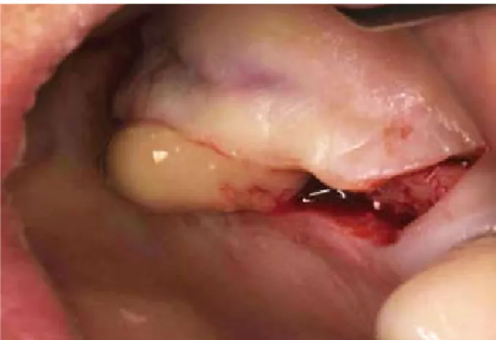
tient after the augmentation procedure. However, the patient visited the clinic 3 weeks later due to swelling of the right pos- terior maxillary area (Figs. 1 and 2). Immediate removal of the graft materials from the sinus was conducted and saline irri- gation was performed several times. Antibiotics (Augmentin 375 mg, Ilsung Pharmaceuticals, Seoul, Korea) were prescribed three times a day for 7 days and 0.12% chlorhexidine solution (Hexamedine, Bukwang Pharmaceutical, Seoul, Korea) was also prescribed twice a day for the first 2 weeks. After removal of the graft materials and prescription of the antibiotics, the infected sinus was successfully treated. The right maxillary si- nus was then re-augmented 9 months after postoperative in- fection due to the patient’s schedule. At that time, the inner portion of the buccal flap had fused with the sinus membrane.
The membrane had also clinically hardened and thickened, with some portions containing residual graft particles. The removal of the graft was impossible because the membrane and the residual graft had fused together. Therefore, the si- nus membrane was separated from the inner portion of the flap by sharp dissection and the stiffened membrane was then carefully elevated. Due to the thickened and hardened condition of the sinus membrane, membrane perforation seldom occurred during re-augmentation. Dental implants were then placed 7 months after sinus re-augmentation. No further complications were observed following re-operation and final restorations were made 10 months later. The im- plants placed in the re-augmented sinus were clinically healthy and the implant–supported restorations have been functioning successfully for 26 months (Fig. 3).
Case 2
A 49-year-old female visited the clinic for reconstruction of the left edentulous posterior maxilla. Radiographic examina-
Figure 1. Postoperative infection following sinus augmentation. Figure 2. The discharge of pus and graft materials after the inci- sion.
4-5 mm (Fig. 4). The dental implants were placed simultane- ously with sinus augmentation because there was sufficient residual bone for the primary stability of the dental implants.
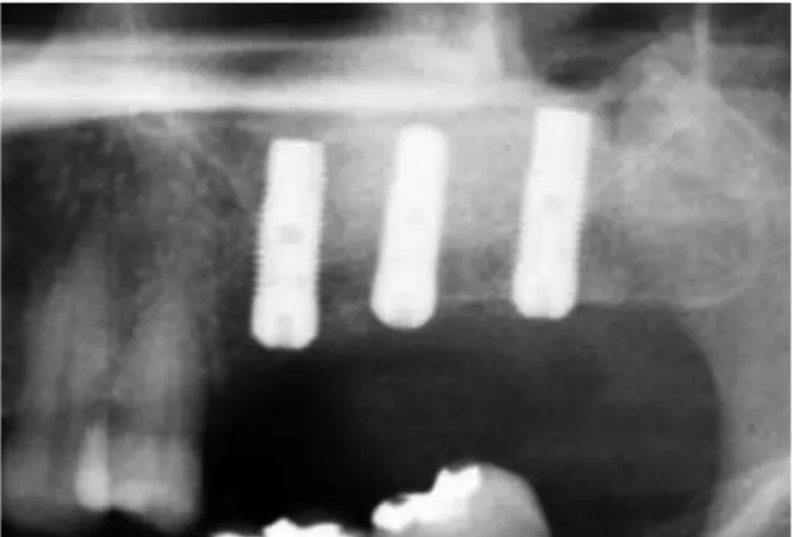
The maxillary sinus was then grafted with deproteinized bo- vine bone and three Astra Tech implants (Tioblast®, Astra Tech Dental Implant System, Mo_lndal, Sweden) 4 mm in diameter and 13 mm in length were placed at the site of #25i-#27i (Fig.
5). Eight days after the procedure, the patient complained of swelling and pain at the surgical area. Evaluation revealed that the left buccal vestibule was swollen due to pus forma- tion. Under local anesthesia, an incision was made at the sur- gical area to drain the pus and infected grafted materials from the maxillary sinus, after which antibiotics (Augmentin 375 mg, Ilsung Pharmaceuticals, Seoul, Korea) were prescribed three times a day for 7 days and 0.12% chlorhexidine solution (Hexamedine, Bukwang Pharmaceutical, Seoul, Korea) was also prescribed twice a day for the first 2 weeks. The infected
the graft material was completely removed from the sinus.
Although the implant at the #26i site was explanted to facili- tate the removal of the graft material, the implants at #25i and
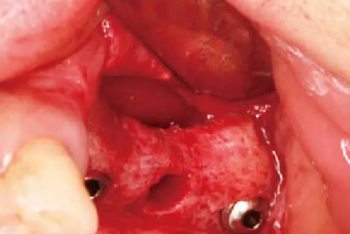
#27i were left in place (Fig. 6). Sinus re-augmentation was then conducted 3 months later, at which time the implant was re- placed simultaneously at position #26i (Figs. 7 and 8). When compared to case 1, the sinus membrane was less stiffened and hardened. This difference may have been due to the dif- ference in waiting period before the sinus re-augmentation.
Consequently, membrane elevation in this case was easier to perform than in case 1. After exposing portions of the dental implants at the site of #25i and #27i, the site was rinsed with tetracycline-HCl solution (50 mg/mL) several times to steril- ize the area, after which deproteinized bovine bone mixed with tetracycline (50 mg/mL) was grafted simultaneously with the placement of the implant at #26i. No further com- plications occurred following sinus re-augmentation, and the
Figure 3. Postoperative panoramic radiograph following sinus re- augmentation along with the placement of dental implants.
Figure 5. Implant placed on positions #25-27 simultaneously with the sinus augmentation.
Figure 4. Pneumatization on the left maxillary sinus was observed in the preoperative panoramic radiograph.
Figure 6. Infected graft material was surgically removed and #26i was removed to ease removal of the infected graft.
implants were loaded 8 months later. The dental implants that were placed in the re-grafted sinus have been functioning very well for 33 months (Fig. 9).
DISCUSSION
This case report demonstrates that surgical procedures that involve total removal of the grafted material, prescription of antibiotics, and sinus re-augmentation can be used to success- fully treat postoperative sinus infection. Although sinus aug- mentation is very predictable [8] and complications caused by sinus graft are very rare, clinicians have experienced various types of complications, such as perforation of the sinus mem- brane, excessive bleeding, infection of the grafted sinuses, and failure of the formation of bone during and after sinus aug- mentation procedures [9-17]. Among these complications, in- fection of the grafted sinuses is less common, but if it occurs,
the infection can spread quickly to the adjacent areas result- ing in brain abscess, infraorbital abscess, and orbital cellulites [14]. For the above reasons, infected sinuses should be treated immediately. According to Anavi et al. [18], postoperative com- plications often occur due to a poor preoperative clinical sta- tus. Misch [19] reported that factors contributing to the de- velopment of the sinus infection included perforation of the sinus membrane, inoculation of the graft with saliva, dehis- cence of the incision line, and lack of aseptic conditions.
Barone et al. [15] described seven maxillary sinus augmenta- tion procedures performed in 7 patients who exhibited suppu- ration 3 to 5 weeks after surgical treatment. Five of those seven patients were smokers. The sinus infections in those patients were treated by draining the area through the bony window and subsequent administration of systemic antibiotics. Five patients received additional sinus augmentation 4 to 6 months after the sinus suppuration. Schwartz-Arad et al. [16] investi- gated 81 cases involving 70 patients and found only 1 report of infection. In the infected case, curettage and H2O2 irriga- tion were used to treat the infected area. Membrane perfora- tions appear to be associated with postoperative complica- tions following sinus augmentation.
Zijderveld et al. [17] reported infection after sinus augmen- tation along with a purulent discharge in 2 patients with local wound dehiscence. These patients were treated with antibi- otics and local debridement. Lindhe et al. [20] found that sur- gical removal of all of the graft material from the sinus cavity and subsequent administration of high doses of antibiotics was essential to the successful treatment of infection. Based on a review of the studies presented above, there are two pos- sible methods of treating such infections. These include: 1) drainage and the administration of systemic antibiotics; 2) total removal of the infected graft and the administration of systemic antibiotics. In this case report, complete removal of Figure 9. Panoramic radiograph of the dental implants placed in
the re-grafted sinus after successfully functioning for 33 months.
Figure 7. The inner portion of the buccal flap and the sinus mem- brane were fused. The membrane and the residual graft material were inseparably fused together.
Figure 8. Elevation of the sinus membrane and ostectomy were performed.
sidual source of infection and treat the infected sinus imme- diately.
Bravetti et al. [21] reported that the membrane remained healthy after sinus augmentation procedures conducted us- ing graft materials. Similarly, Sul et al. [22] reported that sur- gical procedures had little effect on the histologic character- istics of the sinus membrane. However, to the best of the au- thor’s knowledge, there have been few reports conducted to evaluate clinical and histological changes in the infected si- nus membrane following sinus augmentation. In the cases described here, the sinus membrane was clinically different during re-augmentation when compared to the membrane that was subjected to the first sinus augmentation procedure.
This was because the sinus membrane that had gone through sinus infection had fused with the inner portion of the buccal mucoperiosteal flap. These findings indicate that, when si- nus re-augmentation was applied, reflection of the mucope- riosteal flap should be performed very carefully. If needed, sharp dissection of the fused portion should be conducted for the reflection of the buccal flap without tearing the sinus membrane. Additionally, in the case described here, the mem- brane elevation procedure was difficult because the sinus membrane had thickened and hardened and some portions contained residual graft particles. It was impossible to re- move the residual particles because they had fused with the healed membrane. However, it is believed that these particles played no role in the infection because no further complica- tions were reported following sinus re-augmentation. The differences in the membrane following augmentation are believed to play a role in the negative effects on the mucocili- ary function of the respiratory epithelium. However, in this case report, the patients did not show any postoperative com- plications, and the implant-supported restorations have func- tioned properly for more than 2 years.
Removal of the dental implant in cases of infection should be seriously considered when the infection occurs in the case of simultaneous augmentation and implantation. In case 2, the implant placed at position #26i was explanted to facilitate removal of the infected graft material, but the implants at #25i and #27i were retained. When the infection subsided, the ex- posed implant surfaces at #25i and #27i were rinsed with tetra- cycline-HCl solution to sterilize the area. Deproteinized bo- vine bone mixed with tetracycline was then grafted and the implant was simultaneously placed on #26i. The dental im- plants at positions #25i and #27i that were exposed to the postoperative infection have been functioning successfully for 33 months without any complications. Therefore, the re- moval of the dental implants has to be performed selectively, for example, to ease of the removal of an infected graft.
tation are uncommon, they can be very problematic to both patients and clinicians when they do occur. To minimize the occurrence of postoperative infection, the possible causes should be removed prior to sinus augmentation. However, uncontrollable postoperative infection can occur for unknown reasons. The treatments described in this case report con- sisted of surgical removal of the infected graft material from the sinus and the administration of high doses of antibiotics.
These clinical approaches can be used to successfully treat an infected sinus following sinus augmentation.
CONFLICT OF INTEREST
No potential conflict of interest relevant to this article was reported.
REFERENCES
Albrektsson T. A multicenter report on osseointegrated 1.
oral implants. J Prosthet Dent 1988;60:75-84.
Engquist B, Bergendal T, Kallus T, Linden U. A retrospec- 2.
tive multicenter evaluation of osseointegrated implants supporting overdentures. Int J Oral Maxillofac Implants 1988;3:129-34.
Jemt T, Lekholm U, Adell R. Osseointegrated implants in 3.
the treatment of partially edentulous patients: a prelimi- nary study on 876 consecutively placed fixtures. Int J Oral Maxillofac Implants 1989;4:211-7.
Boyne PJ, James RA. Grafting of the maxillary sinus floor 4.
with autogenous marrow and bone. J Oral Surg 1980;38:
613-6.
Misch CE. Maxillary sinus augmentation for endosteal 5.
implants: organized alternative treatment plans. Int J Oral Implantol 1987;4:49-58.
Summers RB. The osteotome technique: Part 3--Less in- 6.
vasive methods of elevating the sinus floor. Compendium 1994;15:698-710.
Fugazzotto PA, De PS. Sinus floor augmentation at the 7.
time of maxillary molar extraction: success and failure rates of 137 implants in function for up to 3 years. J Perio- dontol 2002;73:39-44.
Wallace SS, Froum SJ. Effect of maxillary sinus augmenta- 8.
tion on the survival of endosseous dental implants. A sys- tematic review. Ann Periodontol 2003;8:328-43.
Mardinger O, Nissan J, Chaushu G. Sinus floor augmenta- 9.
tion with simultaneous implant placement in the severely atrophic maxilla: technical problems and complications. J Periodontol 2007;78:1872-7.
Hernandez-Alfaro F, Torradeflot MM, Marti C. Prevalence 10.
tions during sinus-lift procedures. Clin Oral Implants Res 2008;19:91-8.
Pikos MA. Maxillary sinus membrane repair: update on 11.
technique for large and complete perforations. Implant Dent 2008;17:24-31.
Ardekian L, Oved-Peleg E, Mactei EE, Peled M. The clini- 12.
cal significance of sinus membrane perforation during augmentation of the maxillary sinus. J Oral Maxillofac Surg 2006;64:277-82.
Lockhart R, Ceccaldi J, Bertrand JC. Postoperative maxil- 13.
lary cyst following sinus bone graft: report of a case. Int J Oral Maxillofac Implants 2000;15:583-6.
Misch CE. Contemporary implant dentistry. 3rd ed. St.
14.
Louis: Mosby/Elsevier; 2008.
Barone A, Santini S, Sbordone L, Crespi R, Covani U. A 15.
clinical study of the outcomes and complications associ- ated with maxillary sinus augmentation. Int J Oral Maxill- ofac Implants 2006;21:81-5.
Schwartz-Arad D, Herzberg R, Dolev E. The prevalence of 16.
surgical complications of the sinus graft procedure and their impact on implant survival. J Periodontol 2004;75:
Zijderveld SA, van den Bergh JP, Schulten EA, ten Bruggen- 17.
kate CM. Anatomical and surgical findings and complica- tions in 100 consecutive maxillary sinus floor elevation procedures. J Oral Maxillofac Surg 2008;66:1426-38.
Anavi Y, Allon DM, Avishai G, Calderon S. Complications 18.
of maxillary sinus augmentations in a selective series of patients. Oral Surg Oral Med Oral Pathol Oral Radiol En- dod 2008;106:34-8.
Misch CM. The pharmacologic management of maxillary 19.
sinus elevation surgery. J Oral Implantol 1992;18:15-23.
Lindhe J, Lang NP, Karring T. Clinical periodontology and 20.
implant dentistry. Oxford: Blackwell Munksgaard; 2008.
Bravetti P, Membre H, Marchal L, Jankowski R. Histologic 21.
changes in the sinus membrane after maxillary sinus aug- mentation in goats. J Oral Maxillofac Surg 1998;56:1170-6.
Sul SH, Choi BH, Li J, Jeong SM, Xuan F. Histologic chang- 22.
es in the maxillary sinus membrane after sinus membrane elevation and the simultaneous insertion of dental implants without the use of grafting materials. Oral Surg Oral Med Oral Pathol Oral Radiol Endod 2008;105:e1-5.