I. Introduction
Periodontal ligament(PDL) cells are major fibrob- lastic cells in periodontal connective tissue. They serve in maintenance and remodeling of the peri- odontal ligament. Much is known regarding the reg- ulation of proliferation, differentiation, and activity of the cells in the periodontal ligament during nor- mal function. But less is known about alterations on their phenotypic expression under specific condi- tions like a mechanical stress. It is likely that PDL cells stimulated by the mechanical forces produce some important local factors that participate in the maintenance and remodeling of the ligament itself.
It has been reported that PDL cells increase the lev- els of prostaglandin E1,2), interleukin-1β3,4), cyclic adenosine monophosphate(cAMP)2,5), DNA synthe- sis6), and collagen synthesis6) in response to mechanical stress. PDL cells also activate the plas- minogen activator in response to tension force7). In contrast, alkaline phosphatase activity and its mRNA level were decreased by tension force8). So it is like- ly that mechanical stress evokes biochemical
responses in PDL cells.
In order to understand both the development and the differentiation of normal tissues, it is necessary to study the nature and regulation of the normal cell cycle. Increasing evidences suggest that the growth of tissues in both health and disease is a balance between cell proliferation and cell death. Most reproductive cells begin the process that lead to cell division whose objectives are to produce a pair of identical daughter cells. There are two closely relat- ed families of proteins that are involved in regulating the cell cycle: the cyclin-dependent kinases(cdks) and the cyclins themselves9,10). The cdks are serine/threonine protein kinases. They are function- al only when combined with one of the cyclins, and each mammalian cyclin can interact with multiple cdks. Cyclin function is primarily controlled by changes in cyclin levels while cdks are activated by phosphorylation of a conserved threonine residue.
The cyclins regulate the activities of the cdks and control their ability to modulate the enzymes involved in driving the cell cycle11). Hence, the assembly, activation, and disassembly of the cyclin-
Effect of Mechanical Stress on the Proliferation and Expression of Cell Cycle Regulators in Human Periodontal Ligament Cells
Hyung-Keun You*·Hyung-Shik Shin*·Gene Lee**·Byung-Moo Min**
*Department of Periodontology, School of Dentistry, Wonkwang University,
**Department of Oral Biochemistry, College of Dentistry, Seoul National University
대한치주과학회지 : Vol. 29, No. 3, 1999
*This paper was supported by Wonkwang University in 1998
cdk complex are the critical events in controlling the cell cycle. The cyclins undergo synthesis and degra- dation during each cycle of the cell. There are two main classes of cyclins. One is the G1 cyclins that bind cdks during the G1 phase and are required for entry into the S phase. The other is the mitotic cyclins that bind cdks during the G2 to permit the entry of the cell into M phase12). The G1 cyclin com- plexes exist as the quaternary structures of cyclins, cdks, the universal cdk inhibitor p21WAF1/CIP1, and proliferating cell nuclear antigen(PCNA)13-20). Progression from G1 to S phase requires coordinat- ed and independent activation of both cyclin D complexes in early G1 and cyclin E complexes in later G121-24). D-type cyclins(D1, D2, D3) are associ- ated with either cdk 4 or cdk 6, while cyclin E is associated with cdk 2. These cyclins play distinct roles in the regulation of the G1-S phase transition.
Both cdk and cyclin are once associated with each other and activated by cyclin-activating kinase25-27).
Cyclin dependent kinase inhibitors are proteins that bind and inhibit the activity of cdks. One of them to be identified is p21WAF1/CIP1which binds and inhibits G1 cyclin-cdks complexes28). p21WAF1/CIP1 is a constant part of G1 cyclin complexes, but only acts as a cdk inhibitor when its stoichiometric quantities increase from one to several/unit complexes18-20). In addition to its kinase inhibitor activity, p21WAF1/CIP1 is able to inhibit DNA replication in the S phase of the cell cycle by interaction with PCNA, blocking the ability of PCNA to activate DNA polymerase29,30).
The p53 tumor suppressor protein is a transcrip- tion factor required for the transactivation of a num- ber of genes involved in growth control31,32). Inactivation of wild-type p53 function can lead a growth advantage. It has been demonstrated that DNA damage activates p21WAF1/CIP1 transcription in a p53-dependent manner in human fibroblasts and epithelial cells33). In contrast, several studies on p53-
independent induction of p21WAF1/CIP1 following DNA damage have also been reported34). It has also been shown that p53 can inhibit cell cycle progres- sion without induction of p21WAF1/CIP1 expression35), thus, p21WAF1/CIP1 may arrest cell cycle progression following p53-independent activation, and at the same time p53 may promote cell cycle arrest by acti- vation of genes other than p21WAF1/CIP1.
PCNA is an essential component of the DNA repli- cation machinery, functioning as the accessory pro- tein for DNA polymerase δ, required for processive chromosomal DNA synthesis, and DNA polymerase ε, required for repair of nuclear DNA. PCNA is also required for DNA recombination and repair36). In addition, PCNA is shown to interact with cell cycle regulatory proteins, cdks, cyclin D, and p21WAF1/CIP1
13,27,37).
Mechanical stress is known to be associated with proliferation of PDL cells. Though detailed mecha- nisms for the proliferation of PDL cells by mechani- cal stress remain largely unknown, it may be due to increased expression of cell cycle regulatory pro- teins from the cells. The purpose of this study is to investigate on the growth pattern and expression of p53, p21WAF1/CIP1, cdks, cyclins, and PCNA in PDL cells exposed to mechanical stress.
II. Materials and Methods
1. Cell culturesHuman PDL cells were cultured from periodontal ligament explants of healthy premolar teeth extract- ed for orthodontic reason. The periodontal ligament tissues were teased from the mid-root surface, washed in Hanks' balanced salt solution, and plated onto 60-mm culture dish. Separated tissues were cultured in Dulbecco's Modified Eagle's Medium(DMEM, Gibco/BRL, Grand Island, NY,
U.S.A.) supplemented with 10% FBS, 100 U/ml penicillin and 100㎍/ml streptomycin. Confluent cells were subcultured up to six passages by trypsinization. The cultures with the passage num- ber of 6 were used in this experiment.
2. Mechanical stress
Confluent cell monolayers in 100-mm Petri dishes were trypsinized. The cells were suspended in cul- ture medium and 1×10⁴ cells were plated onto 55- mm Petriperm dishes with a hydrophilic flexible plastic growth surface(Heraeus Instruments, South Plainfield, NJ, USA). The bottom of the dish was stretched by placing a template with a convex sur- face under the dish and placing a stainless steel weight(1kg/dish) on the top of the dish. Because the convex surface of dish is uniformly curved, stretching occurs evenly over bottom of the dish.
The bottom surface curvature of the template is an arc of 36oand a radius of 7.6cm38).
3. Determination of Cell proliferation rate
Confluent cell monolayers in 100-mm Petri dishes were trypsinized. The cells were suspended in cul- ture medium, and 1×10⁴cells were plated onto 55- mm Petriperm dishes. The number of viable cells were counted after 0, 2, 4, 6, 8, 10 and 12 days of incubation under mechanical stress at 37℃ by try- pan blue exclusion. There were six cultures in each group at each time.
4. Western blot analysis
Confluent cell monolayers in 100-mm Petri dishes were trypsinized, and 1×10⁴cells were plated onto 55-mm Petriperm dishes. These cells were exposed to mechanical stress for 0, 2, 6, and 10 days. Protein
was isolated from cells grown in 55-mm Petriperm dishes using Tri-reagent solution(Molecular Research Center, Inc., Cincinnati, OH, U.S.A.) under the conditions recommended by the manu- facturer. Protein concentration was determined by Lowry method39). The denatured supernatant con- taining 50 ?g of protein was electrophoresed in a 12.5% SDS-polyacrylamide gel and transferred onto a nitrocellulose membrane(BioRad, Hercules, CA, U.S.A.). To reduce nonspecific antibody binding, the membrane was incubated in a blocking solu- tion(Zymed, San Francisco, CA, U.S.A.) for 1 h at room temperature. The membrane was exposed to a mouse anti-human monoclonal antibody for p53(Ab-2; Oncogene Science, Uniondale, NY, U.S.A.), a mouse anti-human monoclonal antibody for p21WAF1/CIP1(Santa Cruz Biotechnology, Santa Cruz, CA, U.S.A.), a rabbit anti-human polyclonal antibody for cdk 2(Santa Cruz Biotechnology), a rabbit anti-human polyclonal antibody for cdk 4(Oncogene Science), a rabbit anti-human polyclon- al antibody for cdk 6(Santa Cruz Biotechnology), a mouse anti-human monoclonal antibody for cyclin D1(Santa Cruz Biotechnology), a mouse anti-human monoclonal antibody for cyclin E(Oncogene Science) and a mouse anti-human monoclonal anti- body for PCNA(Oncogene Science) at room temper- ature for 90 min. After washing with PBS, the mem- brane was treated with anti-mouse or anti-rabbit IgG-horse raddish peroxidase conjugated secondary antibody for 1 h, and washed again with PBS. The membrane was then incubated in ECL western blot- ting detection reagent(Amersham, Buckingham- shire, U.K.), and was exposed to Hyperfilm- MP(Amersham) for a few min. The membrane was also stained with 1× Ponceau S. The relative levels of proteins were determined by densitometric scan- ning of autoradiograms.
5. Statistical analysis
Values were calculated as the mean ± standard deviation(S.D.). Statistical significance was evaluat- ed by one way analysis of variance(ANOVA) using SAS program of computer.
III. Results
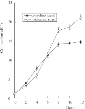
1. Cell proliferation rateHuman PDL cells proliferated well in DMEM sup- plemented with 10% FBS. There was a remarkable increase in the number of cells in the mechanically
stressed cultures as compared to the non-stressed controls. Cell numbers of stressed and non-stressed periodontal ligament cells exhibited different prolif- eration rates in culture during a 12-day period(Figure 1). Before day 6, the number of non- stressed PDL cells was slightly higher than that of stressed PDL cells. But after day 6, the proliferation rate of stressed PDL cells was notably higher than that of non-stressed PDL cells.
2. Expression of PCNA
Because PCNA is a molecular marker of cell pro- liferation, the level of PCNA was studied. Expression of PCNA was increased after day 6 as compared to that of control in the same way as the cell prolifera- tion rate discussed above(Figure 2). On day 6, the
control(no stress) mechanical stress 25
20
15
10
5
0
0 2 4 6 8 10 12
Days Cell number( 10-4)
Figure 1. Growth curves of human PDL cells exposed to mechanical stress. Cells were plated at 1×10⁴cells per 55-mm Petriperm dish, and cultured for 0, 2, 4, 6, 8, 10 or 12 days in the presence of mechanical stress(1 kg/dish). Viable cells were counted with a hemocytome- ter by trypan blue exclusion. Values represent averages from six independent experiments and standard devia- tions.
Figure 2. Western blot analysis for the intracellular PCNA level in human PDL cells exposed to mechanical stress for 0, 2, 6, or 10 days. Cell extract equivalent to 50㎍ of total cellular protein of PDL cells was electrophoresed by 12.5% SDS-PAGE and transferred to a nitrocellulose membrane. The intracellular protein level of PCNA in PDL cells was probed with antibody diluted by 1:1000. After probing, the membrane was stained with 1X Ponceau S stain for 10 min to reveal the total cellular protein loaded per each lane. The densitometric ratio of the Ponceau S stain per each lane to that of the first lane(non-stressed cells) is indicated at the bottom of the figure.
0 2 6 10(days)
kDa 49
33
24
PCNA
Ponceau S
1.0 1.0 1.0 1.0
expression level was notably increased as compared to that of control. And on day 10, it was also increased as compared to that of control, but it was decreased when compared to that of day 6.
3. Expression of p53 and p21WAF1/CIP1
Because p53 can regulate the expression of genes involved in cell cycle, and because the cdk inhibito- ry protein p21WAF1/CIP1can bind cyclin-cdk complex-
es and thereby inhibit their activities, the expression level of both proteins was investigated(Figure 3).
The level of p53 and p21WAF1/CIP1protein in stressed
Figure 3. Western blot analysis for the intracellular p53 and p21WAF1/CIP1 levels in human PDL cells exposed to mechanical stress for 0, 2, 6, or 10 days. Cell extract equivalent to 50㎍ of total cellular protein of PDL cells was electrophoresed by 12.5% SDS-PAGE and trans- ferred to a nitrocellulose membrane. The intracellular pro- tein levels of p53 and p21WAF1/CIP1in PDL cells were probed with respective antibodies diluted by 1:1000.
After probing, the membrane was stained with 1X Ponceau S stain for 10 min to reveal the total cellular pro- tein loaded per each lane. The densitometric ratio of the Ponceau S stain per each lane to that of the first lane(non-stressed cells) is indicated at the bottom of the figure.
0 2 6 10(days)
1.0 1.0 1.0 0.9
1.0 1.0 1.0 0.9 kDa
75 49 A
B 33
24
17 p21WAF1/CIP1
p53
Ponceau S
Ponceau S
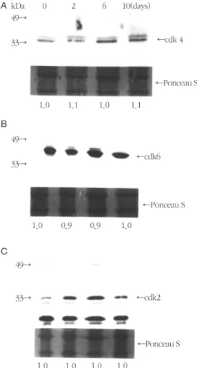
Figure 4. Western blot analysis for the intracellular levels of cdks in human PDL cells exposed to mechanical stress for 0, 2, 6, or 10 days. Cell extract equivalent to 50
㎍ of total cellular protein of PDL cells was elec- trophoresed by 12.5% SDS-PAGE and transferred to a nitrocellulose membrane. The intracellular protein levels of cdks in PDL cells were probed with respective anti- bodies diluted by 1:1000. After probing, the membrane was stained with 1X Ponceau S stain for 10 min to reveal the total cellular protein loaded per each lane. The densit- ometric ratio of the Ponceau S stain per each lane to that of the first lane(non-stressed cells) is indicated at the bot- tom of the figure.
kDa 49
33
49
33
49
33
cdk 4
cdk6
cdk2 Ponceau S
Ponceau S Ponceau S
0 2 6 10(days)
1.0 1.1 1.0 1.1
1.0 0.9 0.9 1.0
A
B
C
1.0 1.0 1.0 1.0
PDL cells was consistent throughout all the experi- mental periods.
4. Expression of cyclins and cdks
Because the proteins involved in the middle G1 phase were cyclin D1 and cdk 4 or 6, and because regulatory proteins involved in the late G1 phase were cyclin E and cdk 2 in normal cells, western
blot analysis was carried out to determine the nature of this complex in periodontal ligament cells. It revealed that cdk 4 was present at a low level on day 2, but it was notably increased on day 6 and 10(Figure 4A). The increase rate was slightly reduced on day 10 when compared to day 6. It might be due to plateau of cell proliferation. The expression of cdk 6(Figure 4B) and cyclin D1(Figure 5A) was shown a slightly different pattern from that of cdk 4. There was a slightly increasing tendency on day 6 and 10 when compared to non-stressed cells. The level of cdk 2 was increased during the experimental periods when compared to that of control cells(Figure 4C), especially the expression on day 6 was notably increased as compared to that of any other experimental period. The degree of expression of cyclin E showed no change on day 6 and 10(Figure 5B).
IV. Discussion
In the present study, when PDL cells were stressed by mechanical stimulation, cell proliferation and the levels of cell cycle progressing proteins, cyclin D1, cdk 4, 6 and 2, and PCNA, were increased after day 6. It was shown that increased expression of cdks caused cell proliferation in PDL cells by mechanical stress. p53, a archetypal check- point regulator of cell cycle progression, and p21WAF1/CIP1, a universal negative regulator of cell cycle, however, had little effect on cell cycle pro- gression in this experiment.
When cells are stimulated by mechanical stress, there are many changes in biochemical characteris- tics as well as in cell number. The effect of stretch- ing on cellular proliferation appears to vary with cell type. Epithelial cells which were derived from the cell rests of Malassez exhibited a 92% increase in the labeling index40), but Leung et al. showed no consis- Figure 5. Western blot analysis for the intracellular levels
of G1 cyclins(cyclin D1 and E) in human PDL cells exposed to mechanical stress for 0, 2, 6, or 10 days. Cell extract equivalent to 50 ㎍ of total cellular protein of PDL cells was electrophoresed by 12.5% SDS-PAGE and transferred to a nitrocellulose membrane. The intracellular protein levels of G1 cyclins in PDL cells were probed with respective antibodies diluted by 1:1000. After probing, the membrane was stained with 1X Ponceau S stain for 10 min to reveal the total cellular protein loaded per each lane. The densitometric ratio of the Ponceau S stain per each lane to that of the first lane(non-stressed cells) is indicated at the bottom of the figure.
kDa 49
33
83 49
28
cyclin D1
cyclin E
Ponceau S Ponceau S
0 2 6 10(days)
1.0 1.0 1.1 1.0
1.0 1.0 1.1 1.0 A
B
tent increase in DNA systhesis as a result of cyclic stretching of smooth muscle cells41). Somjen et al. reported that stretching caused a 45% increase in the amount of DNA synthesis in bone cell cultures42). And some investigators have shown an increase in the level of cAMP after mechanical stress was applied to bone cells43,44), but Harell et al. showed that a prolonged decrease followed an initial increase in the level of cAMP45). In the present study, an increase of PDL cells was observed as a whole, but there was a decrease in cell number at early time period. It might be because the number of cells was not large enough to be influenced by mechanical stress or because there was no direct effect of mechanical stress on PDL cells.
The biochemical mechanisms are poorly under- stood which were involved in the conversion of mechanical stimuli into biological response by signal transduction in cells. Although the mechanism for the detection and conversion of mechanical force into a biochemical signal has yet to be determined, several pathways have been proposed: i)one possi- ble transduction pathway is the extracellular matrix - integrin - cytoskeleton machinery, ii)mechanosensi- tive channels are candidates since no second mes- senger is required for channel activation, iii)another possible mechanism for mechanotransduction involves guanine nucleotide binding proteins, iv) specific regulation may occur at receptor tyrosine kinase by growth factor, v) and mechanical stress may induce a conformational change in a nonrecep- tor protein tyrosine kinase receptor46-50). The bind- ing of ligand to receptor activates phospholipase C that hydrolyzes phosphatidyl inositol 4,5- biphos- phate into inositol 1,4,5-triphosphate(IP3) and dia- cylglycerol(DAG). IP3causes the rapid release of Ca2+from intracellular stores of calcium, and DAG activates protein kinase C(PKC). The increased con- centration of cytosolic Ca2+and PKC activates cAMP
synthesis, and then causes the increase of cellular division and proliferation51,52).
Cylin D and its major catalytic partners, cdk 4 or cdk 6, are an early event in cell cycle initiation, and cyclin E and cdk 2 are involved in late G1 phase. In almost all of the studies, the expression level of cyclin D and E was associated with cdk 4 or 6, and 2. In the present study, the degree of expression of cyclin was changed along that of cdks. The increase in cdk 4 and cdk 6 was comparable to the increase in cyclin D1 as days go by. Although the expression level of cyclin E was slightly increased during the experimental period, especially the expression of cdk 2 under mechanical stress condition was remarkably increased compared to that of control.
The most abundant expression was shown on day 6 in cyclin and cdks. It might be due to the fact that contact inhibition of confluent cells occured on day 10.
PCNA plays an important role in nucleic acid metabolism. The best understood function of PCNA is in DNA replication. PCNA plays an essential role in DNA replication as the auxillary protein. PCNA is isolated as a protein with elevated levels during S phase. Studies have shown that the protein is local- ized to the nucleus only in cells that are in the S phase of the cell cycle53,54). In normal cells, PCNA exists in multiple quaternary complexes, each con- taining a cdk, cyclin, and p21WAF1/CIP1 13-20). In the present study, the increase in the expression of PCNA is regarded as an essential component of cell cycle progression as well as a molecular marker of cell cycle progression.
p53 is associated with cell proliferation, DNA repair, maintenance of DNA integrity, and regulation of apoptosis. Transcription of the p21WAF1/CIP1gene can be activated by p53 and several p53-indepen- dent mechanisms. In some cases p53 did not require induction of p21WAF1/CIP1 to proliferate and
differentiate cell. p53-independent induction of p21WAF1/CIP1 expression at the transcriptional level has been observed as an immediate early response to a variety of physiological and chemical inducers of differentiation. In the present study, p53 level of expression showed little change for 2 to 10 days.
This result indicates that p53 did not affect the pro- gression of the mechanically stressed PDL cell cycle under the condition reported here. Previous studies have established that cdk inhibitor protein p21WAF1/CIP1has a role in regulating the G1-S phase progression by inhibition of cyclin-cdk catalytic activity55,56). cdks inhibitors function by binding themselves to cyclin D-cdk complexes to inhibit their kinase activities. However, p21WAF1/CIP1does not seem to always function as a cyclin-cdk inhibitor because several findings showed that in proliferating cells the majority of p21WAF1/CIP1protein was found in active cyclin-cdk complexes20,57), and that despite the induction of cdk inhibitor p21WAF1/CIP1, cyclin D1 associated with cdk kinase remained activated and the cells grew essentially like that of their parent cells58). In the present study, western blot analysis of p21WAF1/CIP1 in the mechanically stressed PDL cells illustrated that expressions of p21WAF1/CIP1showed little change, but the expression of cyclin-cdk com- plexes was slightly increased in the experimental period by mechanical stress. This result seems to be explained by the fact that the absolute ratio between p21WAF1/CIP1 and the cyclin-cdk complexes deter- mines the cell cycle inhibitory behavior of p21WAF1/CIP1. In more detail, although p21WAF1/CIP1 level is increased in the stoichiometry between the cyclin D1-cdk complex, but if p21WAF1/CIP1does not change, and a stable and active cyclin D1-cdk- p21WAF1/CIP1 complex is sustained, a progression of cell cycle may result under these conditions. If, however, the increased level of p21WAF1/CIP1protein overcomes that of cyclin D1-cdk, the ratio of the
cyclin D1-cdk complexes to p21WAF1/CIP1 will be biased toward p21WAF1/CIP1. This situation would suppress the cell cycle progression58). In the present study, the results indicate that mechanical stress on human PDL cells causes the increase of expression of cdks without change of level of p53 and p21WAF1/CIP1, and the increase of cell proliferation rate.
V. Conclusion
Mechanical stress is known to be associated with proliferation of periodontal ligament(PDL) cells.
Though detailed mechanisms for the proliferation of PDL cells by mechanical stress remain largely unknown, it may be due to the increased expres- sion of cell cycle regulatory proteins from the cells.
To investigate this possibility, growth pattern and expression of p53, p21WAF1/CIP1, cyclin-dependent kinases(cdks), cyclins, and proliferating cell nuclear antigen(PCNA) were determined in PDL cells exposed to mechanical stress(1 kg/55-mm Petriperm dish). Mechanical stress notably increased cell prolif- eration rate and expression of PCNA in the PDL cells exposed to stress for 6-10 days when compared to normal cells. Mechanical stress also slightly increased expression of cdks and cyclin D1 in these cells, but levels of p53 and p21WAF1/CIP1 were not changed. These results indicate that the increase of cell proliferation by mechanical stress may be due to the increased expression of cdks without the change of p53 and p21WAF1/CIP1 levels in human PDL cells.
VI. References
1. Yamaguchi, M., Shimizu, N., Goseki, T., Shibata, Y., Takiguchi, H., Iwasawa, T. and Abiko, Y. Effect of different magnitudes of tension force on prostaglandin E2 production
by human periodontal ligament cells. Archs Oral Biol 39:877-884, 1994.
2. Ngan, P., Saito, S., Saito, M., Lanese, R., Shanfeld, J. and Davidovitch, Z. The interac- tive effects of mechanical stress and inter- leukin-1? on prostaglandin E and cyclic AMP production in human periodontal ligament fibroblasts in vitro:comparison with cloned osteoblastic cells of mouse(MC3T3-E1). Archs Oral Biol 35(9):717-725, 1990.
3. Shimizu, N., Yamaguchi, M., Goseki, T., Ozawa, Y., Saito, K., Takiguchi, H., Iwasawa, T. and Abiko, Y. Cyclic-tension force stimu- lates interleukin-1β production by human periodontal ligament cells. J Periodont Res 29:328-333, 1994.
4. Saito, M., Saito, S., Ngan. P., Shanfeld, J. and Davidovitch, Z. Interleukin-1β and prostaglandin E are involved in the response of periodontal cells to mechanical stress in vivo and in vitro. Am J Orthod Dentofac Orthop 99:226-240, 1991.
5. Yousefian, J., Firouzian, F., Shanfeld, J., Ngan, P., Lanese, R. and Davidovitch, Z. A new experimental model for studying the response of periodontal ligament cells to hydrostatic pressure. Am J Orthod Dentofac Orthop 108:402-409, 1995.
6. Kunz, J., Plascke, C. and Duncker, M. Cell proliferation and 3H-proline incorporation in periodontal ligament exposed to mechanical stress. Exp Pathol 34:51-58, 1988.
7. Yamaguchi, M., Shimizu, N., Ozawa, Y., Saito, K., Miura, S., Takiguchi, H., Iwasawa, T. and Abiko, Y. Effect of tension-force on plasmino- gen activator activity from human periodontal ligament cells. J Periodont Res 32(3):308-314, 1997.
8. Yamaguchi, M., Shimizu, N., Shibata, Y. and
Abiko, Y. Effects of different magnitudes of tension-force on alkaline phosphatase activity in periodontal ligament cells. J Dent Res 75(3):889-894, 1996.
9. Nurse, P. Ordering S phase and M phase in the cell cycle. Cell 79:547-550, 1994.
10. Sherr, C. G1 phase progression:cycling on cue. Cell 79:551-555, 1994.
11. Kato, J.A., Matsuoka, M., Strom, D. and Sherr, C. Regulation ofcyclin D-dependent kinase 4(cdk4) by cdk4-activating kinase. Mol Cell Biol 14:2713-2721, 1994.
12. Leake, R. The cell cycle and regulation of can- cer cell growth. Ann N Y Aca Sci 253-262, 1996.
13. Xiong, Y., Zhang, H. and Beach, D. D type cyclins associate with multiple protein kinases and the DNA replication and repair factor PCNA. Cell 71:505-514, 1992.
14. El-Deiry, W.S., Tokino, T., Velculescu, V.E., Levy, D.B., Parson, R., Trent, J.M., Lin, D., Mercer, W.E., Kinzler, K.W. and Vogelstein, B. WAF1, a potential mediator of p53 tumor suppression. Cell 75:817-825, 1993.
15. Gu, Y., Turck, C.W. and Morgan, D.O.
Inhibition of CDK2 activity in vivoby an asso- ciated 20K regulatory subunit. Nature (London) 366:707-710, 1993.
16. Harper, J.W., Adami, G.R., Wei, N., Keyomarsi, K. and Elledge, S.J. The p21 cdk-interacting protein Cip1 is a potent inhibitor of G1 cyclin- dependent kinases. Cell 75:805-816, 1993.
17. Noda, A., Ning, Y., Venable, S.F., Pereira- Smith, O.M. and Smith, J.R. Cloning of senes- cent cell-derived inhibitors of DNA synthesis using an expression screen. Exp Cell Res 211:90-98, 1994.
18. Xiong, Y., Hannon, G.J., Zhang, H., Casso, D., Kobayashi, R. and Beach, D. p21 is a uni-
versal inhibitor of cyclin kinases.
Nature(London) 366:701-704, 1993.
19. Zhang, H., Xiong, Y. and Beach, D.
Proliferating cell nuclear antigen and p21 are components of multiple cell cycle kinase com- plexes. Mol Cell Biol 4:897-906, 1993.
20. Zhang, H., Hannon, G.J. and Beach, D. p21- containing cyclin kinases exist in both active and inactive states. Genes Dev 8:1750-1758, 1994.
21. Lukas, J., Muller, H., Bartkova, J., Spitkovsky, D., Kjerulff, A.A., Jansen-Durr, P., Strauss, M.
and Bartek, J. DNA tumor virusoncoproteins and retinoblastoma gene mutations share the ability to relieve the cell's requirement for cyclin D1 function in G1. J Cell Biol 125(3):625-638, 1994.
22. Ohtsubo, M., Theodoras, A.M., Schumacher, J., Roberts, J.M. and Pagano, M. Human cyclin E, a nuclear protein essential for the G1- to-S phase transition. Mol Cell Biol 15(5):2612- 2624, 1995.
23. Tam, S.W., Theodoras, A.M., Shay, J.W., Draetta, G.F. and Pagano, M. Differential expression and regulation of cyclin D1 protein in normal and tumor human cells:association with cdk4 is required for cyclin D1 function in G1 progression. Oncogene 9:2663-2674, 1994.
24. Resnitzky, D. and Reed, S.I. Different roles for cyclins D1 and E inregulation of the G1-to-S transition. Mol Cell Biol 15(7): 3463-3469, 1995.
25. Fisher, R.P. and Morgan, D.O. A novel cyclin associates with MO15/CDK7 to form the CDK- activating kinase. Cell 78(8):713- 724, 1994.
26. Makela, T.P., Tassan, J.P., Nigg, E.A., Frutiger, S., Hughes, G.J. and Weinberg, R.A. A cyclin associated with the cdk- activating kinase MO15. Nature(London) 371:254-257, 1994.
27. Matsuoka, M., Kato, J.A., Fisher, R.P., Morgan D.O. and Sherr, C. Activation of cyclin-depen- dent kinase-4(CDK4) by mouse MO15-associ- ated kinase. Mol Cell Biol 14(11):7265-7275, 1994.
28. Gartel, A.L., Serfas, M.S. and Tyner, A.L. p21- negative regulator of the cell cycle. Proc Soc Exp Biol Med 138-149, 1996.
29. Flores-Rozas, H., Kelman, Z., Dean, F.B., Pan, Z.Q., Harper, J.W., Elledge, S.J., O'Donnell, M. and Hurwitz, J. Cdk- interacting protein 1 directly binds with proliferating cell nuclear antigen and inhibits DNA replication catalyzed by the DNA polymerase δ holoenzyme. Proc Natl Acad Sci 91(8):8655- 8659, 1994.
30. Waga, S., Hannon, G.J., and Beach, D. and Stillman B. The p21 inhibitor of cyclin-depen- dent kinases controls DNA replication by interaction with PCNA. Nature(London) 369:574- 578, 1994.
31. Clarke, A.R., Purdie, C.A., Harrison, D.J., Morris, R.G., Bird, C.C., Hooper, M.L. and Wyllie, A.H. Thymocyte apoptosis induced by p53 dependent and independent pathways.
Nature(London) 362:849-852, 1993.
32. Lowe, S.W., Schmitt, E.M., Smith, S.W., Osborne, B.A. and Jacks, T. p53 is required for radiation-induced apoptosis in mouse thy- mocytes. Nature(London) 362:847-849, 1993.
33. Dulic, V., Kaufmann, W.K., Wilson, S.J., Tlsty, T.D., Lees, E., Harper, J.W., Elledge, S.J. and Reed, S.I. p53-dependent inhibition of cyclin- dependent kinase activities in human fibrob- lasts during radiation-induced G1 arrest. Cell 76:1013- 1023, 1994.
34. Akashi, M., Hachiya, M., Osawa, Y., Spirin, K., Suzuki, G. and Koeffler, H.P. Irradiation induces WAF1 expression through a p53-inde- pendent pathway in KG-1 cells. J Biol Chem
270: 19181-19187, 1995.
35. Hirano, Y., Yamato, K. and Tsuchida, N. A temperature sensitive mutant of the human p53, Val138, arrests rat cell growth without induced expression of cip1/waf1/sdi1 after temperature shift-down. Oncogene 10:1879- 1885, 1995.
36. Kelman, Z. PCNA:structure, functions and interactions. Oncogene 14:629-640, 1997.
37. Pagano, M., Theodoras, A.M., Tam, S.W. and Draetta, G.F. Cyclin D1 mediated inhibition of repair and replicative DNA synthesis in human fibroblasts. Genes Dev 8:1627-1639, 1994.
38. Hasegawa, S. , Sato, S., Saito, S., Suzuki, Y.
and Brunette, D. M. Mechanical stretching increases the number of cultured bone cellssynthesizing DNA and alters their pattern of protein synthesis. Calcif Tissue Int 37:431- 436, 1985.
39. Lowry, O.H., Rosebrough, N.J., Lewis Farr, A.
and Randall, R. J.Protein measurement with the folin phenol reagent. J Biol Chem 193:265- 275, 1951.
40. Brunette, D.M. Mechanical stretching increas- es the number of epithelial cells synthesizing DNA in culture. J Cell Sci 69:35-45, 1984.
41. Leung, D.Y.M., Glagov, S. and Mathews, M.B. A new in vitro system for studying cell response to mechanical stimulation. Exp Cell Res 109:285-298, 1977.
42. Somjen, D., Binderman, I., Berger, E. and Harell, A. Bone remodelling induced by phys- ical stress is prostaglandin E2 mediated.
Biochim Biophys Acta 627:91-100, 1980.
43. Binderman, I., Shimshoni, Z. and Somjen, D.
Biochemical pathways involved in the transla- tion of physical stimulus into biological mes- sage. Calcif Tissue Int 36(supplement):82-85, 1984.
44. Binderman, I., Zor, U., Kaye, A.M., Shimshoni, Z., Harell, A. and Somjen, D. The transduction of mechanical force into bio- chemical events in bone cells may involve activation of phospholipase A2. Calcif Tissue Int 42:261-266, 1988.
45. Harell, A., Dekel, S. and Binderman, I.
Biochemical effect of mechanical stress on cul- tured bone cells. Calcif Tissue Res 22(supple- ment):202-207, 1977.
46. Brighton, C.T., Fisher, J.R., Levine, S.E., Corsetti, J.R., Reilly, T., Landsman, A.S. and Williams, J.L. The biochemical pathway medi- ating the proliferative response of bone cells to a mechanical stimulus. J Bone and Joint Surg 78-A(9):1337-1347, 1996.
47. Duncan, R.L. and Turner, C.H.
Mechanotransduction and the functional response of bone to mechanical strain. Calcif Tissue Int 57:344-358, 1995.
48. Carvalho, R.S., Scott, J.E., Suga, D.M. and Yen, E.H.K. Stimulation of signal transduction pathways in osteoblasts by mechanical strain potentiated by parathyroid hormone. J Bone Min Res 9(7):999-1011, 1994.
49. Banes, A.J., Tsuzaki, M., Yamamoto, J., Fischer, T., Brigman, B., Brown, T. and Miller, L. Mechanoreception at the cellular level:the detection, interpretation, and diversity of responses to mechanical signals. Biochem Cell Biol 73:349-365, 1995.
50. Karin, M. Signal transduction from cell surface to nucleus in development and disease.
FASEB J 6:2581-2590, 1992.
51. Sekar, M.C. and Hokin, L.E. The role of phos- phoinositides in signal transduction. J Memb Biol 89:193-210, 1986.
52. Vergara, J., Tsien, R.Y. and Delay, M. Inositol 1,4,5- trisphosphate: a possible chemical link
in excitation-contraction coupling in muscle.
Proc Nat Acad Sci 82:6352-6356, 1985.
53. Bravo, R. and Macdonald-Bravo, H. Changes in the nuclear distribution of cyclin(PCNA) but not its synthesis depend on DNA replication.
EMBO J 4:655-661, 1985.
54. Bravo, R. and Macdonald-Bravo, H. Existence of two populations of cyclin/proliferating cell nuclear antigen during the cell cycle:associa- tion with DNA replication sites. J Cell Biol 105(10):1549-1554, 1987.
55. Harper, J.W., Elledge, S.J., Keyomarsi, K., Dynlacht, B., Tsai, L.H., Zhang, P., Dobrowolski, S., Bai, C., Connel-Crowley, L., Swindell, E., Fox, M.P. and Wei, N.Inhibition of cyclin- dependent kinases by p21. Mol Biol
Cell 6:387-400,1995.
56. Xiong, Y., Zhang, H. and Beach, D. Subunit rearrangement of the cyclin-dependent kinas- es is associated with cellular transformation.
Genes Dev 7:1572-1583, 1993.
57. Chen, X., Bargonetti, J. and Prives, C. p53, through p21(WAF1/CIP1), induces cyclin D1 synthesis. Cancer Res 55(10):4257-4263, 1995.
58. Hiyama, H., Iavarone, A., LaBaer, J. and Reeves, S.A. Regulated ectopic expression of cyclin D1 induces transcriptional activationof the cdk inhibitor p21 gene without altering cell cycleprogression. Oncogene 14:2533-2542, 1997.
- 국문초록 -
기계적 응력이 치주인대세포의 세포증식 및 세포주기 조절인자들의 발현에 미치는 영향
유형근*, 신형식*, 이 진**, 민병무**
*원광대학교 치과대학 치주과학교실
**서울대학교 치과대학 구강생화학교실
치주인대세포는 치주인대의 유지와 개조에 있어서 중요한 역할을 담당하는 섬유아세포성 세포로서, 세포에 가해진 여러가지 조건에 따라 다양한 표현형의 변화를 나타내는 것으로 알려져 있다. 기계적 응력은 치주인대 세포의 세포증식과 밀접히 연관되어 있는 것으로 알려져 있으며, 이는 세포주기 조절인자들의 발현을 증가시 킴으로써 이루어질 것으로 생각되나 그 자세한 작용기전은 알려져 있지 않다. 그러므로 이 연구의 목적은 기계 적 응력이 사람 치주인대세포의 세포증식과 세포주기 조절인자의 발현에 미치는 영향을 연구하기 위하여 사람 치주인대세포에 기계적 응력을 가한 후 세포증식을 관찰하고, 세포주기 조절인자들인 p53, p21WAF1/CIP1, cyclin- dependent kinases(cdks), cyclins 및 proliferating cell nuclear antigen(PCNA)의 단백질 발현 변화를 연구하였 다. 본 연구에 사용한 사람 치주인대세포는 교정치료를 목적으로 발거한 건전한 사람 소구치의 치주인대로부 터 explantation culture하여 얻은 후 계대배양을 시행하여 제6계대의 세포를 사용하였다. 배양한 사람 치주인 대세포를 55-mm Petriperm dish당 1×10⁴개를 분주하고, dish당 1 kg의 기계적 응력을 가하면서 12일동안 세 포배양을 시행하였다. 사람 치주인대세포의 세포증식은 기계적 응력을 가한 후 8-12일 사이에 현저히 증가하 였으며, PCNA 단백질의 발현은 기계적 응력을 가한 후 6-10일 사이에 현저히 증가하였다. 또한 기계적 응력은 사람 치주인대세포의 cdk4, cdk6, cdk2 및 cyclin D1 단백질의 발현을 다소 증가시켰으나, p53 및 p21WAF1/CIP1
단백질의 발현은 큰 변화가 없었다. 이상의 결과에서 기계적 응력은 사람 치주인대세포의 p53 및 p21WAF1/CIP1
단백질 발현의 변화 없이 cdks 단백질 발현을 증가시킴으로써 세포증식을 증가시키는 것으로 생각된다.
주요어 : 치주인대세포, 기계적 응력, cyclin, cyclin-dependent kinase, p53, p21WAF1/CIP1