Received:September 30, 2019, Revised:October 22, 2019, Accepted:October 31, 2019 Corresponding to:Ji Hong Yoon http://orcid.org/0000-0001-8498-2352
Department of Pediatrics, Seoul St. Mary’s Hospital, College of Medicine, The Catholic University of Korea, 222 Banpo-daero, Seocho-gu, Seoul 06591, Korea. E-mail:sirbe@naver.com
Copyright ⓒ 2020 by The Korean College of Rheumatology. All rights reserved.
This is an Open Access article, which permits unrestricted non-commerical use, distribution, and reproduction in any medium, provided the original work is properly cited.
Impending Cardiac Tamponade and Hemorrhagic Pleural Effusion as Initial Presentations of Incomplete Kawasaki Disease: A Case Report
Ye Ji Kim, M.D.1, Kyung Min Kim, M.D.1, Jae Young Lee, M.D., Ph.D.1, Ji Hong Yoon, M.D., Ph.D.1, Dae Chul Jeong, M.D., Ph.D.1, Woo Young Park, M.D.2, Gi Beom Kim, M.D., Ph.D.2
1Department of Pediatrics, Seoul St. Mary’s Hospital, College of Medicine, The Catholic University of Korea, 2Department of Pediatrics, Seoul National University Children's Hospital, Seoul, Korea
Kawasaki disease (KD) is an acute febrile illness that is characterized by systemic inflammation usually involving medium-sized arteries and multiple organs during the acute febrile phase, leading to associated clinical findings. The diagnosis is based on the principal clinical findings including fever, extremity changes, rash, conjunctivitis, oral changes, and cervical lymphadenopathy. However, KD diagnosis is sometimes overlooked or delayed because other systemic organ manifestations may predominate in acute phase of KD. As a cardiovascular manifestation, an acute pericarditis usually shows a small peri- cardial effusion, but large pericardial effusion showing clinical signs of cardiac tamponade is very rare. Here, we described a case of incomplete KD presenting with impending cardiac tamponade, and recurrent fever and pleural effusion. (J Rheum Dis 2020;27:68-72)
Key Words. Mucocutaneous lymph node syndrome, Pleural effusion, Cardiac tamponade
INTRODUCTION
Kawasaki disease (KD) is an acute, self-limiting febrile illness that is characterized by systemic inflammation in- volving all medium-sized arteries and multiple organs during the acute febrile phase, leading to associated clin- ical findings [1]. The diagnosis of KD is based on the pres- ence of principal clinical findings: fever, extremity changes, rash, conjunctivitis, oral changes, and cervical lymphadenopathy [1]. However, the diagnosis of KD is sometimes overlooked or delayed because other systemic organ manifestations may precede such principal clinical features of KD, especially in early infant and older chil- dren [2]. Common clinical scenarios mistakenly diag- nosed before the diagnosis of KD are urinary tract in- fection (pyuria), aseptic meningitis (irritability and a cul- ture-negative pleocytosis of the cerebrospinal fluid), bac-
terial adenitis (retropharyngeal phlegmon), and bacterial sepsis (septicemic shock) [3]. Cardiovascular manifes- tations may include myocarditis, pericarditis, valvular re- gurgitation, shock, and coronary artery abnormalities [3].
Regarding pericarditis, echocardiographic findings of a small pericardial effusion are common, but pericardial ef- fusion large enough to manifest clinical signs of cardiac tamponade is very rare [4,5]. Here, we describe a case of incomplete KD presenting with impending cardiac tam- ponade and hemorrhagic pleural effusion.
CASE REPORT
A previously healthy 10-month-old girl visited the emer- gency room due to high fever for 6 days, diarrhea, and res- piratory distress. The past medical history was unremarkable. On arrival, the patient looked acutely ill
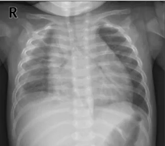
Figure 1. Frontal view of the chest radiograph shows car- diomegaly and pleural effusion at right thorax.
Figure 2. The echocardiographic apical four-chamber view (A) and parasternal short-axis view (B) show a massive pericardial effusion with impending car- diac tamponade.
and showed tachypnea with chest wall retraction. The blood pressure was 72/52 mmHg, heart rate 167/min, respiratory rate 43 breaths/min, and the oxygen satu- ration 86%∼88%. Initial laboratory findings showed white blood cell (WBC) count of 33.6×10⁹/L (neutrophils 84.5%, lymphocytes 13%), erythrocyte sed- imentation rate (ESR) of 43 mm/h (normal range <20), and C-reactive protein of 31.47 mg/dL (normal range
<1.0). The blood urea nitrogen and creatinine were 65 mg/dL and 1.02 mg/dL, respectively. Serum pro-brain na- triuretic peptide level (BNP) was 13,835 pg/mL. Urine analysis showed pyuria (20∼29 WBCs per high-power field and negative nitrate test). Electrocardiogram showed ST elevation in leads V4-6. Chest radiography showed massive cardiomegaly with pleural effusion in the right thoracic cavity (Figure 1). Echocardiography re- vealed a large amount of pericardial effusion with im- pending cardiac tamponade, but normal ventricular sys- tolic function and coronary arteries without giant aneur-
ysm and aneurysmal rupture (Figure 2). Under the im- pression of acute pericarditis with impending cardiac tamponade and sepsis, an emergency pericardiocentesis was performed and serosanguinous fluid was drained.
The results of pericardial fluid analysis were as follows:
red blood cell count 300×106/L, WBC count 570×109/L with 85% of neutrophils, lactate dehydrogenase (LDH) 26,900 IU/L (serum LDH 1,245 IU/L), and protein 3.87 g/dL (serum protein 5.1 g/dL). Exudative pericardial ef- fusion was diagnosed based on pericardial fluid to serum protein ratio >0.5 and pericardial fluid to serum LDH > 0.6 (Light's criteria). In addition, the fluid drained through thoracentesis was hemorrhagic (red blood cell count 9,920×106/L, the ratio of pleural fluid to serum LDH >0.6). Empirical antibiotics (cefotaxime and vanco- mycin) were administrated intravenously. Culture stud- ies from blood, urine, and pericardial and pleural fluid failed to isolate any pathogen. Despite a regimen of multi- ple empirical antibiotics, high fever persisted. On the 19th day of fever, conjunctival injection and desquama- tion of both fingertips suggesting an incomplete KD developed. Follow-up laboratory tests were performed and met the criteria of supplemental laboratory findings supporting a diagnosis of incomplete KD (elevated ESR [25 mm/h] and CRP [20.73 mg/dL], low serum albumin [2.7 g/dL, reference range: 3.5∼5.2], anemia for age [9.2 g/dL], WBC count [38.84×10⁹/L], platelet count [576×109/L] and sterile pyuria) [3]. Peripheral blood cell morphology showed that leukocytosis, neutrophilia, and normocytic normochromic anemia. There was an in- sufficient feature of macrophage activation syndrome (MAS) or systemic onset-juvenile idiopathic arthritis (SoJIA) in clinical course and follow-up laboratory test:
Triglyceride (165 mg/dL, reference range: 40∼200), fer- ritin (210 ng/mL, reference range: 26.1∼287.6), fibri- nogen (243 mg/dL, reference range: 160∼350) (Table 1).
Follow-up echocardiogram also showed a mild dilatation
Table 1. Laboratory values during treatment
Day of fever 1st admission 2nd admission 3rd admission 4th admission
6 13 19* 22 25 28 31 34 39 43 48 53 60 67
WBC count (109/L) 33.6 36.77 38.84 13.56 23.1 17.74 29.78 18.45 35.22 19.85 14.1 29.94 13.33 9.04
Lymphocytes (%) 13 26 25 40 22 24 39.6 41.4 15 31 56 17 50.1 62
Hemoglobin (g/dL) 10 9 9.2 9.6 11.3 10.2 10.1 11.2 11.4 9.4 11 9.8 11.7 11.7 Platelet count (109/L) 208 643 576 415 503 798 948 1,056 563 510 592 519 526 330
ESR (mm/h) 43 5 25 31 76 120 104 93 97 83 41 117 36 11
CRP (mg/dL) 31.47 5.8 20.73 4.38 13.96 27.73 6.32 2.06 8.51 4.82 0.17 23.44 0.13 0.43
Ferritin (ng/mL) 236 210 142.7 348.8
Fibrinogen (mg/dL) >500 225 243 239 465
Triglyceride (mg/dL) 94 165 107 126 80
Albumin (g/dL) 2.6 2.7 2.7 2.9 3.9 3.3 3.3 4.1 4.1 4.1 3.9 4.2 4.1
LDH (IU/L) 1,245 701 586 536 299 203
AST (IU/L) 59 22 22 26 43 22 20 28 28 21 18 23 24
ALT (IU/L) 38 13 17 26 33 22 17 18 19 16 9 8 7
WBC: white blood cell, ESR: erythrocyte sedimentation rate, CRP: C-reactive protein, LDH: lactate dehydrogenase, AST: aspartate aminotransferase, ALT: alanine aminotransferase. *The day of diagnosis with incomplete Kawasaki disease.
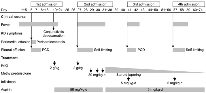
Figure 3. Clinical course and treatment of this patient. KD: Kawasaki disease, PCD: pigtail catheter drainage, IVIG: intravenous immunoglobulin.
of the right coronary artery (2.4 mm in diameter; Z score=2.7) without pericardial effusion. Intravenous im- munoglobulin (IVIG, 2 g/kg) and oral high-dose aspirin (50 mg/kg/day) were administered. Fever subsided on the 2nd day of IVIG treatment. The patient was dis- charged 4 days after IVIG treatment (Figure 3). Three days after the discharge, she was readmitted with a 24-hour history of high fever and pleural effusion.
Despite the second dose of IVIG (2 g/kg), fever persisted (body temperature >38.5oC). Fever and pleural effusion
were resolved after treatment with high dose methyl- prednisolone (Figure 3). After discharge, high fever and pleural effusion redeveloped twice, on the 41st and 57th days of illness, and laboratory findings also showed leu- kocytosis (33.6×10⁹/L of WBC count) and elevated ESR (43 mm/h) and CRP (34.1 mg/dL) on each occasion at the time of admission. After administration of infliximab (5 mg/kg), fever and pleural effusion subsided each time.
After discharge following the 4th admission, the patient is doing well. Echocardiography performed 1 year after
the illness showed normal coronary arteries and ven- tricular function.
DISCUSSION
KD is a systemic vasculitis that can affect various organs [1]. The diagnosis of classic KD is based on the presence of ≥5 of 6 principal clinical criteria. Patients who have in- sufficient principal clinical features (fever ≥5 days and 2 or 3 compatible clinical criteria) may be diagnosed with incomplete KD, based on supportive laboratory findings or a positive echocardiogram [3]. However, KD diagnosis is sometimes delayed because other systemic manifes- tations may precede such principal clinical features of KD.
There are various unusual initial presentations that delay diagnosis of KD, such as acute surgical abdomen [6], aseptic meningitis [7], cervical adenitis [8], and shock syndrome [9]. Zulian et al. [6] reported 10 cases of KD with acute surgical abdomen as an initial presentation, out of which 7 patients were diagnosed after surgical intervention. A prolonged fever and a culture-negative pleocytosis of cerebrospinal fluid suggestive of aseptic meningitis in a neonate may lead to an oversight of the di- agnosis of KD, because enteroviral meningitis is common in this age group [7]. Kanegaye et al. [8] reported that cer- vical adenitis and fever were initial presentations in 57 KD patients and suggested that incomplete KD should be considered in patients with cervical adenitis unresponsive to empirical antibiotics. In this case, incomplete KD was suspected and diagnosed only after development of con- junctivitis and desquamation on the fingertips on the 13th day of hospitalization (the 19th day of fever).
Because clinical criteria are used to diagnosis of KD, pa- tients who have insufficient principal clinical features tend to make early diagnosis difficult [3]. Moreover KD, SoJIA and MAS have similarity that clinical features and laboratory findings tend to overlap [10]. Therefore, the differential diagnosis of incomplete KD presenting espe- cially unusual manifestation has diagnostic dilemma and clinical challenging, which often can be delay in diagnosis [2,10]. In our case, we tried to rule out infectious disease, drug-related polyserositis and connective tissue disease.
According to the laboratory finding including bacterial cultures and virus studies, infectious disease could be excluded. The absence of signs of arthritis during 1 year follow-up, negative study of auto-immune antibodies (anti-nuclear antibody, rheumatoid factor), and other lab- oratory features (ferritin, triglyceride, fibrinogen, WBC
count, platelet, etc.) were insufficient to diagnose of con- nective tissue disease especially SoJIA and MAS [10,11].
KD with massive pericardial effusion showing clinical signs of cardiac tamponade is very rare. There have been several reports on hemorrhagic pleural and/or pericardial effusion in patients with KD [4,5,12,13]. Voynow et al.
[13] reported that 11 (1.83%) out of 602 KD patients pre- sented with predominant pulmonary involvement.
However, pericardial involvement is usually manifested as a small amount of pericardial effusion on the echo- cardiogram, and to our knowledge, only 2 cases with car- diac tamponade have been reported [4,5]. Our case showed an atypical course where massive pleural effusion and pericardial effusion with cardiac tamponade without giant aneurysm and aneurysmal rupture developed as ini- tial presentations in an infant under 12 months.
Currently, first-line treatment of KD is a single dose of IVIG (2 g/kg) and second-line treatment for IVIG-re- sistant KD patients may be a repeat dose of IVIG, high-dose methylprednisolone, or infliximab [3]. It is known that the levels of pro-inflammatory cytokines (interleukin [IL]-2, IL-6, IL-8, interferon-γ and tumor necrosis factor [TNF]-α) and vascular endothelial growth factor (VEGF) are increased in KD [14,15]. Therefore, in- fliximab (anti-TNF-α monoclonal antibody) therapy is preferable to use in patients with KD refractory to IVIG [3]. Other monoclonal antibody therapy (anakinra, a re- combinant, nonglycosylated form of the human IL-1 re- ceptor antagonist ) for treatment of refractory KD was re- ported [16]. Regarding the pathogenesis of pleural and pericardial effusion in KD, it has been speculated that VEGF may play a role by increasing microvascular hyper- permeability [12]. Strunk et al. [17] reported that in- fliximab and glucocorticoids reduced VEGF in patients with rheumatoid arthritis. However, a study in KD showed that infliximab reduced pro-inflammatory cyto- kines but did not suppress VEGF in refractory KD pa- tients [14]. Instead, Hamada et al. [12] reported a case that presented with marked pericardial effusion and pleu- ral effusion with elevated VEGF, which were dramatically resolved after corticosteroid administration. In our case, recurrent fever and pleural effusion developed even dur- ing steroid administration, and showed acute response to infliximab therapy, suggesting its partial role in reducing the hyperpermeable state of KD. Further study is needed to elucidate the possible roles and mechanisms of in- fliximab and corticosteroid in suppressing vascular hy- perpermeability in KD patients.
SUMMARY
KD is included on the list of important causes of MAS in childhood, and the large degree of overlap syndrome should be considered the possibility of similar relation- ships between refractory incomplete KD and MAS [18]. It is important to remember that in infants with un- explained persistent fever and irresponsive to empirical antibiotics, incomplete KD should be suspected. In addi- tion, clinicians should be aware that cardiac tamponade and/or hemorrhagic pleural effusion may be initial clin- ical manifestations of incomplete KD.
CONFLICT OF INTEREST
No potential conflict of interest relevant to this article was reported.
AUTHOR CONTRIBUTIONS
Study concept and design: J.H.Y. Acquisition of data:
K.M.K., D.C.J., W.Y.P., G.B.K. Analysis and interpretation of data: J.H.Y., Y.J.K. Writing manuscript: J.H.Y., Y.J.K., J.Y.L.
REFERENCES
1. Kawasaki T, Kousaki F. Fabrile oculo-oro-cutaneo-acro- desquamatous syndrome with or without acute non-suppu- rative cervical lymphadenitis in infancy and childfood: clin- ical observations of 50 cases. Jpn J Allergol 1967;16:
178-222, 225.
2. Minich LL, Sleeper LA, Atz AM, McCrindle BW, Lu M, Colan SD, et al.; Pediatric Heart Network Investigators.
Delayed diagnosis of Kawasaki disease: what are the risk factors? Pediatrics 2007;120:e1434-40.
3. McCrindle BW, Rowley AH, Newburger JW, Burns JC, Bolger AF, Gewitz M, et al.; American Heart Association Rheumatic Fever, Endocarditis, and Kawasaki Disease Committee of the Council on Cardiovascular Disease in the Young; Council on Cardiovascular and Stroke Nursing;
Council on Cardiovascular Surgery and Anesthesia; and Council on Epidemiology and Prevention. Diagnosis, treat- ment, and long-term management of Kawasaki disease: a scientific statement for health professionals from the American Heart Association. Circulation 2017;135:e927- 99.
4. Dahlem PG, von Rosenstiel IA, Lam J, Kuijpers TW. Pulse methylprednisolone therapy for impending cardiac tampo- nade in immunoglobulin-resistant Kawasaki disease.
Intensive Care Med 1999;25:1137-9.
5. Ozdogu H, Boga C. Fatal cardiac tamponade in a patient with Kawasaki disease. Heart Lung 2005;34:257-9.
6. Zulian F, Falcini F, Zancan L, Martini G, Secchieri S, Luzzatto C, et al. Acute surgical abdomen as presenting manifestation of Kawasaki disease. J Pediatr 2003;142:731-5.
7. Rosenfeld EA, Corydon KE, Shulman ST. Kawasaki disease in infants less than one year of age. J Pediatr 1995;126:
524-9.
8. Kanegaye JT, Van Cott E, Tremoulet AH, Salgado A, Shimizu C, Kruk P, et al. Lymph-node-first presentation of Kawasaki disease compared with bacterial cervical adenitis and typical Kawasaki disease. J Pediatr 2013;162:1259-63, 1263.e1-2.
9. Thabet F, Bafaqih H, Al-Mohaimeed S, Al-Hilali M, Al-Sewairi W, Chehab M. Shock: an unusual presentation of Kawasaki disease. Eur J Pediatr 2011;170:941-3.
10. Ravelli A, Minoia F, Davì S, Horne A, Bovis F, Pistorio A, et al.; Paediatric Rheumatology International Trials Organisation;
Childhood Arthritis and Rheumatology Research Alliance;
Pediatric Rheumatology Collaborative Study Group; Histiocyte Society. 2016 Classification criteria for macrophage activa- tion syndrome complicating systemic juvenile idiopathic ar- thritis: a European League Against Rheumatism/American College of Rheumatology/Paediatric Rheumatology International Trials Organisation Collaborative Initiative. Ann Rheum Dis 2016;75:481-9.
11. Mizuta M, Shimizu M, Inoue N, Kasai K, Nakagishi Y, Takahara T, et al. Serum ferritin levels as a useful diagnostic marker for the distinction of systemic juvenile idiopathic ar- thritis and Kawasaki disease. Mod Rheumatol 2016;26:
929-32.
12. Hamada H, Terai M, Honda T, Kohno Y. Marked pleural and pericardial effusion with elevated Vascular Endothelial Growth Factor production: an uncommon complication of Kawasaki disease. Pediatr Int 2005;47:112-4.
13. Voynow JA, Schanberg L, Sporn T, Kredich D. Pulmonary complications associated with Kawasaki disease. J Pediatr 2002;140:786-7.
14. Hirono K, Kemmotsu Y, Wittkowski H, Foell D, Saito K, Ibuki K, et al. Infliximab reduces the cytokine-mediated in- flammation but does not suppress cellular infiltration of the vessel wall in refractory Kawasaki disease. Pediatr Res 2009;65:696-701.
15. Jinkawa A, Shimizu M, Nishida K, Kaneko S, Usami M, Sakumura N, et al. Cytokine profile of macrophage activa- tion syndrome associated with Kawasaki disease. Cytokine 2019;119:52-6.
16. Kone-Paut I, Cimaz R, Herberg J, Bates O, Carbasse A, Saulnier JP, et al. The use of interleukin 1 receptor antago- nist (anakinra) in Kawasaki disease: a retrospective cases series. Autoimmun Rev 2018;17:768-74.
17. Strunk J, Bundke E, Lange U. Anti-TNF-alpha antibody Infliximab and glucocorticoids reduce serum vascular endo- thelial growth factor levels in patients with rheumatoid ar- thritis: a pilot study. Rheumatol Int 2006;26:252-6.
18. Latino GA, Manlhiot C, Yeung RS, Chahal N, McCrindle BW. Macrophage activation syndrome in the acute phase of Kawasaki disease. J Pediatr Hematol Oncol 2010;32:527- 31.