Ⅰ. 서 론
상악동골이식을 위한 측방 접근법 시 골절단과 이식부위 에 차폐막 사용여부를 놓고 많은 논쟁이 존재한다
1. 차폐막 을 사용할 경우 장점으로는 상피성결합조직의 개제 없이 이식재입자를 유지할 수 있고,연조직의 이주를 방지하고,
골형성을 증가시키고, 임플란트의 생존율을 증가시키는 것 등이다
2. 그러나 많은 임상가들은 Schneider 막의 손상 없이 골이식이 완료된 경우 차폐막의 이점과 비용을 비교 하며 차폐막 사용 여부에 대하여 고민한다. 만일 차폐막이 비흡수성이거나 고정태그를 사용할 경우, 이들의 제거를 위한 추가 수술이 필요하며 이로 인한 환자의 불편함이 가 중될 수 있어 그 고민은 더욱 커진다
3.
상악동골이식술의 측방 접근법 시 본 교실에서는 기존에 흔히 사용하여 온 상부경첩술식(superior hinge technique) 또는 내측골절술식(infracturing technique) 대신 360�골절제 후 Schneider 막으로부터 분리된 골창을 떼어내는 이른바
“off-the-wall”술식 또는 외측골절술식(outfracture tech- nique)을 사용하였다
4,5. 보통 이러한“off-the-wall”술식을
박 창 주133-791 서울특별시 성동구 행당동17번지
한양대학교 의과대학 본관421호 치과학교실 구강악안면외과 Chang-Joo Park
Room 421, Division of Oral and Maxillofacial Surgery, Department of Dentistry, College of Medicine, Hanyang University
17 Haengdang-dong, Seongdong-gu, Seoul, 133-791, Korea TEL: +82-2-2290-8646 FAX: +82-2-2290-8673 E-mail: fastchang@hanyang.ac.kr
상악동측방접근법시 차폐막을 사용하지 않는 골창재위치술의 유용성 평가
전승환
1∙조용석
2∙이병하
1∙임태윤
1∙황경균
1∙박창주
11
한양대학교 의과대학 치과학교실 구강악안면외과,
2앞선치과병원 구강악안면외과
Evaluation of the feasibility of bony window repositioning without using a barrier membrane in sinus lateral approach
Seung-Hwan Jeon
1, Yong-Seok Cho
2, Byung-Ha Lee
1, Tae-Yun Im
1, Kyung-Gyun Hwang
1, Chang-Joo Park
11
Division of Oral and Maxillofacial Surgery, Department of Dentistry, College of Medicine, Hanyang University,
2
Private Practice of Oral and Maxillofacial Surgery, Apsun Dental Hospital, Seoul, Korea
Introduction:In the lateral window approach for a maxillary sinus bone graft, there has been considerable controversy regarding the placement of a barrier membrane over the osteotomy site. In particular, when there is no damage to the Schneiderian membrane, clinicians should decide whether to use a barrier membrane or not, considering the benefits and costs. This study presents the clinical cases to demonstrate that only repositioning the detached window can lead to satisfactory bony healing of the grafted material without using a barrier membrane in the lateral approach for a maxillary sinus bone graft.
Materials and Methods:Five consecutive patients were treated with the same surgical procedures. After performing the antrostomy on the lateral maxillary wall using a round carbide bur and diamond bur, the bony window was detached by a gentle levering action. After confirming no perfora- tion of the Schneiderian membrane, the grafting procedure was carried out the detached window of the lateral maxillary wall was repositioned over the grafted material without using a barrier membrane. A gross examination was carried out at the postoperative 6 month re-entry, and the the preopera- tive and postoperative dental computed tomography (CT) at re-entry were compared.
Results:All the procedures in the 5 patients went on to uneventful healing with no complications associated with the bone graft. Satisfactory bone regeneration without the interference of fibrous tissue on the gap between the repositioned window and lateral wall of the maxillary sinus was observed in the postoperative 6 month re-entry. The CT findings at re-entry revealed the, reconstruction of the external cortical plate including reposi- tioned bony window. In addition, the loss of the discontinuity of the lateral maxillary wall was confirmed.
Conclusion:This preliminary report showed that the detached window, which was just repositioned on the grafted material, could function as a bar- rier membrane in the lateral approach for a maxillary sinus bone graft. Therefore additional morphometric and histologic studies will be needed.
Key words:Bone graft, Dental implants, Membrane
[paper submitted 2010. 5. 12 / revised 2011. 1. 14 / accepted 2011. 3. 21]
Abstract (J Korean Assoc Oral Maxillofac Surg 2011;37:122-6)
통해 얻어진 골창을 부수어 골이식재와 섞어 사용하였지만 양이 너무 적은 관계로 그 골생성능력은 매우 의심스러웠 다.
이에 이번 연구에서는 차폐막을 사용하지 않고 분리된 골창을 골절제 부위 위에 단순히 재위치 시키는 술식을 소 개하고, 이 술식을 통해 수술받은 5명의 환자에서 술후 6개 월째의 2차 수술 시 이식 부위의 임상적 관찰을 통하여 재 위치된 골창이 일종의 차폐막으로 작용할 수 있는지 그 가 능성에 대하여 평가하고자 하였다.
Ⅱ. 연구 대상 및 방법
모든 환자는 전신적 병력과 국소적 금기증 즉, 비조절성 당뇨병, 이갈이, 상악동병변, 비조절성 치주염을 가지고 있 지 않았다. Computed tomography (CT)을 포함한 술전 방사 선사진을 통하여 상악동의 후구치 부위의 잔존골이 임플 란트를 식립하기에 불충분함을 확인하였다. 환자들은 임 상적 및 방사선학적 정보를 통하여 가능한 모든 치료방법 들에 대하여 충분히 설명을 들었으며 술전고지에 입각한 동의서를 작성하였다.
0.12% 클 로 르 헥 시 딘 용 액 (헥 사 메 딘 액 , 부 광 약 품 , Gyeonggi-do, Korea)으로 2분간 함수 후 1:100,000 에피네프 린을 포함한 2% 리도케인(2%염산리도카인∙에피네프린 주사, 유한양행, Seoul, Korea)을 사용하여 국소마취를 시행 한 뒤 상악후구치 부위에 통상적인 방법으로 절개와 점막 박리를 시행하였고 4번이나 2번 라운드카바이드 버를 사 용하여 골창을 형성하였다. Schneider 막이 천공되지 않은 것을 확인한 뒤 천공 가능성을 최소화하기 위해 필요에 따 라 2번 다이아몬드 버나 piezo tip을 추가적으로 사용하였 다. 골창이 움직이기 시작하면 끝이 둔한 골막 기자나 프리 어 기자를 골 절단부경계에 조심스럽게 삽입한 뒤 건전한 상악동 벽에 기대어 지렛대를 올리듯 골창을 Schneider 막 으로부터 조심스럽게 분리하였다.
이후 Schneider 막의 거상을 조심스럽게 시행하고 동결건 조 이종골(SureOss, HansBiomed, Seoul, Korea)을 식염수에 적셔 상악동 안으로 신중하게 채워넣었다. 임플란트(GSIII, Osstem Implant Co., Ltd., Seoul, Korea) 식립은 잔존골의 양 에 따라 동시에 시행하거나 또는 시행하지 않았다. 골이식
을 마친 뒤 떼어냈던 골창을 골이식 부위에 재위치 시킨 후 부드럽게 눌렀다. 견고고정(rigidfixation)은 시행하지 않았 다. 보통 상악동측벽과 재위치된 골창 사이에 1-2 mm의 간 격이 존재하였으나 차폐막을 사용하여 이식부위를 덮어주 지 않았다. 수술부위를 조심스럽게 식염수로 세척한 후 봉 합하였다. 만약 추가적으로 골막에 이완절개가 필요한 경 우 이완된 골막 절개부가 직접적으로 골창 위에 위치하지 않도록 하였다. 술후에 amoxacillinclavulanated (Augmentin, Ilsung Pharmaceuticals Co., Ltd., Seoul, Korea) 375 mg과 ace- clofenac (Clanza, Korea United Pharm,Inc.,Seoul, Korea) 100 mg을 10일간 하루 3번 경구로 투약하였다. 클로르헥시딘 용액과 비충혈완화제 역시 10일간 하루 2번 사용하도록 처 방하였다. 봉합사는 술후 10일 뒤에 제거하였다.
환자는 술후 1개월간 매주 검진을 받았고 2차 수술까지 매달 점검을 받았다. 술후 6개월째 국소마취로 임플란트를 식립하거나 치유지대주를 장착하기 위한 2차 수술, 또는 기타 필요한 시술을 시행하였다. 일차 수술과 유사한 판막 을 열어 골이식 부위에 재위치된 골창 주변의 골재생 정도 에 대한 임상적 평가를 위한 재접근(re-entry)을 시도하였으 며 수술이 완료된 후에는 CT를 포함한 술후 방사선사진도 촬영하였다.
Ⅲ. 결 과
다섯 명의 환자를 2008년 5월부터 2008년 7월까지 연속 적으로 평가하였다. 남성 4명과 여성 1명으로 평균나이는 53세(34-65세)였다. 각 임상증례들은 Table 1에 요약하였 다. 만약 잔존 치조골의 높이가 5 mm 이상일 경우 상악동 골이식 시 임플란트식립을 동시에 진행하였고, 그렇지 못 한 경우에는 단계적으로 술식을 진행하였다. 상악동 안에 이식되는 골이식재는 오직 동결건조 이종골만을 사용하였 으나 네 번째 증례의 경우 상악결절에서 채취한 자가골을 첨가하였다.
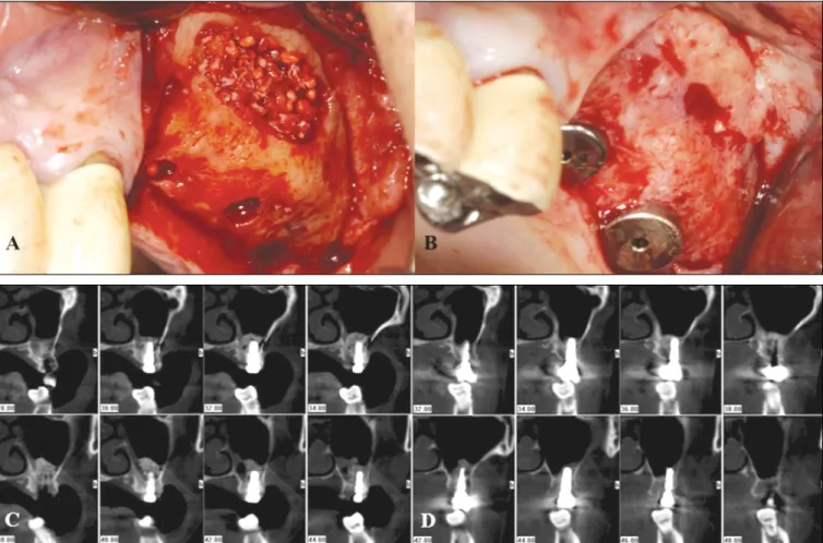
모든 환자는 골이식과 관련된 특별한 합병증 없이 정상 적인 치유과정을 보였다. 술후 6개월에 행해진 2차 수술 시 상악동 측벽과 재위치된골창 사이 틈에 상대적으로 얇은 재생골을 발견할 수 있었다.(Figs. 1.A, 1.B) 모든 증례에서 재위치된 골창 주변으로 섬유성 조직의 개재는 없었고 골
Table 1.Summary of the clinical cases
Case No. Sex Age
No. of missing teeth Mean residual alveolar
Implant placement Grafted material
(yr) bone height (mm)
1 M 34 1 4 Simultaneous SureOss
2 M 55 3 2 Staged SureOss
3 M 52 2 5 Simultaneous SureOss
4 M 65 4 5 Simultaneous SureOss+autogenous
5 F 59 2 2 Staged SureOss
창을 골막 기자로 건드려 보아도 육안적인 동요도를 보이 지 않았다. 또한 재위치된 골창의 흡수나 생활력 상실 역시 보이지 않았다. 술후 6개월 당시 촬영된 CT에서도 재위치 된 골창 주변의 불연속성이 사라지며 상악동 측벽의 외측 피질골판의 재건이 명확히 관찰되었다.(Figs. 1.C, 1.D)
Ⅳ. 고 찰
측방접근법 시 자주 사용하는 골절제술은 상부경첩술식 또는 내측골절술식이다
6-10. 이번“off-the-wall”술식 또는 외측골절술식은 360�로 골절제술을 시행한 후 골창을 들 어올려 떼어내는 술식으로 얇은 골창을 Schneider 막으로 부터 쉽게 분리할 수 있었다. 특히 상악동의 측벽이 두껍거 나 상악동 내 골성중격이 존재할 때기존의 방법에 비하여 Schneider 막의 손상을 줄일 수 있도록 훌륭한 시야를 제공 하는 장점을 가진다
4,5. 처음에는 분리된 골창을 부수어 골 이식재에 섞어 상악동 안으로 이식하였다. 분쇄된 골창을
골이식재와 섞어 사용하여 좋은 결과를 보고한 논문도 있 지만
11이번 실험은 분리된 골창의 자가골이식재로서의 용 도보다는 골창을 골이식 부위에 재위치 시켜 일종의 차폐 막 구실을 할 수 있음을 증명하는데 초점을 맞추어 진행하 였다.
상악동골이식을 위한 측방 접근법에서 골창을 형성하고 이식을 시행한 부위에 차폐막을 동시에 사용하는 것에 대 한 많은 논란이 있다. 차폐막으로 덮어주지 않은 부위에 비 해 차폐막을 사용한 골창 형성 부위의 높은 골형성율은 많 은 연구에서 보고되어 있다
12,13. 이는 골의 성장방향이 상악 동을 둘러싸는 골벽에서 시작하여 중심을 향하기 때문에 상악동 측벽의 골창 형성 부위는 골화가 이루어지는 마지 막 부위들 중 한 부위이므로
13, 상악동 측벽의 외측 피질골 을 확실히 재건해 주는 것이 임플란트의 성공과 실패 여부 를 임상적으로 예측 가능하게 해줄 수 있다는 주장을 뒷받
침한다
1,14. 그러나 실제로는 측방 접근법 시 골창 형성 부위
에 차폐막의 사용 여부가 임플란트 성공률의 관점에서는
Fig. 1. A: Intraoperative view of a bony window repositioned over the graft material. B: Postoperatively 6 month view of bone regeneration around the bony window in Case 3. Reconstruction of the external cortical plate and gap bone regeneration was clinically acceptable. C: Immediately postoperative serial coronal views of CT. D: Postoperatively 6 month serial coronal views of CT. Reconstruction of the external cortical plate including repositioned bony window was shown. Also, loss of the discontinuity of lateral maxillary wall was confirmed. (CT: computed tomography)
유의할만한 차이를 찾을 수 없었다는 보고도 있다
3. 차폐막의 설치는 외측 피질골의 치유를 향상시키고 이는 차폐막이 위치한 부위의 골의 생존률이 크게 증가시킬 수 있음을 의미한다
3,15. 창위에 차폐막이 위치하지 않았을 경 우 이식 부위의 피질골화의 결핍을 어렵지 않게 관찰할 수 있으며 창의 크기가 증가할수록 이런 현상은 증가한다. 이 와 같은 현상은 골표면에서 한번 거상되어진 골막이 본디 섬유성이라는 이유와 치유 시 결합조직의 개제 등의 이유 로 설명된다
2.
비록 우리의 연구가 임상적 관찰에만 초점을 맞추었지만 차폐막 없이도 단순히 재위치된 골창 주변에 골형성이 만 족스럽게 일어났고 외측 피질골판의 치유도 정상적으로 진행되었음을 확인할수 있었다. 이는 그 적은 양을 고려하 였을 때 최고의 자가골이식재로서 기능하였다고도 할 수 있다. 첫 번째와 두 번째 증례에서 우리는 골절제시 4번 라 운드카바이드버를 사용하였고 이 경우 재위치된 골창과 건전한 상악동측방골 사이의 간격이 꽤 넓었다. 이후 우리 는 세 번째, 네 번째, 그리고 다섯 번째 증례를 통해 보다 작 은 2번 라운드카바이드버를 사용, 골간격을 줄이고자 시도 하였고 이는 골재생 능력을 증가시키고 골창의 적합성과 안정성을 용이하게 할 수 있었다.
수술 부위에서 결합조직합입을 방지하기 위해 많은 비흡 수성 또는 흡수성 차폐막, 예를 들어 expanded polytetrafluo- roethylene (e-PTFE) 막, 티타늄 메쉬, 교차결합 교원질막, 그리고 calcium sulphate 막 등이 상악동측방 골창 위에 사 용되었다
2. Schneider 막의 손상이 있을 경우 Schneider 막의 치유를 위해 차폐막을 동시에 사용하는 것이 좋다는 것은
확실하다
16,17. 그러나 Schneider 막의 거상과 골이식과정이
별다른 문제없이 끝났을 경우 항상 구강악안면외과의는 차폐막의 비용문제로 그 사용을 망설인다. 만일 차폐막이 비흡수성 이거나 고정태그를 사용하면 이들의 제거를 위 한 추가 수술이 필요하여 이로 인한 환자의 불편함이 가중 되므로 그 고민은 더욱 커질 수 밖에 없다. 이러한 점들을 고려하였을 때 이번 연구에서 제시된 방법은 임상적으로 큰 의미를 가질 수 있을 것이다.
그러나 이번 연구는 단지 5명의 환자에서 시행되었고 술 후 6개월의 임상적인 관찰에 한정되므로 술자는 반드시 잠 재적 합병증을 염두에 두어야 하며, 술식의 모든 내용은 충 분히 환자에게 설명해야 할 것이다. 향후 이 술식을 일반적 으로 사용하기 위해서는 임플란트의 성공률과 관련하여 임상적, 방사선학적 그리고 조직학적 연구를 추가적으로 수행하여야 할 것이다.
Ⅴ. 결 론
이번 연구는“off-the-wall”술식을 통해 얻어진 골창을 골 절제와 골이식 부위 상에 단지 재위치시키는 것만으로도 일종의 차폐막 역할을 충분히 수행할 수 있음을 보여준다.
술후 6개월의 재접근을 통한 임상적 관찰 시에도 골창과 상악동측방골 사이의 간격에 골재생이나 외부 피질골의 치유양상 역시 만족스러웠다. 이에“off-the-wall”술식 또 는 외측골절 술식이 상악동골이식을 위한 측방접근법 시 매우 유용하게 사용될 수 있다고 할 수 있다.
References
1. Tawil G, Mawla M. Sinus floor elevation using a bovine bone mineral (Bio-Oss) with or without the concomitant use of a bilay- ered collagen barrier (Bio-Gide): a clinical report of immediate and delayed implant placement. Int J Oral Maxillofac Implants 2001;16:713-21.
2. Wallace SS, Froum SJ, Tarnow DP. Use of barrier membranes in sinus augmentation. In: Jensen OT, ed. The sinus bone graft. 2nd ed. Chicago: Quintessence Publishing Co.; 2006:229-39.
3. Froum SJ, Tarnow DP, Wallace SS, Rohrer MD, Cho SC. Sinus floor elevation using anorganic bovine bone matrix (OsteoGraf/N) with and without autogenous bone: a clinical, his- tologic, radiographic, and histomorphometric analysis-Part 2 of an ongoing prospective study. Int J Periodontics Restorative Dent 1998;18:528-43.
4. Song SI, Jeong HR, Kim HM, Lee JK. Clinical investigation on the feasibility of outfracture osteotomy sinus graft technique. J Korean Assoc Oral Maxillofac Surg 2009;35:367-71.
5. Lee JK. Outfracture osteotomy on lateral maxillary wall as a modified sinus graft technique. J Oral Maxillofac Surg 2010;68:
1639-41.
6. Boyne PJ, James RA. Grafting of the maxillary sinus floor with autogenous marrow and bone. J Oral Surg 1980;38:613-6.
7. Wallace SS, Froum SJ, Cho SC, Elian N, Monteiro D, Kim BS, et al. Sinus augmentation utilizing anorganic bovine bone (Bio- Oss) with absorbable and nonabsorbable membranes placed over the lateral window: histomorphometric and clinical analyses. Int J Periodontics Restorative Dent 2005;25:551-9.
8. Jurisic M, Markovic A, Radulovic M, Brkovic BM, Sa′ndor GK.
Maxillary sinus floor augmentation: comparing osteotome with lateral window immediate and delayed implant placements. An interim report. Oral Surg Oral Med Oral Pathol Oral Radiol Endod 2008;106:820-7.
9. Lorenzoni M, Pertl C, Wegscheider W, Keil C, Penkner K, Polansky R, et al. Retrospective analysis of Frialit-2 implants in the augmented sinus. Int J Periodontics Restorative Dent 2000;20:255-67.
10. Zijderveld SA, Schulten EA, Aartman IH, ten Bruggenkate CM.
Long-term changes in graft height after maxillary sinus floor ele- vation with different grafting materials: radiographic evaluation with a minimum follow-up of 4.5 years. Clin Oral Implants Res 2009;20:691-700.
11. de Vicente JC, Herna′ndez-Vallejo G, Braña-Abascal P, Peña I.
Maxillary sinus augmentation with autologous bone harvested from the lateral maxillary wall combined with bovine-derived hy- droxyapatite: clinical and histologic observations. Clin Oral Implants Res 2010;21:430-8.
12. Wallace SS, Froum SJ. Effect of maxillary sinus augmentation on the survival of endosseous dental implants.A systematic re- view. Ann Periodontol 2003;8:328-43.
13. Zitzmann NU, Scha¨rer P. Sinus elevation procedures in the re- sorbed posterior maxilla. Comparison of the crestal and lateral approaches. Oral Surg Oral Med Oral Pathol Oral Radiol Endod 1998;85:8-17.
14. Margolin MD, Cogan AG, Taylor M, Buck D, McAllister TN, Toth C, et al. Maxillary sinus augmentation in the non-human primate: a comparative radiographic and histologic study be- tween recombinant human osteogenic protein-1 and natural bone
mineral. J Periodontol 1998;69:911-9.
15. Tarnow DP, Wallace SS, Froum SJ, Rohrer MD, Cho SC.
Histologic and clinical comparison of bilateral sinus floor eleva- tions with and without barrier membrane placement in 12 pa- tients: part 3 of an ongoing prospective study. Int J Periodontics Restorative Dent 2000;20:117-25.
16. Vlassis JM, Fugazzotto PA. A classification system for sinus membrane perforations during augmentation procedures with op- tions for repair. J Periodontol 1999;70:692-9.
17. Proussaefs P, Lozada J, Kim J, Rohrer MD. Repair of the perfo- rated sinus membrane with a resorbable collagen membrane: a human study. Int J Oral Maxillofac Implants 2004;19:413-20.