Copyright © 2016 Journal of Rhinology
70
A Case of Nasal Swell Body (Septal Turbinate) Combined
with Pneumatization of Perpendicular Plate of the Ethmoid Bone
Soo Kweon Koo, MD, PhD1, Sung Hoon Jung, MD1, Ji Seung Moon, MD1 and Hyuni Son, MD2
1Department of Otolaryngology-Head and Neck Surgery, Busan St. Mary’s Hospital, Busan; and
2Department of Pathology, Busan Saint Mary’s Hospital, Busan, Korea
The “nasal swell body” (NSB) or septal turbinate is a distinct structure of the anterior nasal septum that is observed on endoscopic and radiographic examination. It is primarily a glandular rather than a venous formation that is comprised of septal cartilage, bone, and thick mucosal lining. It is commonly found in patients with symptoms of chronic sinusitis and allergic rhinitis, and is linked to septal deviation. Space occupying lesions of the septum such as tumors, mucoceles, and pneumatization of the septum can lead to anatomical and functional disorders such as nasal obstruction and sinusitis, while more serious clinical conditions can develop when these lesions are combined with the NSB. Recently, there has been emphasis on the functional aspects of the NSB. It is especially being emphasized for clinicians to pay attention to the NSB and its connection with the stuffy nose. We report an interesting case of the NSB combined with pneumatization of the perpendicular plate of the ethmoid bone causing se- vere nasal obstruction and repetitive sinusitis along with a literature review.
KEY WORDS: Nasal ObstructionㆍTurbinateㆍNasal SeptumㆍSinusitis.
INTRODUCTION
Anatomical abnormality such as septal deviation is rela- tively common; it causes nasal obstruction and limitation of the access as well as leads to frequent recurrence after sinus surgery. In most cases, septal deviation is simple bony or cartilaginous septal deviation, but sometimes it is also observed in the case of septal mucosa swelling or septal deviation caused by space occupying lesions. “The nasal swell body (NSB, Septal turbinate)” is a distinct structure of ant nasal septum that is observed endoscopic examination and radiographic study. It is primarily a glandular formation and not a venous one and comprised of septal cartilage, bone and thick mucosal lining. It is common in patients with symptoms of chronic sinusitis, allergic rhinitis and is linked to septal deviation.1)2) Space occupying lesions of the sep-
tum such as tumor, mucocele, and pneumatization of sep- tum show anatomical and functional disorder such as nasal obstruction, sinusitis etc. It may develop more serious clin- ical conditions in the case of combined NSB. Recently, there has been emphasis on functional aspects of the NSB. It plays an important role in nasal airflow regulation and humidifi- cation.2)3) and in connection with the stuffy nose, it is being emphasized to pay attention to the NSB.
We report interesting case of NSB combined with peuma- tization of perpendicular plate of the ethmoid bone causing severe nasal obstruction and repetitive sinusitis with a lit- erature review.
CASE REPORT
A 23-year-old female visited our otolaryngology depart-
Received: January 28, 2016 / Revised: March 16, 2016 / Accepted: May 9, 2016
Address for correspondence: Soo Kweon Koo, MD, PhD, Department of Otolaryngology-Head and Neck Surgery, Busan St. Mary’s Hospital, 25- 14 Youngho-ro 232beon-gil, Nam-gu, Busan 48575, Korea
Tel: +82-51-933-7214, Fax: +82-51-956-1956, E-mail: [email protected]
pISSN 1229-1498 / eISSN 2384-4361 www.ksrhino.or.kr CASE REPORT
J Rhinol 2016;23(1):70-73 http://dx.doi.org/10.18787/jr.2016.23.1.70
Koo et al : A Case of Nasal Swell Body (Septal Turbinate) 71
ment with a history of nasal obstruction and profuse mu- copurulent rhinorrhea. In her past history, she had received endoscopic sinus surgery with polypectomy three times because of recurrent sinusitis. Rigid nasal endoscopy showed bulging of both antero-superior portions of the septum, and middle turbinates on both sides and previous surgical site could not be observed (Fig. 1). Non contrast computed tomography (CT) of the ostiomeatal unit (OMU) also showed pneumatization of perpendicular plate of the ethmoid bone and mucosal thickening in both the antero-superior por- tions of the septum. Total opacity was observed in the right middle turbinate and the left frontal, ethmoid, and maxil- lary sinuses (Fig. 2). The patient underwent septoplasty and revision endoscopic sinus surgery under general anesthesia.
We found a mucosal swelling and huge round mass on the bony septum, and we performed volume reduction of na- sal septal mucosa swelling(NSB) using microdebrider and removed huge round mass using a septum forceps (Fig. 3).
The round mass was hard and it had a honeycomb appear- ance. The right middle turbinate showed polyposis and mu-
copurulent discharge and polyps were identified in the pre- vious surgical site. Histological findings included an osseous nasal septum showing focal fibrous changes and fatty re-
A B
Fig. 1. Pre-operative nasal endoscopic findings. It showed bulg- ing of both antero-superior portions of the septum and middle turbinates on both sides and previous surgical site could not be observed. Right side of the nasal cavity (A), left side of the nasal cavity (B). S: nasal septum, IT: inferior turbinate, ST: septal turbi- nate.
Fig. 2. CT images of the patient. Coronal view (A), Axial view (B).
These CT images showed pneumatization of perpendicular plate of the ethmoid bone and mucosal thickening in both the antero-superior portions of the septum (Black arrow). Total opacity was observed in the right middle turbinate and the left frontal, ethmoid, and maxillary sinuses.
A B
Fig. 3. Intra-operative finding. This figure shows a huge round mass on the bony septum. Black arrow: Bony cartilage junction of septum. White arrow: Huge round mass.
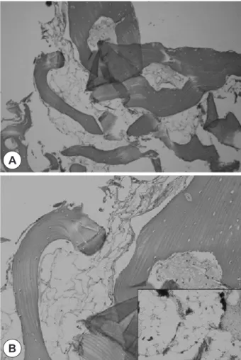
Fig. 4. A: An osseous nasal septum showing focal fibrous chang- es and fatty reduction (upper middle part) in the marrow part (H-E stain, ×40). B: A higher power view of perpendicular plate of the ethmoid bone shows variably-sized empty and collapsed air spaces in the marrow cavity. The spaces replace normal white marrow components. Inset shows comparable normal white marrow interrupted by thin fibrovascular tissue (H-E stain,
×200).
A
B
J Rhinol 2016;23(1):70-73
72
duction in the marrow part (Fig. 4A). A higher power view of perpendicular plate of the ethmoid bone shows variably- sized empty and collapsed air spaces in the marrow cavity.
The spaces replace normal white marrow components. In- set shows comparable normal white marrow interrupted by thin fibrovascular tissue (Fig. 4B). During 8 months of follow-up, recurrence was not observed.
DISCUSSION
Anatomic variations such as concha bullosa, lateral and medial bending of uncinate process, paradoxical middle tur- binate, Haller cell, and septal deviation are responsible for the pathogenesis of chronic paranasal sinusitis.4) Especial- ly, septal deformities formed in both sides at the anterior por- tion of middle turbinate are difficult to access surgically, and they are a cause of frequent recurrence after surgery.
This type of septal deformity appears in the form of a bilat- eral mucosal swelling macroscopically and is called the
“nasal swell body(septal turbinate)”.1)2)
After Morgagni in 1662 described it as a mucosal protu- berance located anteriorly on the septum, Schiefferdecker defined a vascular network on the anterior part of the sep- tum and named it as “septal turbinate”.5) NSB has been de- scribed as several different names, septal turbinate, intumes- centia septi nasi anterior and Kisselbach’s body.1)
NSB plays an important role in nasal airflow regulation due to its close proximity to the nasal valve region and ex- pansile venous sinusoids.6) In addition, NSB humidifies the inspired air current with its seruminous gland and protect mucosal dryness and crust formation based on anatomical histological studies.7)
Pneumatization can occur in the sinonasal area such as the uncinate process, middle nasal concha, inferior nasal concha, crista galli, and clivus. Pneumatization of the mid- dle turbinate is the most common, with incidence rates of 8-20% in normal persons and 33-43.9% in patients with rhinosinusitis.8) Septal pneumatization can occur in both the cartilage septum and bony septum. It is often detected on CT and some cases have clinical significance such as nasal obstruction and sinusitis.9) Pneumatization of the bony sep- tum can appear in the form of pneumatization of perpendic- ular plate of the ethmoid bone; it is categorized into two types, one is this type and the other is pneumatization of the sphenoidal nasal septum.
Pneumatization of the frontal nasal septum originates
from the frontal spinal junction at the anterior part of per- pendicular plate, whereas pneumatization of the sphenoi- dal nasal septum originates from the sphenoidal spinal junc- tion at the posterior part of the perpendicular plate, which is closely related to the origin of the sphenoid sinus.9) Our case shows pneumatization of the frontal nasal septum. The diagnosis is made on the basis of findings of nasal endosco- py and CT imaging and surgical removal is the mainstay of treatment.
NSB has a relatively strong link to allergic rhinitis10) and septal deviation11) and it may play a role in regulating airflow and causing nasal obstruction. Our case relapsed several times, and repeated surgery was performed due to igno- rance of the presence of the NSB and lack of treatment.
The treatment of NSB and peumatization of septum is sur- gical removal. Some researchers have suggested that ag- gressive mucosal destructive procedures analogous to tur- binate surgery are not considered appropriate for the NSB, considering seromucinous gland.6)12) So, we reduced NSB effectively in submucosal fashion using microdebrider. It is important to establish a tailored treatment plan based on a thorough diagnosis of the uncommon anatomical variation.
Recently, there has been emphasis on functional aspects of the NSB. In connection with the stuffy nose, it is being emphasized to pay attention to the NSB. We report interest- ing case of NSB combined with peumatization of perpen- dicular plate of the ethmoid bone causing severe nasal ob- struction and repetitive sinusitis with a literature review.
REFERENCES
1) Yenigun A, Ozturan O, Buyukpinarbasili N. Pneumatized septal turbinate. Auris Nasus Larynx 2014;41:310-2.
2) Wotman M, Kacker A. Should otolaryngologists pay more attention to nasal swell bodies? Laryngoscope 2015;125(8):1759-60.
3) Wexler D, Braverman I, Amar M. Histology of the nasal septal swell body(septal turbinate). Otolaryngol Head Neck Surg 2006;134(4):
596-600.
4) Chao TK. Uncommon anatomic variations in patients with chronic paranasal sinusitis. Otolaryngol Head Neck Surg 2005;132:221-5.
5) Arslan M, Muderris T, Muderris S. Radiological study of the intu- mescentia septi nasi anterior. J Laryngol Otol 2004;118:199-201.
6) Costa DJ, Sanford T, Janney C, Cooper M, Sindwani R. Radiograph- ic and anatomic characterization of the nasal septal swell body. Arch Otolaryngol Head Neck Surg 2010;136(11):1107-10.
7) Elwany S, Salam SA, Soliman A, Medanni A, Talaat E. The septal body revisited. J Laryngol Otol 2009;123(3):303-8.
8) Unlu HH, Altuntas A, Aslan A, Eskiizmir G, Yucel A. Inferior con- cha bullosa. J Otolaryngol 2002;31:62-4.
9) Lei L, Wang R, Han D. Pneumatization of perpendicular plate of the ethmoid bone and nasal septal mucocele. Acta Otolaryngol 2004;
Koo et al : A Case of Nasal Swell Body (Septal Turbinate) 73
124:221-2.
10) Setlur J, Goyal P. Relationship between septal body size and septal deviation. Laryngoscope 2010;120 Suppl 4:S246.
11) Arslan M, Muderris T, Muderris S. Radiologic stusy of the intumes-
centia septi nasi anterior. J Laryngol Otol 2004;118(3):199-201.
12) Wexler D, Braverman I, Amar M.Histology of the nasal septal swell body (septal turbinate). Otolaryngol Head Neck Surg 2006;134(4):
596-600.