Korean J Radiol 8(2), April 2007 173
Transarterial Embolization of an Inferior Genicular Artery Pseudoaneurysm with Arteriovenous Fistula after Arthroscopy
Arthroscopic meniscectomy of the knee is generally a safe and effective proce- dure with a low rate of vascular complications. We report here on a unique case of a 55-year-old man with a lateral inferior genicular artery pseudoaneurysm and a concomitant arteriovenous fistula that developed after arthroscopic meniscecto- my; this was successfully treated with selective angiographic embolization. This case illustrates the effectiveness of an endovascular approach as a minimally invasive treatment for this uncommon complication that occurs after an arthro- scopic procedure.
rthroscopy meniscectomy of the knee has become a routine therapeutic procedure. The worldwide clinical experience has proven this technique to be safe, effective and well tolerated by patients, with low rates of morbidity and mortality (1, 2). The overall complication rate for knee arthroscopy ranges from 0.56% to 8.2%. Vascular injuries are quite rare and they represent less than 1% of all the complications (1 3), and most have involved the popliteal artery or vein, or both. There are only a few reports about injuries to the genicular artery during arthroscopic procedures. In this report, we describe a unique case of iatrogenic lateral inferior genicular artery pseudoaneurysm and concomitant arteriovenous fistula that were detected one week after arthroscopic meniscectomy; these lesions were successfully treated with superselective percutaneous transarterial embolization.
CASE REPORT
A 55-year-old man in good general health had pain and intermittent locking of the left knee of two months duration. Clinical examination revealed tenderness at the lateral joint line and moderate effusion. Magnetic resonance imaging (MRI) showed a displaced bucket-handle tear of the lateral meniscus and so arthroscopic lateral meniscectomy was performed. Increasing pain and swelling in the left popliteal fossa developed postoperatively over the following five days. A 65-mL hematoma was aspired and repeat arthroscopy of the knee detected only remnants of blood. The limb showed no signs of ischemia. The same symptomatology was displayed four days later and so the patient was referred for endovascular management. Selective arteriography was performed via the right femoral artery, and a 4-Fr catheter was placed into the left proximal popliteal artery. After contrast injection (30 mL at 8 mL/sec; Iopromida, Shering, Madrid, Spain), a pseudoaneurysm measuring 2.5 1.6 0.9 mm and arising from the lateral inferior genicular artery was detected; there was early venous drainage via the hypertrophied genicular veins, forming an arteriovenous (AV) fistula (Fig. 1). A hydrophilic 3-Fr catheter (Terumo, Leuven, Belgium) was selectively Josep Puig, MD
Joan Perendreu, MD Jose Ramon Fortuno, MD Jordi Branera, MD
Joan Falco, MD
Index terms :
Arteriovenous malformations Interventional procedures Interventional procedures,
technology Knee
Korean J Radiol 2007 ; 8 : 173-175 Received January 7, 2006; accepted after revision February 28, 2006.
Department of Radiology, Dr. Josep Trueta University Hospital, Av. França s/n, 17007 Girona, Spain
Unitat de Diagnostic per la Imatge d’Alta Tecnologia (UDIAT-CD), Corporacio Sanitaria del Parc Tauli, Parc-Tauli s/n, 08208 Sabadell, Spain.
Address reprint requests to : J. Puig, MD, Department of Radiology, Dr. Josep Trueta University Hospital, Av.
França s/n 17007 Girona, Spain.
Tel. +34-972-486020 Fax. +34-972-483085
e-mail: [email protected]
A
advanced over a guide wire into the injured genicular artery. The pseudoaneurysm was embolized with two small (2 mm in diameter, 20 mm in length) fibered platinum coils (Cook Europe, Bjaeverskov, Denmark).
Follow-up angiography demonstrated complete occlusion of the AV fistula, with no filling of the pseudoaneurysm (Fig. 2). Clinically, the patient became free of symptoms a few days after the embolization and further clinical follow- up at six months was uneventful.
DISCUSSION
Vascular injuries arising from artroscopy of the knee are very rare, and several large series have reported an incidence of less than 1% (1, 2). Most vascular injuries involve the popliteal artery or vein, or both (2).
Pseudoaneurysms of the popliteal and lateral genicular arteries after knee surgery are rare and only a few, isolated case reports have been published (3 6). Pseudoaneurysms are more likely to form when a vessel is incompletely divided and blood dissects into the surrounding soft tissues.
The susceptibility of the popliteal artery and its branches to injury during arthroscopic meniscectomy is due to several factors, including the employed surgical technique, and the operator’s experience and knowledge of their anatomic location (3, 4). The popliteal artery is close to the posterior capsule of the joint and it is moved forward during knee flexion. However, visualization of this region during knee arthroscopy is limited and any injury is often not immedi- ately recognized (4).
Nevertheless, the formation of combined pseudoa- neurysm and AV fistula after knee arthorscopy is
extremely rare. Only two such cases have been previously reported on, and both of these occurred in the popliteal artery (5, 6). Karkos et al. reported a case of AV fistula of the lateral superior and inferior geniculate arteries, and the etiology of this was unclear (7). In our case, the mechanism to injury was probably direct trauma by cutting all three layers of both the artery and vein with scissors, which resulted in an AV fistula. Alternatively, hematoma formation around the artery and vein with degradation of the enclosed vessels may have also resulted in a combined pseudoaneurysm with AV fistula.
When a patient has popliteal swelling, mass, bruit, thrill, recurrent hemarthosis, pain, calf edema, and/or a
neurologic deficit after a knee arthroscopy, the possibility of a vascular injury should be considered early on in the diagnostic process (2, 4). The diagnosis can be confirmed by conventional arteriography, as in our case, by Color Doppler sonography, or with using three-dimensional CT or MRI arteriography techniques (4 8).
Surgical exploration with vessel ligation is the “gold standard” of treatment because it preserves distal flow if collateral circulation has formed (3, 5 8). Other treatment options include correction of the vascular tear by vascular patch or direct suture (8). Sonographically guided compres- sion to occlude the postcatherterization pseudoaneurysm of the femoral and brachial arteries has been reported on (9). However, with the advances in selective endovascular techniques, percutaneous embolization has been success- fully performed for the management of popliteal pseudoa- neurysms and also for the delayed combined pseudoa- neurysm and AV fistula of the anterior tibial artery after an open tibial fracture (10). In our case, we embolized the Puig et al.
174 Korean J Radiol 8(2), April 2007
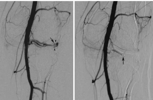
Fig. 1. A. Digital subtraction intraarterial angiography reveals a pseudoaneurysm originating from the inferior genicular artery (arrow) with shunting of contrast into the venous system (arrowheads).
B. Postembolization digital subtraction intraarterial angiography demonstrating the embolized pseudoaneurysm; note the coils that were used and complete occlusion of the aneurismatic fistula (arrow).
A B
lesion with using microcoils to definitely occlude the pseudoaneurysm and AV fistula.
This case demonstrates that superselective percutaneous angiographic embolization with coils offers the advantages of a minimally invasive approach (no surgical incision, a reduced risk of infection and a shortened hospital stay);
therefore, this technique may represent an effective and safe therapeutic alternative for this uncommon complica- tion of knee arthroscopy.
In summary, we report here on a unique case of a lateral inferior genicular artery pseudoaneurysm with concomi- tant arteriovenous fistula as a complication of knee arthroscopy. This was successfully treated with superselec- tive percutaneous transarterial embolization.
References
1. Committee on Complications of the Arthroscopy Association of North America. Complications in arthroscopy: the knee and other joints. Arthroscopy 1986;2:253-258
2. Small NC. Complications in arthroscopic surgery of the knee and shoulder. Orthopedics 1993;16:985-988
3. Hilborn M, Munk PL, Miniaci A, MacDonald SJ, Rankin RN, Fowler PJ. Pseudoaneurysm after therapeutic knee arthroscopy:
imaging findings. AJR Am J Roentgenol 1994;163:637-639 4. Tozzi A, Ferri E, Serrao E, Colonna M, De Marco P,
Mangialardi N. Pseudoaneurysm of the descending genicular artery after arthroscopic meniscectomy: report of a case. J Trauma 1996;41:340-341
5. Vassallo P, Reiser MF, Strobel M, Peters PE. Popliteal pseudoa- neurysm and arteriovenous shunt following arthroscopic meniscectomy: case report. Cardiovasc Intervent Radiol 1989;12:142-144
6. Mullen DJ, Jabaji GJ. Popliteal pseudoaneurysm and arteriove- nous fistula after arthroscopic meniscectomy. Arthroscopy 2001;17:E1
7. Karkos CD, Sampath SA, Bury R, Mohandas P, Forrest L.
Arteriovenous fistula of the lateral superior and inferior genicu- late arteries. A unique cause of a “recurrent prepatellar bursa.
Int Angiol 2002;21:280-283
8. Kiss H, Drekonja T, Grethen C, Dorn U. Postoperative aneurysm of the popliteal artery after arthroscopic meniscec- tomy. Arthroscopy 2001;17:203-205
9. Skibo L, Polak JF. Compression repair of a postcatheterization pseudoaneurysm of the brachial artery under sonographic guidance. AJR Am J Roentgenol 1993;160:383-384
10. Wolford H, Peterson SL, Ray C, Morgan SJ. Delayed arteriove- nous fistula and pseudoaneurysm after an open tibial fracture successfully managed with selective angiographic embolization.
J Trauma 2001;51:781-783
Transarterial Embolization of Inferior Genicular Artery Pseudoaneurysm
Korean J Radiol 8(2), April 2007 175