Clinical Manifestations and Risk Factors of Anaphylaxis in Pollen–Food Allergy Syndrome
Minji Kim 1,2 *, Youngmin Ahn 3 *, Young Yoo 4 , Dong-Kyu Kim 5 , Hyeon-Jong Yang 6 , Hae-Sim Park 7 , Hyun Jong Lee 8 , Mi-Ae Kim 9 , Yi Yeong Jeong 10 , Bong-Seong Kim 11 , Woo Yong Bae 12 , An-Soo Jang 13 , Yang Park 14 , Young-Il Koh 15 , Jaechun Lee 16 , Dae Hyun Lim 17 , Jeong Hee Kim 17 , Sang Min Lee 18 , Yong Min Kim 19 , Young Joon Jun 20 , Hyo Yeol Kim 21 , Yunsun Kim 22 , Jeong-Hee Choi 2,23 ; and Work Group for Rhinitis, the Korean Academy of Asthma, Allergy and Clinical Immunology
1
Department of Pediatrics, Hallym University Dongtan Sacred Heart Hospital, Hallym University College of Medicine, Hwaseong;
2
Allergy and Clinical Immunology Research Center, Hallym University College of Medicine, Chuncheon;
3
Department of Pediatrics, Eulji Hospital, Eulji University, Seoul;
4
Department of Pediatrics, Korea University Anam Hospital, Korea University College of Medicine, Seoul;
5
Department of Otorhinolaryngology-Head and Neck Surgery, Hallym University Chuncheon Sacred Heart Hospital and Nano-Bio Regenerative Medical Institute, Hallym University College of Medicine, Chuncheon;
6
Department of Pediatrics, Pediatric Allergy and Respiratory Center, Soonchunhyang University Seoul Hospital, Soonchunhyang University College of Medicine, Seoul;
7
Department of Allergy and Clinical Immunology, Ajou University School of Medicine, Suwon;
8
Lee & Hong ENT, Sleep and Cosmetic Center, Seongnam;
9
Department of Pulmonology, Allergy and Critical Care Medicine, CHA Bundang Medical Center, CHA University, Seongnam;
10
Department of Internal Medicine, Gyeongsang National University College of Medicine, Jinju;
11
Department of Pediatrics, Gangneung Asan Hospital, University of Ulsan College of Medicine, Gangneung;
12
Department of Otorhinolaryngology, Head and Neck Surgery, Dong-A University College of Medicine, Busan;
13
Department of Internal Medicine, Soonchunhyang University Bucheon Hospital, Bucheon;
14
Department of Pediatrics, Wonkwang University Sanbon Hospital, Wonkwang University College of Medicine, Gunpo;
15
Department of Internal Medicine, Chonnam National University Hospital, Chonnam National University Medical School, Gwangju;
16
Department of Internal Medicine, Jeju National University School of Medicine, Jeju;
17
Department of Pediatrics, Inha University Hospital, Inha University College of Medicine, Incheon;
18
Division of Pulmonology and Allergy, Department of Internal Medicine, Gil Medical Center, Gachon University College of Medicine, Incheon;
19
Department of Otorhinolaryngology-Head and Neck Surgery, Chungnam National University College of Medicine, Daejeon;
20
Department of Otorhinolaryngology-Head & Neck Surgery, Soonchunhyang University Gumi Hospital, Soonchunhyang University College of Medicine, Gumi;
21
Department of Otorhinolaryngology-Head and Neck Surgery, Samsung Medical Center, School of Medicine, Sungkyunkwan University, Seoul;
22
SCH Biomedical Informatics Research Unit, Soonchunhyang University Seoul Hospital, Seoul;
23
Department of Pulmonology, Allergy and Critical Care Medicine, Hallym University Dongtan Sacred Heart Hospital, Hallym University College of Medicine, Hwaseong, Korea.
Purpose: Many studies have reported that pollen-food allergy syndrome (PFAS) can cause anaphylaxis. No comprehensive inves- tigations into anaphylaxis in PFAS have been conducted, however. In this study, we investigated the clinical manifestations and risk factors for anaphylaxis in PFAS in Korean patients with pollinosis.
pISSN: 0513-5796 · eISSN: 1976-2437
Received: May 21, 2019 Revised: July 27, 2019 Accepted: August 9, 2019
Co-corresponding authors: Jeong-Hee Choi, MD, PhD, Department of Pulmonology, Allergy and Critical Care Medicine, Hallym University Dongtan Sacred Heart Hospital, Hallym University College of Medicine, 7 Keunjaebong-gil, Hwaseong 18450, Korea.
Tel: 82-31-8086-2928, Fax: 82-31-8086-2482, E-mail: [email protected] and
Young Yoo, MD, PhD, Department of Pediatrics, Korea University Anam Hospital, Korea University College of Medicine, 73 Goryeodae-ro, Seongbuk-gu, Seoul 02841, Korea.
Tel: 82-2-920-5090, Fax: 82-2-922-7476, E-mail: [email protected]
*Minji Kim and Youngmin Ahn contributed equally to this work.
•The authors have no potential conflicts of interest to disclose.
© Copyright: Yonsei University College of Medicine 2019
This is an Open Access article distributed under the terms of the Creative Commons Attribution Non-Commercial License (https://creativecommons.org/licenses/by-nc/4.0) which permits unrestricted non-commercial use, distribution, and reproduction in any medium, provided the original work is properly cited.
Yonsei Med J 2019 Oct;60(10):960-968
https://doi.org/10.3349/ymj.2019.60.10.960
INTRODUCTION
Pollen-food allergy syndrome (PFAS) is an immunoglobulin E (IgE)-mediated allergic manifestation to fruits and vegetables due to cross-reactivity with prior sensitization to plant inhal- ant allergens. PFAS is an emerging public health issue, with a number of studies reporting an increased prevalence of pol- len allergies over the last decade due to changes in atmo- spheric CO
2, climate, and pollen counts.
1-3PFAS is the most common food allergy in adults, and the prevalence of PFAS varies in the literature from 5–8%.
4,5PFAS, called oral allergy syndrome previously, was thought to be restricted to oropharyngeal symptoms; however, extra- oral symptoms and systemic symptoms in PFAS have been re- ported.
6-8Moreover, one study reported that 3% of patients ex- perienced systemic reactions without oral symptoms, and 1.7%
experienced anaphylaxis.
6,9Because the systemic symptoms reported by researchers have increased and because symp- toms are not limited to the oral cavity, the use of the term PFAS is more relevant than oral allergy syndrome:
10This historical background has led to confusion among allergists concerning the diagnosis of PFAS. In a US study investigating the percep- tion of PFAS, allergists estimated that 5–8% of patients with pollinosis had PFAS.
4However, PFAS has been reported in 20–
70% of patients with a pollen allergy, and anaphylaxis was re- ported in 1–2%, which is more prevalent than what allergists had estimated.
7,8,11Although the importance of PFAS is increasing and trigger- ing foods differ from geographic regions and dietary habits, there are only a few studies on PFAS in Korea.
12,13The first na- tionwide study of PFAS in Korea recently reported that the prevalence of PFAS is 41.7% in Korean patients with pollinosis and that 8.9% of patients with PFAS manifest with anaphylaxis, which is a substantial proportion among patients with PFAS.
14Several allergen components from plant foods are known to contribute to the development of anaphylaxis. Lipid transfer
protein and cross-reactive carbohydrate determinants are known as pan-allergens of the plant, and they exhibit thermo- stability and resistance to proteolysis, which enables the food allergen to reach intestinal absorption in its intact form.
15Al- though several studies on anaphylaxis-inducing allergenic components have been investigated, studies on detailed clini- cal manifestations and risk factor analysis of anaphylaxis in PFAS have been rarely conducted worldwide. Therefore, in this study, we investigated the clinical characteristics and risk factors of anaphylaxis in PFAS among Korean patients with pol- linosis using data from a previous nationwide survey of PFAS in 2016.
14MATERIALS AND METHODS
Study design and participants
The nationwide, cross-sectional study on PFAS was conducted in South Korea between March and December 2016.
14Data were collected from patients diagnosed with pollinosis at 21 institutes (19 university hospitals and two allergy clinics). The protocol was reviewed and approved by the institutional re- view boards of each institute (Hallym University Dongtan Sa- cred Heart Hospital, HDT-2016-04-155-003, etc.). Written in- formed consent was obtained from each patient or their parents.
Pollinosis was diagnosed according to 1) one or more aller- gic disease, including allergic rhinitis, allergic conjunctivitis, and/or bronchial asthma; 2) sensitization to the pollen of ≥one tree, grass, and/or weed; and 3) aggravated allergic symptoms when exposed to sensitized pollens. Sensitization to pollen was diagnosed by positive results to allergy skin tests and/or high serum-specific IgE levels using multiple allergen simul- taneous tests [class ≥2+, Polycheck Allergy (Biocheck Co., Mun- ster, Germany), AdvanSure Allergy Screen (LG Life Science, Seoul, Korea), AllergyScreen (Mediwiss Analytic GmbH, Mo- Materials and Methods: Data were obtained from a nationwide cross-sectional study that previously reported on PFAS in Korean patients with pollinosis. Data from 273 patients with PFAS were collected, including demographics, list of culprit fruits and vege- tables, and clinical manifestations of food allergy. We analyzed 27 anaphylaxis patients and compared them with patients with PFAS with oropharyngeal symptoms only (n=130).
Results: The most common cause of anaphylaxis in PFAS was peanut (33.3%), apple (22.2%), walnut (22.2%), pine nut (18.5%), peach (14.8%), and ginseng (14.8%). Anaphylaxis was significantly associated with the strength of sensitization to alder, hazel, willow, poplar, timothy, and ragweed (p<0.05, respectively). Multivariable analysis revealed that the presence of atopic dermatitis [odds ratio (OR), 3.58; 95% confidence interval (CI), 1.25–10.23; p=0.017]; sensitization to hazel (OR, 5.27; 95% CI, 1.79–15.53;
p=0.003), timothy (OR, 11.8; 95% CI, 2.70–51.64; p=0.001), or ragweed (OR, 3.18; 95% CI, 1.03–9.87; p=0.045); and the number of culprit foods (OR, 1.25; 95% CI, 1.15–1.37; p<0.001) were related to the development of anaphylaxis in PFAS.
Conclusion: The most common culprit foods causing anaphylaxis in PFAS were peanut and apple. The presence of atopic derma- titis; sensitization to hazel, timothy, or ragweed; and a greater number of culprit foods were risk factors for anaphylaxis in PFAS.
Key Words: Pollen-food allergy syndrome, pollen, food allergy, anaphylaxis
ers, Germany)] or the ImmunoCAP
®system (≥0.35 kU/L, Ther- moFisher Scientific, Uppsala, Sweden). A positive skin prick test was defined as wheal size equal or greater to that of hista- mine [allergen/histamine (A/H) ratio ≥3+, Allergopharma (Reinbek, Germany), Lofarma (Milan, Italy), Bencard (Brefod, UK)] or a mean allergen wheal diameter of at least 3 mm.
16The investigated pollens included tree pollens (birch, alder, hazel, beech, oak, willow, poplar, pine, and tree mix), grass pollens (bermuda, meadow, orchard, rye, timothy, and grass mix), and weed pollens (mugwort, ragweed, and Hop Japani- cus). Data were collected using a questionnaire and medical record review, including demographic characteristics, under- lying allergic diseases, and allergy test results. For further analysis, we categorized subjects according to the number of systemic symptoms: 1) group 0 (G0), only oropharyngeal symp- toms; 2) group 1 (G1), patients with any one systemic symp- tom; and 3) group 2 (G2) patients with more than two system- ic symptoms, that is anaphylaxis.
Questionnaires about PFAS
The list of culprit foods included apple, pear, peach, apricot, plum, cherry, watermelon, melon, Korean melon, banana, kiwi, orange, mandarin, pineapple, strawberry, mango, avo- cado, grape, carrot, potato, sweet potato, celery, crown daisy, perilla leaf, lettuce, kale, chicory, taro/taro stem, ginseng, de- odeok (Codonopsis lanceolata), bellflower root, kudzu, lotus root, Chinese yam, eggplant, zucchini, cucumber, tomato, ju- jube, chestnut, peanut, walnut, pine nut, and soy.
In addition to oropharyngeal symptoms (tingling/itching sense or edema of the lips, oral cavity, and/or throat), systemic symptoms were categorized according to the organ in which the symptoms developed: dermatologic symptoms (itching, urticaria, or angioedema), respiratory symptoms (rhinorrhea, cough, dyspnea, wheezing, cyanosis, or hypoxia), cardiovas- cular symptoms (chest pain, hypotension, pale, sweating, or cardiac arrest), gastrointestinal symptoms (nausea or vomit- ing, diarrhea, or abdominal pain), neurologic and systemic symptoms (dizziness, unconsciousness, anxiety, change of sense, or death), and anaphylaxis. The diagnosis of anaphy- laxis was confirmed by a physician using the criteria proposed in the second symposium on the definition and management of anaphylaxis by the National Institute of Allergy and Infec- tious Disease/Food Allergy and Anaphylaxis Network.
17Ana- phylaxis was defined by two or more of the following symptoms occur after exposure to a likely allergen, including involvement of skin tissue, respiratory compromise, cardiovascular symp- toms, gastrointestinal symptoms, and neurologic symptoms.
Statistical analysis
All statistical analyses were performed using R version 3.4.0 (R Foundation, Vienna, Austria). Comparison between groups was performed using chi-squared analysis and Kruskal–Wallis test (>two independent groups) for discrete and continuous
variables, respectively. Because the use of separate univariate tests leads to an inflated type 1 error, Bonferroni correction was applied to each pollen analysis. Multivariate logistic re- gression was utilized to determine potential predictors of ana- phylaxis in PFAS. p<0.05 was considered statistically signifi- cant.
RESULTS
Baseline characteristics of the study subjects
Of 273 patients with PFAS, 130 (47.6%) reported oropharyn- geal symptoms only, 88 (32.2%) showed oropharyngeal symp- toms and one systemic symptom, and 27 (9.9%) showed ana- phylaxis. Of 27 patients with anaphylaxis, 21 (77.8%) had oropharyngeal symptoms, and 6 (22.2%) experienced system- ic reactions without oral symptoms. The patients showed cu- taneous manifestations (84.0%), such as pruritus, urticaria and angioedema; respiratory (84.0%); gastrointestinal (48.0%);
neurologic (36.0%); and cardiovascular (36.0%) symptoms.
Twelve patients (44.4%) were younger than 18 years, and 15 were older than 18 years (Supplementary Table 1, only online).
Clinical characteristics of anaphylaxis patients with PFAS
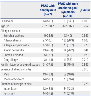
The clinical characteristics of the patients with anaphylaxis in PFAS are described in Table 1. No differences were found be-
Table 1. Clinical Characteristics of the Study Subjects with Anaphylaxis in PFAS
PFAS with anaphylaxis
(n=27)
PFAS with only oropharyngeal
symptoms (n=130)
p value
Sex (male) 14 (51.9) 69 (53.1) 1.000
Age (yr) 27.2±18.7 26.2±16.7 0.767
Allergic diseases
Bronchial asthma 9 (33.3) 52 (40) 0.667
Allergic rhinitis 27 (100)� 126 (96.9) 1.000 Allergic conjunctivitis 17 (63.0) 75 (57.7) 0.770 Atopic dermatitis 13 (48.1) 34 (26.2) 0.041 Chronic urticaria 5 (18.5) 6 (4.6) 0.023
Drug allergy 3 (11.1) 11 (8.5) 0.710
Family history of allergic diseases 21 (77.8) 98 (75.4) 0.986
Severity of allergic rhinitis 0.613
Mild 13 (48.1) 52 (40.6)
Moderate/severe 14 (51.9) 76 (59.4)
Duration of allergic rhinitis 0.723
Intermittent 13 (48.1) 54 (42.2)
Persistent 14 (51.9) 74 (57.8)
PFAS, pollen-food allergy syndrome.
Data are presented as mean±standard deviation or number (%) unless oth-
erwise indicated.
tween PFAS with anaphylaxis and only oropharyngeal symp- toms groups in terms of sex and age (p>0.05, respectively).
The anaphylaxis group had a higher prevalence of developing chronic urticaria than those in the only oropharyngeal symp- tom group (18.5% vs. 4.6%, p=0.023), as well as a higher prev- alence of atopic dermatitis (AD) (48.1% vs. 26.2%, p=0.041).
Patients with PFAS with anaphylaxis showed no significant dif- ferences in the presence of other allergic diseases, such as bron- chial asthma, allergic rhinitis, allergic conjunctivitis, or drug allergy, and family history of allergic diseases, compared with only oropharyngeal symptoms (p>0.05, respectively). The se- verity and duration of allergic rhinitis symptoms were also not significantly different between the anaphylaxis and only oro- pharyngeal symptom groups. The patients with PFAS with anaphylaxis had significantly higher sensitization rates to ha- zel (66.7% vs. 41.5%, p=0.030) and willow (29.6% vs. 11.5%, p=
0.031) than those with only oropharyngeal symptoms (Fig. 1).
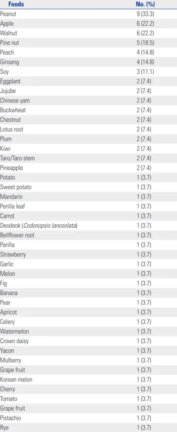
Causative foods of anaphylaxis in PFAS
Twenty-seven anaphylaxis patients had 84 cases with 44 cul- prit foods (Table 2). Peanut (33.3%) was the most common of- fending food, followed by apple (22.2%), walnut (22.2%), pine nut (18.5%), peach (14.8%), ginseng (14.8%), soy (11.1%), etc.
Most of the anaphylaxis patients (n=18, 66.7%) developed anaphylaxis to one food item. Two patients (7.4%) experi-
enced anaphylactic reactions to two food items, three patients to three (11.1%), one patient to four (3.7%), and three patients to more than five food items (11.1%).
Risk factors for the development of anaphylaxis in PFAS After analysis of the association of systemic symptoms in PFAS with the strength of sensitization to pollen (A/H ratio by skin prick test), a significant association was found between anaphylaxis (G2) and strength of sensitization to alder, birch, hazel, beech, oak, willow, poplar, timothy, ragweed, and Hop Japanicus (Fig. 2). Moreover, the anaphylaxis patients (G2) had a significantly higher number of causative foods (p<0.05), whereas the number of sensitized pollens was not associated with anaphylaxis in PFAS (Fig. 3).
Multivariate regression analysis revealed that the presence of AD [odds ratio (OR), 3.58; 95% confidence interval (CI), 1.25–
10.23; p=0.017], sensitization to hazel (OR, 5.27; 95% CI, 1.79–
15.53; p=0.002), sensitization to timothy (OR, 11.8; 95% CI, 2.70–51.64; p=0.001), sensitization to ragweed (OR, 3.18; 95%
CI, 1.03–9.87; p=0.045), and number of culprit foods of PFAS (OR, 1.25; 95% CI, 1.15–1.37; p<0.001) were potential risk fac- tors for the development of anaphylaxis in PFAS (Table 3).
Fig. 1. Comparison of allergen sensitization profiles between anaphylaxis (n=27) and only oropharyngeal symptom patients (n=130) with pollen-food allergy syndrome (PFAS). More patients with anaphylaxis were sensitized to hazel and willow than patients with only oropharyngeal symptoms.
*p<0.05. D, Dermatophagoides.
100
90
80
70
60
50
40
30
20
10
0
Alder Birch Beech Hazel Oak Willow Poplar Japanese cedar
Pine
BermudaMeadow Orchard
Rye TimothyGrass mixMugwortRagweed Hop japanicus D. pteronyssinus
D. farinae Cat Dog
AspergillusAlternaria
Sensitization (%)
PFAS with an aphylaxis
PFAS with only oropharyngeal symptoms
*
*
Inhalant allergens
Online COLOR
DISCUSSION
In this study, the most commonly offending foods of anaphy-
laxis in PFAS were peanut, followed by apple, walnut, pine nut, peach, ginseng, and soy in order of frequency. Anaphylaxis was significantly associated with the strength of sensitization to al- der, birch, hazel, willow, timothy, and ragweed pollen. Pres- ence of AD; sensitization to hazel, timothy, and ragweed; and the increased number of culprit foods for PFAS would more likely lead to the development of anaphylaxis in PFAS.
Approximately 20% to 70% of patients with pollinosis report- ed symptoms of PFAS after ingesting causative foods,
7,8,11and the prevalence of anaphylaxis in patients with PFAS is estimated to be 1–2%.
6In our previous reports, the prevalences of PFAS and anaphylaxis in PFAS were 41.7% and 8.9% in Korea, re- spectively.
14The prevalence of PFAS in our country was similar to that in previous reports for other countries, while the preva- lence of anaphylaxis was much higher. This might be due to several reasons, such as 1) anaphylaxis in PFAS might be over- looked as class I food allergy by physicians; 2) the definition of anaphylaxis was broadly defined, including more than two organ involvements; and 3) the number of patients with PFAS might be increased when considering the increasing pollen al- lergic populations due to global climate changes and increases in pollen counts.
17,18In the present study, the most common anaphylaxis-trigger- ing foods in PFAS were peanut, followed by apple, walnut, pine nut, peach, ginseng, and soy, whereas the most common caus- ative foods of PFAS in Korea were peach, apple, kiwi, peanut, and plum.
14Although common foods that are causative of ana- phylaxis in PFAS have not been reported in other countries, ap- ple and hazelnut are the most common causative foods of PFAS because of the high rate of birch sensitization in Europe, and apple and peach are known as the most common causative foods in relation to oak sensitization in Japan.
19-21In Korea, the most important sensitized pollens are oak, followed by birch and alder, which have cross-allergenicity with the Rosaceae family (apple, peach, plum, pear, cherry, apricot, almond, etc.), Apiaceae family (cantaloupe, honeydew, watermelon, zucchi- ni, and cucumber), Fabaceae family (soybean and peanut), Jug- lans family (walnut), and Betulaceae family (hazelnut).
9,18,22,23Accordingly, cross-reactivity patterns between pollens and foods vary depending on regional distribution of inhaled pol- lens and dietary habits. Relatedly, hazelnut anaphylaxis has not been reported in Korea, potentially in reflection of dietary Table 2. Causative Foods of Anaphylaxis in Pollen-Food Allergy Syn-
drome (n=27)
Foods No. (%)
Peanut 9 (33.3)
Apple 6 (22.2)
Walnut 6 (22.2)
Pine nut 5 (18.5)
Peach 4 (14.8)
Ginseng 4 (14.8)
Soy 3 (11.1)
Eggplant 2 (7.4) �
Jujube 2 (7.4) �
Chinese yam 2 (7.4) �
Buckwheat 2 (7.4) �
Chestnut 2 (7.4) �
Lotus root 2 (7.4) �
Plum 2 (7.4) �
Kiwi 2 (7.4) �
Taro/Taro stem 2 (7.4) �
Pineapple 2 (7.4) �
Potato 1 (3.7) �
Sweet potato 1 (3.7) �
Mandarin 1 (3.7) �
Perilla leaf 1 (3.7) �
Carrot 1 (3.7) �
Deodeok (Codonopsis lanceolata) 1 (3.7) �
Bellflower root 1 (3.7) �
Perilla 1 (3.7) �
Strawberry 1 (3.7) �
Garlic 1 (3.7) �
Melon 1 (3.7) �
Fig 1 (3.7) �
Banana 1 (3.7) �
Pear 1 (3.7) �
Apricot 1 (3.7) �
Celery 1 (3.7) �
Watermelon 1 (3.7) �
Crown daisy 1 (3.7) �
Yacon 1 (3.7) �
Mulberry 1 (3.7) �
Grape fruit 1 (3.7) �
Korean melon 1 (3.7) �
Cherry 1 (3.7) �
Tomato 1 (3.7) �
Grape fruit 1 (3.7) �
Pistachio 1 (3.7) �
Rye 1 (3.7) �
Data are presented as number (%).
Table 3. Multivariate Analysis of Risk Factors for the Development of Anaphylaxis in Pollen-Food Allergy Syndrome
Predictor OR 95% CI p value
Presence of atopic dermatitis 3.58 1.25–10.23 0.017 Sensitization to hazel 5.27 1.79–15.53 0.002 Sensitization to timothy 11.8 2.70–51.64 0.001 Sensitization to ragweed 3.18 1.03–9.87 � 0.045 The number of culprit foods 1.25 1.15–1.37 � <0.001 OR, odds ratio; CI, confidence interval.
Sensitization was diagnosed by allergy skin tests.
6 5 4 3 Neg
6 5 4 3 Neg
6 5 4 3 Neg
6 5 4 3 Neg
6 5 4 3 Neg
6 5 4 3 Neg
6 5 4 3 Neg
6 5 4 3 Neg
6 5 4 3 Neg
6 5 4 3 Neg Number of systemic symptoms other than
oral symptoms
Number of systemic symptoms other than oral symptoms
Number of systemic symptoms other than oral symptoms
Number of systemic symptoms other than oral symptoms
Number of systemic symptoms other than oral symptoms
Number of systemic symptoms other than oral symptoms
Number of systemic symptoms other than oral symptoms
Number of systemic symptoms other than oral symptoms
Number of systemic symptoms other than oral symptoms
Number of systemic symptoms other than oral symptoms
G0 G1 G2
G0 G1 G2
G0 G1 G2
G0 G1 G2
G0 G1 G2
G0 G1 G2
G0 G1 G2
G0 G1 G2
G0 G1 G2
G0 G1 G2 Alder
Birch
Hazel
Beech
Oak
Willow
Poplar
Timothy
Ragweed
Hop Japanicus
A/H ratio of skin prick test A/H ratio of skin prick test A/H ratio of skin prick test A/H ratio of skin prick test A/H ratio of skin prick test A/H ratio of skin prick test A/H ratio of skin prick test A/H ratio of skin prick test A/H ratio of skin prick test A/H ratio of skin prick test
*
*
†
†