DOI 10.3349/ymj.2008.49.5.735
Purpose: Melatonin, the most potent scavenger of toxic free radicals, has been found to be effective in protecting against pathological states due to the release of reactive oxygen species. This study was performed to establish the effect of high dose melatonin on protection against ischemia- reperfusion (I/R) injury in rat hearts. Materials and Methods:
Forty male Sprague-Dawley rats were used in this study. They were separated into four groups of ten rats each. A left coronary artery occlusion was induced in the rats by ligating the artery for 20 minutes and then releasing the ligation (reperfusion) afterwards. The control group was Group A.
Group B was subjected to myocardial ischemia-reperfusion without any treatment, while Group C underwent myocardial ischemia-reperfusion with a melatonin treatment before the ischemia. Group D was subjected to myocardial ischemia- reperfusion with a melatonin treatment before the reperfusion.
After 20 minutes of reperfusion, blood samples were obtained from each group for biochemical studies, and the animals were sacrificed for histological and, immunohistochemical examinations of the myocardial tissue. Results: We found that the cardiac troponin T(cTn-T) levels were significantly increased in Group B when all groups were compared. In the Group C rats treated with melatonin, the cTn-T values were significantly lower than those in Groups B and D. In addition, malondialdehyde (MDA) and antioxidant enzymes including, superoxide dismutase (SOD) and myeloperoxidase (MPO) were lower than those in Group B in the melatonin treated groups. The differences were statistically significant (p < 0.05).
Histopathologic and immunohistopathologic studies also supported the effectiveness of melatonin. Conclusion: Our study suggests that high dose melatonin, appears to offer protection against cardiac ischemia-reperfusion injuries in rats by scavenging the free radicals and could have a potential clinical use in the management of myocardial ischemia.
Key Words: Melatonin, myocardial ischemia-reperfusion, antioxidant, Fas and Bcl-2 expression
INTRODUCTION
Cell injury occurring after ischemia-reperfusion (I/R) in the heart is attributed to necrosis caused by calcium overload, acidosis, and oxidative stress.
1After I/R, myocardial cells die by necrosis and/or apoptosis. Apoptosis is a process for disposing of injured or redundant cells through self-destruction.
2Free radicals and antioxidants play an important role in cellular damage that can cause atherosclerosis and myocardial infarction.
Both neutrophils and free radicals, such as super- oxide anions, hydroxyl radicals and hydrogen peroxide, can cause oxidative damage to cell lipids, proteins, and nucleic acids.
3Melatonin (N-acetyl-5-methoxytryptamine) is secreted by the pineal gland, and has been confirmed to be potent scavenger of hydroxyl and peroxyl radicals.
3,4Many researchers have con- sidered the antioxidant role of melatonin and it is ability to trap cellular free radicals.
5In addition, melatonin has also been shown to act as an immune system modulator.
6Although there is a large amount of consistent data available that shows the potential protective effect of melatonin in cells, a consensus still exists that reactive oxygen species play an important role in the pathogenesis of cardiac reperfusion injury.
7Several previous studies have examined melatonin and its effect on cell death following ischemia-reperfusion. What differentiates our study is the combination of
The Effect of High Dose Melatonin on Cardiac Ischemia- reperfusion Injury
Hakan Ceyran,
1Figen Narin,
2Nazmi Narin,
3Hülya Akgün,
4A. Bahar Ceyran,
5Figen Öztürk,
4and Yiğit Akçalı
1Departments of 1Cardiovascular Surgery, 2Biochemistry, 3Pediatric Cardiology, and 4Pathology, Erciyes University Medical Faculty, Kayseri; 5Pathologist of National Hospital, Kayseri, Turkey.
Received March 29, 2004 Accepted June 14, 2004
Reprint address: requests to Dr. Hakan Ceyran, Department of Cardiovascular Surgery, Erciyes University Medical Faculty, 38030 Kayseri, Turkey. Tel: 90-532-2575846, Fax: 90-352-2229613, E- mail: [email protected]
histological parameters, immunohistopathologic studies and biochemical parameters that support the hypothesis that high-dose melatonin provides effective protection following I/R.
MATERIALS AND METHODS
This study was performed in the Hakan Çetinsaya Clinical and Experimental-Research Center at Erciyes University. The Ethics Com- mittee approved this study. All animals involved received humane care in compliance with the European Convention on Animal Care.
Animals
Male Sprague-Dawley rats (n = 40) weighing 310 to 340 g (average 321 g) were used in this study. The rats were anesthetized with ketamine hydrochloride (20 mg/kg), intraperitoneally (i.p.), and heparin (500 IU/kg, i.p) was also admini- stered.
A left thoracotomy was performed, and the pericardium was incised. The left coronary artery was surgically occluded through ligation with a suture (monoflamant polypropylene, size 6.0) and than followed with a coronary reperfusion through the release of the tie.
Rats were separated in to four groups
Group A (n = 10) was the control group and no procedures were carried out.
Group B (n = 10) was subjected to cardiac is- chemia (20 minutes) - reperfusion (20 minutes) without any treatment.
Group C (n = 10) was treated with melatonin (50 mg/kg i.p, Sigma, M-5250, St. Louis, MO, USA) 30 minutes before ischemia (20 minutes) - reperfusion (20 minutes).
Group D (n = 10) uderwent left coronary artery occlusion (ischemia time of 20 minutes) and then melatonin (50 mg/kg i.p.) was administered just before reperfusion.
Rats that developed ventricular fibrillation or cardiac arrest during I/R were excluded from the study, so only the 40 rats that did not have any complications were used. For biochemical testing blood samples were taken from all rats via the
atrium using a 22 G intravenous cannula (Polyflon) after 20 minutes reperfusion. Hepar- inized blood obtained from the rats was cen- trifuged at 2,000 rpm for 15 minutes at 4°C. After separating the plasma, the samples were stored at -20°C until analysis. Tissue samples were taken from the left ventricle and bathed in a 10%
formalin solution for pathologic study. The tissue samples were stained with Hematoxylin and Eosin (H & E) and were evaluated by light micro- scopy.
Immunohistochemical analysis of Fas and Bcl-2 expression
Over the last few years, compelling evidence has established an important physiologic role for apoptotic cell death in maintaining optimal cell numbers in multicellular organisms. Proteins such as Bcl-2 or Fas and its ligand, have been shown to regulate this process.
Representative sections of the heart tissue were fixed in 10% neutral buffered formalin for 24 hours. After standard histological processing the sections were embedded in paraffin, then 5- m μ paraffin sections were deparaffinized with three rinses of xylene, and than followed by rehyded with ethanol. To unmask any antigenic deter- minants, slides were pretreated in a microwave for 10 minutes in a 0.01 M. citrate buffer, treated for 30 minutes in 0.5% H
2O
2, then washed with PBS (phosphate buffered saline: SIGMA #1000-3).
Next the sections were incubated with the
primary antibodies (1 : 10 dilution overnight for
Fas, 1 : 50 dilution overnight for Bcl-2). Both
mouse monoclonal antibodies against Fas, and
mouse monoclonal antibodies against Bcl-2 (both
from DAKO Corporation, Carpenteria, CA, USA)
were used in this study. The immunohistochemical
staining was performed using the streptoavidin-
biotin kit (DAKO Corporation, Carpenteria, CA,
USA) by the avidin-biotin-peroxidase method,
according to the manufacturer's instructions. The
peroxidase reaction was developed with 3,3'-dia-
minobenzidine, and the slides were counterstained
with hematoxylin. Formalin-fixed, paraffin-
embedded sections of the colon were used as
positive controls for Fas, and tonsil sections were
used as controls in Bcl-2 staining.
The intensity of immunostaining was evaluated by repeated staining of the same specimens, and an observer who had no knowledge of the experimental group examined the staining. The immunostaining was graded as "0" for no immu- nostaining or as, "1" if immunostaining was definitely detectable.
Determination of myeloperoxidase (MPO) activity The assay mixture was composed of a 0.3 mL 100 mM phosphate buffer (pH 6.0; 0.3 mL 10 mM H
2O
2, 0.5 mL 20 mM o-dionisidine (freshly pre- pared) in deionized water; and 10 mL PMNL homogenate in a final volume of 3.0 mL. The absorbance at 460 nm was followed for 10 minutes.
All measurements were carried out in duplicate.
One unit of MPO was defined as that which degrade 1 mmol H
2O
2/min at 25°C; specific activity was given as U/mg of protein. A molar extinction coefficient of 11,300 for oxidized o-dianisidine was used for the calculation.
Determination of malondialdehyde (MDA) activity The determination of MDA, was measured using the spectrophotometrical method defined by Ohkawa et al. in 1979. MDA couples to thiobar- bituric acid to form a pink chromogen compound, which has a maximum absorbance at 540 nm in wavelength. Plasma MDA levels were expressed as micromoles per liter ( mol MDA/liter). μ Determination of superoxide dismutase (SOD) activity
The enzymatic activity of SOD was measured according to the inhibition of nitroblue tetrazo- lium (NBT) reduction with xanthine-xanthine ox- idase used as a superoxide generator. One unit of SOD activity was defined as the amount of protein that inhibited the rate of NBT reduction by 50%.
Enzyme activity was expressed as U/mg protein.
Statistical analysis
A Kruskal-Wallis non-parametric variance an- alysis was performed to test the significance of the p value obtained between groups. All groups were
analyzed with one another using the Mann- Whitney U Test. A p value lower than 0.05 was considered significant.
RESULTS Troponin
In Group A, cTn-T values were significantly lower in comparison with all other groups. The cTn-T values in Group B were significantly higher compared with all other groups. In Group C, where melatonin treatment was used before ischemia, cTn-T values were significantly lower than Groups B and D, which signified the efficacy of pre-ischemia treatment (Table 1).
MDA
The values of MDA in Group A, were signifi- cantly lower compared with all groups. In both Groups C and D where melatonin treatments were used a significantly lower level of MDAs was seen than that of Group B (Table 1).
MPO
The values of MPO in both Groups C and D, which received melatonin treatments, were significantly lower than in Group B (Table 1).
SOD
The values of SOD in Groups C and D, where melatonin treatments, were used showed signifi- cantly lower levels than Group B. There were no significant differences between Groups C and A for these levels (Table 1).
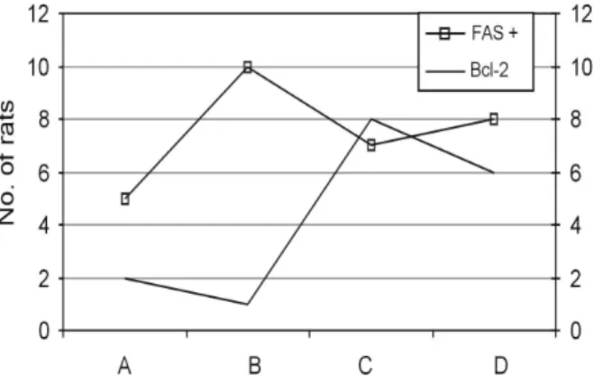
Immunohistochemical studies showed that apoptotic activity in Group B was more intensive than in the other groups (Fig. 1). Melatonin treatment was associated with diminishing Fas expression in ischemic-reperfused hearts in rats.
Bcl-2 expression was significantly higher in Groups C and D (Figs 1 and 2).
The light microscopic study established the
existence of several indications of I/R injuries
such as edema in cardiac myofibrilles, increased
vascularity and increased inflammatory infil- tration in Group B (Fig. 3). These findings were
minimal in the melatonin treated groups (Fig. 4).
DISCUSSION
It is well known that after myocardial ischemia, free radicals, cytokines and antioxidants play a major role in myocardial damage. Early reperfu- sion of the ischemia can save the myocardium before it becomes irreversibly injured. A delay in reperfusion often results in injury to the myo- cardial cells, which has been termed reperfusion injury.
8,9Oxygen-derived free radicals can cause damage to various biological targets, such as proteins, DNA, and lipids, and can also inflict severe membrane damage with consequent cell derangement or death.
10SOD is a metalloenzyme that catalyzes the dismutation of O
2- into O
2and H
2O
2, and affords protection against free radical damage. It can also be associated with increased oxidative stress.
3,11Other important antioxidative enzymes such as MPO and MDA also can result in an increase in ischemia-reperfusion injuries.
12Troponin T is one of the regulatory proteins of the troponin complex, and cardiac troponin is one of the isoforms of this complex. Even minor myo- cardial damage can be detected using cardiac troponin T levels. The importance of cTn-T is its reilablity in identifying perioperative MI and in the assessment of reperfusion therapy of an infarct zone after MI.
13In our study, the levels of cTn-T, SOD, MDA, and MPO were significantly high only in the non-treatment I/R group.
Fig. 2.Strong immunoreactivity with Bcl-2 in myofibrilles in Group C (IHK × 200).
Fig. 3. Severe edema among myofibrilles in non-treated melatonin Group B (H & E × 200).
Fig. 4. Minimal edema among myofibrilles in treated melatonin Group C (H & E × 100).
Fig. 1. Melatonin treatment was associated with decrease Fas expression while Bcl-2 expression were significantly higher in ischemic-reperfused hearts in rats Groups C and D.
Cell death that occurs after I/R in the heart is caused by energy depletion, acidosis, and oxidative stress. Apoptosis occurs during I/R, and it is a significant contributor to myocardial cell death.
Death receptors are cell surface receptors that transmit apoptosis signals initiated by specific ligands. The best characterization of the death receptors is Fas. The Bcl-2 proteins are a family of proteins involved in the response to apoptosis.
Some of these proteins (such as Bcl-2 and Bcl-XL) are anti-apoptotic. The sensitivity of cells to apoptotic stimuli can depend on the balance of pro- and anti-apoptotic Bcl-2 proteins. The pro- apoptotic Bcl-2 proteins are often found in the cytosol, where they act as sensors of cellular damage or stress.
2,8,14In our study, apoptotic activity was intensive in the non-treated I/R group.
Despite the discovery of melatonin more than 40 years ago, it was not recognized as an antioxidant and a free radical scavenger until the last ten years. Several investigators have shown that melatonin is a derivative of the amino-acid tryptophan, and is produced in the pineal gland.
Melatonin is a free radical scavenger that has the ability to neutralize the toxicity of the hydroxyl radical, the singlet oxygen, and possibly the peroxyl radical and the superoxide anion.
15Ianas and colleagues were the first to investigate melatonin as an antioxidant.
16Later, Tan et al.
demonstrated that melatonin detoxifies the hydroxyl radical.
4Sainz et al. were the first to find that both naturally-occurring and induced- apoptosis is slowed down by melatonin.
17As an antioxidant, melatonin is effective in protecting DNA, membrane lipids and, presumably, cytosolic proteins from oxidative damage. In
addition to its direct free radical scavenging and membrane stabilization, melatonin has been reported to alter the activities of enzymes that improve the total antioxidative defense capacity of the organism (including SOD, and nitric oxide synthase).
18The effect of melatonin on myocardial reper- fusion injury has only been studied in recent years. These studies have shown the effect of melatonin on the incidence of myocardial reperfu- sion arrhythmias, stunning, and the limitation of infarct size.
3,19The effect of melatonin on ischemia- reperfusion-induced arrhythmias in the isolated rat heart model has been reported, and it has been found that melatonin is a potent agent in pro- tecting against ischemia-reperfusion injury. This is due to the hydroxyl radical scavenging effect of melatonin and also due to the reduction of the extent of lipid peroxidation.
3In reviewing studies that investigated the effects of antioxidants on the limitation of infarct size, Bolli found that approximately half of the 40 studies published between 1984 - 1989 reported a reduction in infarct size, while the other half did not find any effect.
The only consistent findings were that the antioxidant had to be present during the entire reperfusion period in order to produce positive results.
20The ability of melatonin to act as a peroxyl radical scavenger was compared with that of glutathione and Vitamins E, and C. Melatonin's scavenging ability was about two times higher than that of Vitamins E, and C and almost three times higher than that of glutathione.
21The protective effect of melatonin against oxidative stress during reperfusion of the liver, lungs, intestines and brain after induced ischemia
Table 1. Biochemical Parameters in Group A, B, C, and DGroups A (n = 10) B (n = 10) C (n = 10) D (n = 10) p value
cTn-T 0.64 ± 0.22* 17.36 ± 2.20 2.05 ± 0.93 6.61 ± 1.29 < 0.05
MDA 0.81 ± 0.16* 1.92 ± 0.07 1.42 ± 0.06 1.40 ± 0.06 < 0.05
MPO 61.79 ± 4.73 166.05 ± 14.14 74.11 ± 2.20 83.52 ± 9.35 < 0.05
SOD 0.47 ± 0.04 0.85 ± 0.09 0.57 ± 0.02 0.58 ± 0.01 < 0.05
cTn-T, the cardiac troponin T; MDA, malondialdebhyde; MPO, myeloperoxidase; SOD, superoxide dismutase.
*p < 0.05 (compared with Groups B, C and D), p < 0.01 (compared with Groups A, C and D), p < 0.05 (compared with Group D).
Data presented are mean ± SE.
was examined using both biochemical and mor- phologic measurements
22-25The authors reported that exogenously administered melatonin effec- tively protected the liver, lungs, intestines and brain against oxidative damage. This protection was evident by reduced lipid peroxidation, lowered polymorphonuclear leukocyte infiltration, and reduced antioxidant enzymes.
The observation by Szarszoi et al. that the exogenous administration of a low physiological dose of melatonin does not provide protection does not automatically imply that melatonin is inactive at physiological concentrations.
20In our study, we demonstrated that high doses of melatonin have statistically significant effects on ischemia-reperfusion-induced myocardial injury in rat hearts. The levels of cardiac troponin T and MDA, MPO, and SOD activity in the non-treated I/R groups were higher than in those treated with high doses of melatonin. In the melatonin-treated groups both before ischemia and reperfusion, these values were significantly lower than in the non-treated group. These values were slightly lower in Group C, in which melatonin was given before ischemia, when compared to Group D, in which melatonin was administered before reper- fusion. The effects of melatonin on the expression of Fas and Bcl-2 in the myocardium were deter- mined based on the established death-promoting effect of Fas and the anti-apoptotic effect of Bcl-2.
The high intensity of Bcl-2 in the melatonin treatment groups (particularly before ischemia) indicated that anti-apoptotic activity was present.
All these biochemical results were supported by immunohistochemical and histopathological findings.
Although low-dose melatonin studies seem to have been inconclusive, our study has em- phasized that high-dose melatonin treatment increases antioxidant enzymes and anti-apoptotic activity. In conclusion, our study confirmed that high-dose melatonin treatment acts as a protective agent against cardiac I/R in rats.
REFERENCES
1. Hearse DJ, Bolli R. Reperfusion induced injury:
manifestations, mechanisms, and clinical relevance.
Cardiovasc Res 1992;26:101-8.
2. Yue TL, Ma XL, Wang X, Romanic AM, Liu GL, Louden C, et al. Possible involvement of stress-activated protein kinase signaling pathway and Fas receptor expression in prevention of ischemia /reperfusion-in- duced cardiomyocyte apoptosis by carvedilol. Circ Res 1998;82:166-74.
3. Kaneko S, Okumura K, Numaguchi Y, Matsui H, Murase K, Mokuno S, et al. Melatonin scavenges hydroxyl radical and protects isolated rat hearts from ischemic reperfusion injury. Life Sci 2000;67:101-12.
4. Tan DX, Chen LD, Poeggeler B, Manchester LC, Reiter RJ. Melatonin: a potent, endogenous hydroxyl radical scavenger. Endocr J 1993;1:57-60.
5. Reiter RJ, Poeggeler B, Tan DX, Chen LD, Manchester LC, Cuerrero JM. Antioxidant capacity of melatonin: a novel action not requiring receptor. Neuro Endocrinol Lett 1993;15:103-16.
6. Fjaerli O, Lund T, Osterud B. The effect of melatonin on cellular activation processes in human blood. J Pineal Res 1999;26:50-5.
7. Hearse DJ, Tosaki A. Reperfusion-induced arrhythmias and free radicals: studies in the rat heart with DMPO.
J Cardiovasc Pharmacol 1987;9:641-50.
8. Jeremias I, Kupatt C, Martin-Villalba A, Habazettl H, Schenkel J, Boekstegers P, et al. Involvement of CD95/
Apo1/Fas in cell death after myocardial ischemia.
Circulation 2000;102:915-20.
9. Salie R, Harper I, Cillie C, Genade S, Huisamen B, Moolman J, et al. Melatonin protects against ischaemic- reperfusion myocardial damage. J Mol Cell Cardiol 2001;33:343-57.
10. Basaga HS. Biochemical aspects of free radicals.
Biochem Cell Biol 1990;68:989-98.
11. Rodríguez AB, Nogales G, Ortega E, Barriga C.
Melatonin controls superoxide anion level: Modulation of superoxide dismutase activity in ring dove hetero- phils. J Pineal Res 1998;24:9-14.
12. Cuzzocrea S, Costantino G, Mazzon E, Micali A, De Sarro A, Caputi AP. Beneficial effects of melatonin in a rat model of splanchnic artery occlusion and reperfu- sion. J Pineal Res 2000;28:52-63.
13. Haider KH, Stimson WH. Cardiac myofibrillar proteins:
biochemical markers to estimate myocardial injury. Mol Cell Biochem 1999;194:31-9.
14. Ohno M, Takemura G, Ohno A, Misao J, Hayakawa Y, Minatoguchi S, et al. "Apoptotic" myocytes in infarct area in rabbit hearts may be oncotic myocytes with DNA fragmentation: analysis by immunogold electron microscopy combined with in situ nick end-labeling.
Circulation 1998;98:1422-30.
15. Reiter R, Tang L, Garcia JJ, Muñoz-Hoyos A. Pharma- cological actions of melatonin in oxygen radical patho- physiology. Life Sci 1997;60:2255-71.
16. Ianăs O, Olinescu R, Bădescu I. Melatonin involvement in oxidative processes. Endocrinologie 1991;29:147-53.
17. Sainz RM, Mayo JC, Uría H, Kotler M, Antolín I, Rodriguez C, et al. The pineal neurohormone melatonin
prevents in vivo and in vitro apoptosis in thymocytes.
J Pineal Res 1995;19:178-88.
18. Bettahi I, Pozo D, Osuna C, Reiter RJ, Acuña- Castroviejo D, Guerrero JM. Melatonin reduces nitric oxide synthase activity in rat hypothalamus. J Pineal Res 1996;20:205-10.
19. Szárszoi O, Asemu G, Vanecek J, Ost'ádal B, Kolár F.
Effects of melatonin on ischemia and reperfusion injury of the rat heart. Cardiovasc Drugs Ther 2001;15:251-7.
20. Bolli R. Oxygen-derived free radicals and myocardial reperfusion injury: an overview. Cardiovasc Drugs Ther 1991;5 Suppl 2:249-68.
21. Pieri C, Marra M, Moroni F, Recchioni R, Marcheselli F. Melatonin: a peroxyl radical scavenger more effective than vitamin E. Life Sci 1994;55:PL271-6.
22. Sewerynek E, Reiter RJ, Melchiorri D, Ortiz GG, Lewinski A. Oxidative damage in the liver induced by ischemia-reperfusion: protection by melatonin. Hepato- Gastroenterology 1996;43:898-905.
23. Inci I, Inci D, Dutly A, Boehler A, Weder W. Melatonin attenuates posttransplant lung ischemia-reperfusion injury. Ann Thorac Surg 2000;73:220-5.
24. Kazez A, Demirbağ M, Ustündağ B, Ozercan IH, Sağlam M. The role of melatonin in prevention of intestinal ischemia-reperfusion injury in rats. J Pediatr Surg 2000;35:1444-8.
25. Wakatsuki A, Okatani Y, Izumiya C, Ikenoue N.
Melatonin protects against ischemia and reperfusion- induced oxidative lipid and DNA damage in fetal rat brain. J Pineal Res 1999;26:147-52.