INTRODUCTION
Viscoelastic substances are now widely used in the field of ophthalmologic surgery to maintain anterior chamber depth during surgery and to pro-
tect corneal endothelial cells from mechanical injuries.1The cause of corneal endothelial toxicity was thought to be oxygen free radicals from poly- morphonuclear leukocytes (PMNs). Among the var- ious kinds of viscoelastics, hyaluronic acids (HAs) of various molecular weights have been most fre- quently used. However, not only mechanical contact but also inflammation causes tissue injury by surgery. Inflammation during intraocular surgery may cause cataracts, corneal opacity and retinal edema. Some investigators2 have reported that by using HA instead of an air bubble during glaucoma Vol. 18:23-28, 2004
Effects of Hyaluronic Acid on the Polymorphonuclear Leukocyte (PMN) Release of Active Oxygen and Protection of Bovine Corneal
Endothelial Cells from Activated PMNs
Hyun Soo Lym, MD, Youn Suh, MD, Chan Kee Park, MD*
Department of Ophthalmology, St. Mary’s Hospital,
*Department of Ophthalmology, Kangnam St. Mary’s Hospital, College of Medicine, The Catholic University of Korea, Seoul, Korea
The goal of this study was to evaluate the function of hyaluronic acid (HA) on the active oxygen release from polymorphonuclear leukocytes (PMNs) and the protec- tive effect of bovine corneal endothelial cells (BCEC) from activated PMNs. We used HA with three different molecular weights (MW 700,000, 2,000,000, and 4,000,000) and five different concentrations (0, 0.1, 1, 2, and 3 mg/ml). We evaluated the amount of released superoxide from activated PMNs by using dismutase-inhibitable ferricytochrome C reduction. To compare the property and protective effect of HA with those of other viscoelastic substances, we used the same concentration of methylcellulose. HA suppressed superoxide release from PMNs and protected BCEC from activated PMNs in a dose-dependent, rather than a molecular weight-depen- dent, manner. The effect of HA reached almost a plateau at concentration above 2 mg/ml. However, methylcellulose, another viscoelastic substance, showed a similar effect. Therefore, it seems that the suppression of superoxide released from PMNs is not a property that is unique to HA, but is a general property of viscoelastic sub- stances. Our results indicate that the action mechanism of HA proceeds not only through cell surface HA-receptor. We think that HA also acts as a physical barrier and/or a scavenger of superoxide.
Key words: corneal endothelial cell, hyaluronic acid, polymorphonuclear leukocyte, superoxide
Reprint requests to Chan Kee Park, MD, PhD, Department of Ophthalmology, Kangnam St. Mary’s Hospital, College of Medicine, The Catholic University of Korea, #505 Banpo-dong, Seocho-ku, Seoul 137-701, Korea.
This paper was presented at an ARVO meeting in Fort Lauderdale, Florida in April, 1996.
filtering surgery, they could decrease the rate of cataract development after operation.
HA is a disaccharide polymer of glucuronic acid and N-acetylglucosamine. Because of its mechani- cal protective effect, it is used in cataract, glaucoma and ocular penetrating injury operations.1 Furthermore, it is known that HA inhibits the attach- ment of leukocytes to the intraocular lens and inhibits phagocytosis of macrophages.3,4 However, there is still considerable debate over its actions.
Suzuki and Yamaguchi3 reported that HA sup- pressed active oxygen release and phagocytosis of macrophage in a molecular-dependent manner and Koich et al.4reported that HA inhibited chemotaxis and phagocytosis of macrophage but not active oxy- gen release. However, there have been few studies regarding the possible properties as protectors of the corneal endothelium from the oxidative damage caused by free radicals.
The purpose of this study was to determine whether HA actually inhibits active-oxygen release from the leukocyte and if this effect is molecular- dependent or dose-dependent. We also wanted to determine whether HA would protect cultured corneal endothelial cells from activated PMN by measuring the viability of bovine corneal endothe- lial cells (BCEC) in MTT assay.
MATERIALS AND METHODS
1. Effect of HA on PMN superoxide release Human peripheral blood was obtained in 20 ml quantities with a 20 U/ml heparin-coated syringe and then mixed with Hank’s balanced salt solution (HBSS, GIBCO). Neutrophils were separated from red cells and other lymphocytes using Ficoll-Paque (Pharmacia & Upjohn, Biotech AB Uppsala, Sweden). After that, neutrophils were mixed with dextran (SIGMA, St. Louis, Mo, USA) and then preserved in an ice bag for 45 minutes. The upper fluid was removed and the remaining red blood cells were destroyed with hypotonic solution (0.2%
NaCl). Purified neutrophils were obtained by cen- trifugation of the solution at 4°C and 1000 rpm for 10 minutes with 10% fetal bovine serum (FBS, GIBCO).
Using a 96-well plate, about 1 x 10⁶of the puri-
fied neutrophils were placed into each well along with 50 ul of HBSS. Into each well was then placed one of the three types of HA, molecular weight (MW) 700,000 (Viscoat®, Alcon, USA), MW 2,000,000 (Healon®, Pharmacia & Upjohn, Sweden), and MW 4,000,000 (Healon GV®, Pharmacia & Upjohn, Sweden) at four different concentrations (0.1, 1, 2, 3 mg/ml). We also employed two types of control groups: viscoelastic free wells and 2% hydroxymethylcellulose (Ocucoat®, Storz, USA) wells. The 2% hydrox- ymethylcellulose wells did not contain HA but had similar viscoelasticity to the HA wells. Twenty microliters of a 10 ng/ml phorbol myristate acetate solution (PMA, SIGMA), a chemoactive peptide for neutrophils, was applied into each well. Fifty micro- liters of cytochrome C (SIGMA) was applied into each well and the plates were incubated for 90 minute in a CO2 incubator. After incubation, the reduced form of cytochrome C was measured at 550 nm with a Microplate reader (MR 700, Dynatech laboratories, USA) with spectral filter. To avoid measurement of superoxide that was not derived from PMN, we prepared wells with superoxide dis- mutase (SOD, SIGMA). The inhibitory effect of vis- coelastics was expressed as a percentage of inhibi- tion as follows:
Percentage Inhibition = 100 –
Nanomoles of Superoxide Generated in the Presence of Viscoelastics
–––––––––––––––––––––––––––––––––– X 100 Nanomoles of Superoxide Generated in
the Absence of Viscoelastics
2. Protective effect of HA on BCEC from activat- ed PMN
BCEC were prepared with modification as described by Crawford KM et al.5Briefly, bovine eyes were obtained from a local abattoir, and the corneas were excised with an attached scleral ring and placed, endothelium side up, in a concave plas- tic cup. The endothelial surface was ringed and sub- sequently incubated at 37°C for 90 minutes in a sterile solution of 20mM HEPES buffered Earle’s balanced salt solution (EBSS,GIBCO). Endothelial cells were dislodged from Descemet’s membrane by gentle scraping using a spatula with forceps.
Dislodged cells were aspirated with pipette and
added with RPMI 1640 media with 10% FBS. The cells were gently centrifuged (600g, 2 minutes), resuspended in 5 ml of medium supplement and incubated in 25-cm2 flasks at 37°C in 95% air-5%
CO2. Third-passage cells were inoculated in a 96- well plate, 1 x 10⁵ per well, and cultured for three additional days for confluencing. Into each well, we applied human PMN, 1 x 10⁶ per well, and then supplied one of the three types of HA, or methylcel- lulose, at the four different concentrations (0.1, 1, 2, 3 mg/ml). After PMN activation with PMA, PMNs were incubated for 90 minutes and then washed out with HBSS. The viability of the remaining BCEC was measured with an MTT (3-[4,5-dimethylthia- zol-2-yl]-2,5-diphenyl tetrasodium bromide, SIGMA) assay.6The protective effect of viscoelas- tics was expressed as a percentage of protection as follows:
Percentage Protection = 100 –
MTT score in the Presence of Viscoelastics –––––––––––––––––––––––––––––––––– X 100 MTT score in the Absence of Viscoelastics
3. Statistical analysis of results
All of these procedures were repeated three times.
The results were analyzed with one-way ANOVA according to molecular weight and concentration and Duncan’s pairwise comparison test was per- formed. To analyze for the dual effects of molecular weight and concentration, we used two-way ANOVA. We calculated the above statistics using a
PC-statistical analysis system program (SAS, ver- sion 6.12).
RESULTS
1. Effect of HA on PMN superoxide release The degree of superoxide release decreased in a concentration-dependent manner in all three types of HA and methylcellulose (p < 0.0001) (Table 1).
However, the comparison according to molecular weight at each concentration showed no significant difference (p value of 0.1mg/ml concentration was 0.8495, that of 1 mg/ml concentration was 0.0733) between types of viscoelastics except for methylcel- lulose and 4,000,000 MW HA, both over 2 mg/ml concentration: the percentage inhibition of 2mg/ml concentration of methylcellulose and 4,000,000 MW HA was 78.23 ± 2.08 and 86.69 ± 7.45, respec- tively (p = 0.0001), and of 3mg/ml concentration was 75.00 ± 2.08 and 78.43 ± 6.27, respectively (p = 0.0001) (Table 1). In two-way ANOVA analysis that considered both the effects of molecular weight and concentration of HA, both were found to influ- ence the results. Specifically, HA inhibited superox- ide release dose-dependently at concentrations over 0.1 mg/ml (Fig. 1). The mean values of cytochrome C reduction in SOD wells and viscoelastic-free wells were equal. This revealed that the main source of superoxide was PMN.
Table 1. Inhibitory effect of viscoelastics according to their types and concentrations (percentage, mean ± standard deviation) on PMN superoxide release.
type of viscoelastic substances conc.(mg/ml)
methylcellulose HA70 HA200 HA400
P-value
control 99.19 ± 6.78 99.80 ± 4.35 93.55 ± 2.28 102.2 ± 9.28 0.2416 0.1 96.37 ± 10.73 98.79 ± 6.44 97.18 ± 3.05 95.77 ± 6.79 0.8495 1 81.05 ± 2.03 92.54 ± 13.5 85.08 ± 5.94 94.56 ± 5.02 0.0733 2 78.23 ± 2.08 66.73 ± 2.10 71.77 ± 2.08 86.69 ± 7.45 0.0001 3 75.00 ± 2.08 62.30 ± 3.55 68.55 ± 2.79 78.43 ± 6.27 0.0001 P-value < 0.0001 < 0.0001 < 0.0001 < 0.0001
conc.: concentration, HA70: Hyaluronic Acid, molecular weight 700,000, HA200: Hyaluronic Acid, molecular weight 2,000,000, HA400: Hyaluronic Acid, molecular weight 4,000,000, P-value: results of one-way ANOVA in each type of viscoelastic and its concentration
2. Protective effect of HA on BCEC from activat- ed PMN
In all three types of HA and methylcellulose, the results of the MTT assay showed that the protective effect of viscoelastics on BCEC from activated PMN occurred in a concentration-dependent man- ner. However, there was no significant difference in the types of viscoelastics in the one-way ANOVA analysis: percentage protection of methylcellulose, and HA of MW 700,000, 2,000,000 and 4,000,000, was 112.22 ± 4.19, 113.2 ± 1.83, 108.7 ± 0.26 and
112.2 ± 1.05, respectively (p = 0.3663) (Table 2). In the two-way ANOVA, both type and concentration of viscoelastics influenced the results. In the con- centration analysis, the BCEC survival rate was pos- itively correlated with viscoelastic concentration:
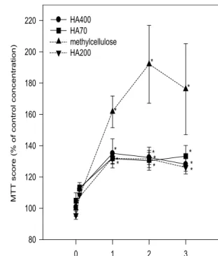
percentage protection of methylcellulose, and HA of MW 700,000, 2,000,000, 4,000,000, at 1 mg/ml, was 161.67 ± 10.12, 131.9 ± 3.23, 131.9 ± 3.67 and 135.1 ± 9.29, respectively (p = 0.0002), and at 2 mg/ml was 192.04 ± 24.90, 130.8 ± 4.86, 131.7 ± 7.35 and 132.5 ± 4.50, respectively (p = 0.0001) (Table 2). However, the curve of survival plateaued at concentrations over 1 mg/ml (Fig. 2). In the type Fig. 1. Amount of superoxide release (percentage
score of reduced cytochrome C) depending on the concentration of viscoelastic substances. The inhibitory effects of superoxide release were influ- enced by both the molecular weight and concentra- tion of hyaluronic acid. Specifically, hyaluronic acid inhibited superoxide release in proportion to its con- centration over 0.1 mg/ml.
HA70: Hyaluronic Acid, molecular weight 700,000 HA200: Hyaluronic Acid, molecular weight 2,000,000 HA400: Hyaluronic Acid, molecular weight 4,000,000
*P < 0.05 compare to control concentration (0 mg/ml) by Duncan’s multiple pairwise comparison test.
Fig. 2. Viability of bovine corneal endothelial cells (percentage score of MTT assay) depending on the concentration of viscoelastic substances. There was no significant difference in BCEC viability except for methylcellulose wells in which more BCEC survived than in other wells.
HA70: Hyaluronic Acid, molecular weight 700,000 HA200: Hyaluronic Acid, molecular weight 2,000,000 HA400: Hyaluronic Acid, molecular weight 4,000,000
*P < 0.05 compare to control concentration (0 mg/ml) by Duncan’s multiple pairwise comparison test.
analysis, there was no significant difference in BCEC viability except for methylcellulose wells in which more BCEC survived than in other wells (Fig. 2).
DISCUSSION
Recently, HA has been actively researched. Some investigators have shown that HA, which is normal- ly present in umbilical cord, vitreous and synovial fluid, was depolymerized in the vitreous cavity by active oxygen,7inhibited arthritis in the synovial cavity,8-10and also inhibited the migration and phagocytosis of macrophages and PMN and super- oxide release from them.3,4In our study, we also found that HA inhibited superoxide released from PMN activated by the chemoactive peptide, PMA.
Furthermore, this phenomenon occurred in a con- centration-dependent manner similar to that reported in previous studies by other investigators. However, unlike other studies,3,4these effects of HA were not proportional to molecular weight and did not occur only with high-molecular-weight HA. The results of our BCEC study also showed that the effect of HA was dependent on concentration, not molecular weight.
The action mechanism of HA’s inhibition of cell migration, phagocytosis and superoxide release was not clearly understood until now. The inhibition probably occurs through the HA receptor that is located in the cell membrane. In addition, it is known that the hyaluronic receptor is a glycoprotein
which penetrates the cell membrane and attaches to cytoskeletal proteins; its structure and molecular weight are similar to the CD44 adhesion glycopro- tein.11Since CD44 is involved with cellular mecha- nisms in T-lymphocyte and monocyte,12,13 some investigators think that HA, especially high-molecu- lar-weight HA, acts like anti-CD44 antibody, inhibiting cell phagocytosis and chemotaxis by influencing the intracellular cytoskeleton through its receptor.4 If the action of HA occurs only through its receptor, HA can only inhibit superoxide release from PMN, but cannot remove pre-existing active oxygen like other oxygen scavengers. However, Artola et al14showed that HA and hydroxypropyl- methylcellulose (HPMC) protected rabbit corneal endothelial cells from hydrogen peroxide which was injected into the anterior chamber. They explained this effect by arguing that either HA formed a mechanical barrier between the corneal endothelium and injected hydrogen peroxide, acted as a chemical scavenger, or some combination of the two.
In our study, although there were statistically sig- nificant effects of inhibition and protection of HA found by two-way ANOVA, a cross-effect between molecular weight and concentration was observed.
One-way ANOVA and Duncan’s multiple pairwise comparison test were therefore performed. Table 1 and 2 show that HA inhibited the release of super- oxide from PMN in a concentration-dependent man- ner and that the protection of BCEC resulted from this inhibition. In Figures 1 and 2, we can see that the above effects were statistically significant at Table 2. Protective effect of viscoelastics according to their types and concentrations (percentage, mean standard deviation) on BCEC from activated PMNs.
type of viscoelastic substances conc.(mg/ml)
methylcellulose HA70 HA200 HA400
P-value
control 100.00 ± 5.10 104.8 ± 5.10 95.56 ± 2.62 100.7 ± 5.76 0.5235 0.1 112.22 ± 4.19 113.2 ± 1.83 108.7 ± 0.26 112.2 ± 1.05 0.3663 1 161.67 ± 10.12 131.9 ± 3.23 131.9 ± 3.67 135.1 ± 9.29 0.0002 2 192.04 ± 24.90 130.8 ± 4.86 131.7 ± 7.35 132.5 ± 4.50 0.0001 3 176.11 ± 29.07 133.3 ± 6.81 126.3 ± 2.62 128.2 ± 6.29 0.0830
P-value 0.0057 0.0009 0.0001 0.0011
conc.: concentration, HA70: Hyaluronic Acid, molecular weight 700,000, HA200: Hyaluronic Acid, molecular weight 2,000,000, HA400: Hyaluronic Acid, molecular weight 4,000,000, P-value: results of one-way ANOVA in each type of viscoelastic and its concentration
concentration over 1 mg/ml. However, we could not find the proportional effect of molecular weight as in Suzuki and Yamaguchi’s study.3 Furthermore, methylcellulose showed nearly the same or even better results in its inhibitory and protective effects as HA. From our results, we think that HA may not play a role through its receptor, but instead may behave as a mechanical barrier between either chemoactive peptide and PMN, or between superox- ide and corneal endothelial, or act as an active-oxy- gen scavenger. We also can not rule out the possi- bility that two or all of these actions occur at the same time. Further studies of HA and its receptor will be necessary to understand its action mecha- nism.
REFERENCES
1. Liesegang TJ. Viscoelastic substances in ophthalmol- ogy. Surv Ophthalmol. 1990;34:268-293.
2. Ayako A, Michael EY. Posttrabeculectomy anterior subcapsular cataract formation induced by anterior chamber air. Ophthalmic Surg. 1993;24:314-319.
3. Suzuki Y, Yamaguchi T. Effect of hyaluronic acid on macrophage phagocytosis and active oxygen release.
Agents Actions. 1993;38:32-37.
4. Koichi T, Masahito T, Sachigy S, Hiromi N, Yoki M.
Effects of high-molecular-weight hyaluronates on the functions of guinea pig polymorphonuclear leuko- cytes. Seminars in Arthritis and Rheumatism. 1993;
2:4-8.
5. Crawford KM, Ernst SA, Meyer RF, MacCallumDK.
Na/K-ATPase pump sites in cultured bovine corneal endothelium of varying cell density at confluence.
Invest Ophthalmol Vis sci. 1995;36:1317-1326.
6. Wagner M, Benson MT, Rennie IG, MacNeil S.
Intracellular regulation of retinal pigment epithelial cell attachment to extracellular matrix proteins. Curr Eye Res. 1995;14:374-384.
7. Ueno N. Changes in vitreous structure caused by oxygen free radicals. Nippon Ganka Gakkai Zasshi.
1995;99:1342-1360.
8. Saari H, Konttinen YT, Friman C, Sorsa T.
Differential effects of reactive oxygen species on native synovial fluid and purified human umbilical cord hyaluronate. Inflammation. 1993;17:403-415.
9. Schenck P, Schneider S, Miehlke R, Prehm P.
Synthesis and degradation of hyaluronate by synovia from patients with rheumatoid arthritis. J Rheumatol.
1995:22:400-405.
10. Fragonas E, Degrassi A, Kvam C, Matulova M, Pollesello P, Zanetti F, Vittur F. Oxygen-derived free radical (ODFR) action on hyaluronic acid (HA), on two HA ester derivatives, and on the metabolism of articular chondrocytes. Exp Cell Res. 1995;218:79- 86.
11. Lacy BE, Underhill CB. The hyaluronate receptor is associated with actin filaments. J Cell Biol.
1987;105:1395-1404.
12. Miyake K, Underhill CB, Lesley J. Hyaluronate can function as a cell adhesion molecule and CD44 par- ticipates in hyaluronate recognition. J Exp Med.
1990;172:69-75.
13. Culty M, Miyake K, Kincade PW. The hyaluronate receptor is a member of the CD44 (H-CAM) family of cell surface glycoproteins. J Cell Biol.
1990;111:2765-2774.
14. Alberto A, Jorge LA, Juan LB, Jose MR. Protective properties of viscoelastic substances (sodium hyaluronate and 2% hydroxymethylcellulose) against experimental free radical damage to the corneal endothelium. Cornea. 1993;12:109-114.