Early Differential Changes in Coronary Plaque Composition According to Plaque Stability Following Statin Initiation in Acute
Coronary Syndrome: Classification and Analysis by Intravascular Ultrasound-Virtual Histology
Dae Seong Hwang, Eun Seok Shin, Shin Jae Kim, Jun Ho Lee, Jong Min Kim, and Sang-Gon Lee
Department of Medicine, Ulsan University Hospital, University of Ulsan College of Medicine, Ulsan, Korea.
Received: June 1, 2012 Revised: July 2, 2012 Accepted: July 4, 2012
Corresponding author: Dr. Sang-Gon Lee, Department of Medicine,
Ulsan University Hospital,
University of Ulsan College of Medicine, 877 Bangeojinsunhwando-ro, Dong-gu, Ulsan 682-714, Korea.
Tel: 82-52-251-8235, Fax: 82-52-250-8238 E-mail: [email protected]
∙ The authors have no financial conflicts of interest.
© Copyright:
Yonsei University College of Medicine 2013 This is an Open Access article distributed under the terms of the Creative Commons Attribution Non- Commercial License (http://creativecommons.org/
licenses/by-nc/3.0) which permits unrestricted non- commercial use, distribution, and reproduction in any medium, provided the original work is properly cited.
Purpose: The aim of this study was to demonstrate the early effects of statin treat- ment on plaque composition according to plaque stability on Intravascular Ultra- sound-Virtual Histology at 6 months after a coronary event. Previous trials have demonstrated that lipid lowering therapy with statins decreases plaque volume and increases plaque echogenicity in patients with coronary artery disease. Materials and Methods: Fifty-four patients (54 lesions) with acute coronary syndrome were prospectively enrolled. We classified and analyzed the target plaques into two types according to plaque stability: thin-cap fibroatheroma (TCFA, n=14) and non-TCFA (n=40). The primary end point was change in percent necrotic core in the 10-mm subsegment with the most disease. Results: After 6 months of statin therapy, no change was demonstrated in the mean percentage of necrotic core (18.7±8.5% to 20.0±11.0%, p=0.38). There was a significant reduction in necrotic core percentage in patients with TCFA (21.3±7.2% to 14.4±8.9%, p=0.017), but not in patients with non-TCFA. Moreover, change in percent necrotic core was significantly correlated with change in high-sensitivity C-reactive protein levels (r=0.4, p=0.003). Changes in low-density lipoprotein cholesterol levels and lipid core percentage demonstrated no significant associations. Conclusion: A clear reduction of lipid core was ob- served only for the TCFA plaque type, suggesting that changes in plaque composi- tion following statin therapy might occur earlier in vulnerable plaque than in stable plaque; the effect may be related to the anti-inflammatory effects of statins.
Key Words: Acute coronary syndromes, statin, IVUS-VH
INTRODUCTION
Histopathologic data indicate that plaque composition is a major determinant of the tendency of atherosclerotic lesions to provoke clinical events. In particular, thin-cap fibroatheroma (TCFA) plaques with macrophage and large necrotic cores are at high risk for rupture and result in epicardial coronary occlusion.1,2 Recent data suggest that spectral analysis of intravascular radiofrequency ultrasound,
lipid-lowering therapy in the 4 weeks prior to enrollment were also excluded. Patients aged 18 to 75 years were eligi- ble for inclusion in the present study if they had been rec- ommended for an intracoronary revascularization procedure and had successfully undergone intracoronary intervention.
The target plaque was qualified for the IVUS study if it had not been influenced by any previous therapeutic interven- tion, and if the diameter of the stenosis was <50% on quan- titative coronary angiography. The plaque had to be >10 mm proximal to the acute intervention site. Another coro- nary artery was imaged if there was no plaque visible in the intervened vessel.3 Bifurcation lesions, lesions with severe angulation, heavily calcified lesions, and lesions with poor image quality were excluded from the present study. ACS was defined as unstable angina, non-ST-segment elevation MI or ST-segment elevation MI. The present study was ap- proved by the hospital’s Ethics Committee, and written in- formed consent was obtained from all patients.
Gray scale and IVUS-VH analyses
Details regarding the validation of the technique, on ex- planted human coronary segments, have previously been reported.3 Briefly, IVUS-VH (Volcano Corp., Rancho Cor- dova, CA, USA) uses spectral analysis of IVUS radiofre- quency data to construct tissue maps that classify plaque into four major components. In preliminary in vitro studies, four histological plaque components (fibrous, fibro-lipid, necrotic core, and calcium) were correlated with a specific spectrum of the radiofrequency signal. These different plaque components were assigned color codes. Calcified, fibrous, fibrolipidic, and necrotic core regions were labeled white, green, greenish-yellow, and red, respectively. IVUS-VH data were acquired after intracoronary administration of nitrates using a continuous pullback (0.5 mm/s) with commercially available mechanical sector scanners (2.9 Fr Eagle Eye 20- MHz catheter, Volcano Corp., Rancho Cordova, CA, USA) by a dedicated IVUS-VH console (Volcano Corp., Rancho Cordova, CA, USA). The IVUS-VH data were stored on a CD-ROM/DVD and sent to the imaging core lab for offline analysis (Asan Medical Center, Seoul, Korea). Manual con- tour detection of both the lumen and the media-adventitia in- terface was performed. IVUS-VH analyses were reported in absolute amounts and as percentages (relative amounts) of each plaque. The target segment was selected and deter- mined with a reproducible index side branch. Geometrical and compositional data were obtained for each cross-sec- tional area (CSA), and an average was calculated for the known as Intravascular Ultrasound-Virtual Histology (IVUS-
VH), offers an opportunity to simultaneously assess the mor- phological and histopathological characteristics of plaque.
This ability allows IVUS-VH to define TCFA plaques in vivo.3-5
Numerous clinical trials have shown that lipid lowering therapy with statins reduces cardiovascular morbidity and mortality, with significant effects evident only after 12 to 24 months of treatment. Although these trials excluded patients with recent unstable angina or acute myocardial infarction (MI),6,7 patients experience the highest rates of death and recurrent cardiovascular ischemic events during the early period after an acute coronary syndrome (ACS).8 Therefore, strategies to stabilize vulnerable plaques during the early high-risk period are of paramount importance. Recently, ear- ly statin treatment in patients with ACS showed a significant advantage at short-term follow-up.9,10 Of note, early aggres- sive lipid-lowering by atorvastatin induced a significant re- duction in coronary plaque volume, assessed by volumetric IVUS analysis at 6 months after the onset of ACS.11 This early benefit in ACS might be explained by the difference in target plaque characteristics between ACS and stable coronary artery disease, and is expected to be more pro- nounced in patients with vulnerable plaque. We therefore hypothesized that target plaques according to plaque stabili- ty may exhibit different susceptibilities to statin therapy during the early period of ACS; specifically, TCFA, repre- sentative of vulnerable plaque, would demonstrate earlier and greater reduction of lipid core compared with non-TC- FA. To test this hypothesis, we prospectively examined IVUS-VH in our patients with ACS at baseline and after 6 months on statin therapy. Because this may also contribute to demonstrating the early benefits of statin following ACS in vivo, the roles of lipid profile and systemic levels of C- reactive protein (CRP) were also investigated.
MATERIALS AND METHODS
Study design
This prospective, single-center study was designed to as- sess the effect of 6 months of treatment with statin on in- duction of plaque composition change in non-PCI sites of the culprit vessel by serial volumetric IVUS-VH analysis.
Patients received statin treatment immediately after PCI.
Use of lipid-lowering medication for >3 months within the previous 12 months was not allowed. Patients who received
RESULTS
Characteristics of the patients
Between January 2007 and May 2008, 64 patients with ACS were enrolled in this study. Six patients were withdrawn be- cause they refused follow-up angiography. Four patients did not take statins during the follow-up period. Thus, a to- tal of 54 patients (54 lesions) completed protocol. The mean patient age was 59±10 years; 38 patients (70%) were men;
and 50 patients were diagnosed with unstable angina. We classified and analyzed the target plaques into two types ac- cording to plaque stability: TCFA (n=14) and non-TCFA (n=40). Demographic characteristics and baseline medica- tions were similar in both groups (Table 1), with the excep- tion of a significant difference in median CRP levels: 2.53 mg/L (IQR; 0.95 mg/L to 8.40 mg/L) in patients with TCFA versus 1.02 mg/L (IQR; 0.42 mg/L to 2.00 mg/L) in patients with non-TCFA (p=0.02).
Laboratory results
Patients were treated with various statins as follows: 40 (74%) atorvastatin, 8 (15%) simvastatin, and 6 (11%) rosu- vastatin. Baseline and follow-up laboratory characteristics are summarized in Table 2. Statin treatment resulted in a 44% reduction in LDL-C level to a mean of 67.3 mg/dL;
CRP level decreased by 47% to a mean of 1.69 mg/L.
Volumetric IVUS-VH and gray-scale IVUS analysis Table 3 shows the findings of IVUS-VH measurements at baseline and follow-up. On the analysis of primary efficacy parameters in the entire patient population (n=54), no change in percent necrotic core (18.7±8.5% to 20.0±11.0%, p=0.38) was demonstrated in the most diseased 10-mm sub- segments. Similarly, there was no change in percent necrotic core at the minimum lumen site (19.8±10.0% to 20.3±
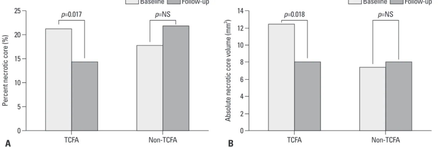
11.8%, p=0.76) and largest necrotic core site (26.6±9.6% to 29.5±12.5%, p=0.1). When we performed the analysis ac- cording to plaque stability, however, there was a significant reduction in necrotic core percentage (21.3±7.2% to 14.4±
8.9%, p=0.017) and absolute volume (12.5±6.7 mm3 to 8.1±8.4 mm3, p=0.018) in patients with TCFA. On the other hand, no changes in absolute volume and necrotic core per- centage were observed in patients with non-TCFA (Fig. 1).
Among 14 patients with TCFA, 13 patients showed a reduc- tion in percent necrotic core at 6 months. However, 18 pa- tients (out of 40) with non-TCFA showed a decrease in ne- most diseased 10-mm subsegments. Baseline and follow-up
IVUS-VH images (6 months) were reviewed side by side on a display. The primary end point of this study was change in percent necrotic core (baseline minus follow-up).
Two experienced, independent IVUS analysts defined IVUS-derived TCFA as a lesion fulfilling the following cri- teria in at least three consecutive frames: 1) necrotic core
≥10% without evident overlying fibrous tissue and 2) per- cent atheroma volume ≥40%.
To assess the reproducibility of VH measurement, base- line images of 10 cases were randomly selected. The in- terobserver correlation coefficient for percent necrotic core was 0.994 and the percentage of errors was 0.36±0.75%.
There was little intra-observer or inter-observer disagree- ment in the diagnosis of VH-derived TCFA (intra-observer;
κ=0.83, 95% CI, 0.74-0.92, inter-observer; κ=0.80, 95%
CI, 0.70-0.89).
Gray-scale IVUS measurements of external elastic mem- brane, plaque and media and lumen CSAs, and plaque bur- den (plaque and media divided by external elastic mem- brane) were performed for every recorded frame; volumetric data were generated by software using Simpson’s method.
Measurements of markers of lipid metabolism and inflammation
CRP levels were measured in serum using a commercially available kit (N High Sensitivity CRP, Dade Behring, Mar- burg, Germany). Plasma concentrations of total cholesterol, high-density lipoprotein cholesterol, and triglycerides were measured at our clinical laboratory. The Friedewald formu- la was used to derive low-density lipoprotein cholesterol (LDL-C) levels. Changes of lipid and inflammatory param- eters were calculated as the difference between baseline and follow-up values.
Statistical analysis
Simple descriptive statistics were used to summarize the data. Categorical variables are described using frequencies and percentages. For continuous variables with a normal distribution, means±SD are reported. For CRP levels, which were not normally distributed, median and inter-quartile ranges (IQR) are reported. Correlations between variables are described with the use of Spearman rank-correlation co- efficients. To assess inter- and intra-observer variability, re- sults were compared using the κ-test of concordance for the categorical data. p-values <0.05 were considered statistical- ly significant.
ly observed in a patient with TCFA only after 6 months of pharmacological intervention, whereas an increase in the necrotic core is observed in a patient with non-TCFA. Table 4 shows the baseline and 6-month follow-up data of volu- metric gray-scale IVUS analysis in the target plaque. Plaque volume was significantly reduced (2.85±10.99% decrease;
p=0.03 for baseline versus follow-up), as well were lumen and vessel volume.
crotic core after statin therapy. The analysis of change in each plaque component in 14 patients with TCFA showed that the percentage of fibrotic plaque was increased (60.6±
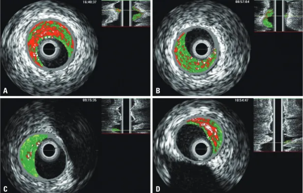
9.6% to 66.4±9.9%, p=0.14), although statistical signifi- cance was not reached (Fig. 2). Fig. 3 shows the IVUS-VH images of a representative patient for each type of plaque.
Baseline and follow-up IVUS-VH images are presented side by side. Marked reduction in the necrotic core is clear- Table 1. Baseline Clinical Characteristics
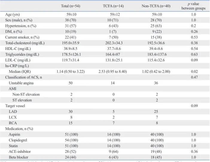
Total (n=54) TCFA (n=14) Non-TCFA (n=40) p value
between groups
Age (yrs) 59±10 59±12 59±10 1.0
Sex (male), n (%) 38 (70) 10 (71) 28 (70) 1.0
Hypertension, n (%) 31 (57) 6 (43) 25 (63) 0.2
DM, n (%) 10 (19) 1 (7) 9 (22) 0.26
Current smoker, n (%) 22 (41) 7 (50) 15 (38) 0.53
Total-cholesterol (mg/dL) 195.0±35.9 202.3±34.3 192.5±36.6 0.38
HDL-C (mg/dL) 38.9±8.5 37.7±8.6 39.4±8.6 0.54
Triglycerides (mg/dL) 178.5±126.1 164.4±87 183.4±137.6 0.63
LDL-C (mg/dL) 119.7±31.4 131.8±25.1 115.4±32.6 0.09
hs-CRP (mg/L)
Median (IQR) 1.14 (0.50 to 3.22) 2.53 (0.95 to 8.40) 1.02 (0.42 to 2.00) 0.02
Classification of ACS, n 0.47
Unstable angina 50 14 36
AMI
Non-ST elevation 2 0 2
ST elevation 2 0 2
Target vessel 0.09
LAD 30 5 25
LCX 8 2 7
RCA 15 7 8
Medication, n (%)
Aspirin 51 (100) 14 (100) 40 (100) 1.0
Clopidogrel 54 (100) 14 (100) 40 (100) 1.0
Statin 51 (100) 14 (100) 40 (100) 1.0
ACE-inhibitor 28 (52) 9 (64) 19 (48) 0.36
Beta blocker 24 (44) 6 (43) 18 (45) 1.0
AMI, acute myocardial infarction; LAD, left anterior descending artery; RCA, right coronary artery; LCX, left circumflex artery; IQR, inter-quartile ranges;
TCFA, thin-cap fibroatheroma; HDL-C, high-density lipoprotein cholesterol; LDL-C, low-density lipoprotein cholesterol; hs-CRP, high-sensitivity C-reactive protein; ACS, acute coronary syndrome.
Values are expressed as mean±SD.
Table 2. Change in Lipid Parameters and CRP Levels during Treatment (n=54)
Baseline Follow-up p value
Total cholesterol (mg/dL) 195.0±35.9 136.1±27.2 <0.001
HDL-C (mg/dL) 38.9±8.5 40.1±10.1 0.13
Triglycerides (mg/dL) 178.5±126.1 118.8±53.0 <0.001
LDL-C (mg/dL) 119.7±31.4 67.3±20.4 <0.001
hs-CRP (mg/L) 3.18±5.29 1.69±5.56 0.08
HDL-C, high-density lipoprotein cholesterol; LDL-C, low-density lipoprotein cholesterol; hs-CRP, high-sensitivity C-reactive protein; SD, standard deviation.
Values are expressed as mean±SD.
Table 3. IVUS-VH Findings at Baseline and Follow-Up (n=54)
Baseline Follow-up p value
10-mm most diseased segment Absolute volume (mm³)
Fibrotic (green) 27.7±15.6 26.5±17.5 0.48
Fibrofatty (yellow-green) 4.5±3.9 4.8±5.3 0.65
Dense calcium (white) 4.2±3.2 4.5±3.9 0.53
Necrotic core (red) 8.7±6.4 8.0±6.0 0.31
Percentages (%)
Fibrotic 62.7±10.5 58.7±12.6 0.03
Fibrofatty 9.3±5.8 10.2±8.2 0.43
Dense calcium 9.5±6.6 11.2±8.3 0.09
Necrotic core 18.7±8.5 20.0±11.0 0.38
Minimum lumen area site Absolute areas (mm²)
Fibrotic 3.8±1.8 3.6±2.2 0.35
Fibrofatty 0.6±0.5 0.6±0.5 0.87
Dense calcium 0.5±0.4 0.6±0.5 0.53
Necrotic core 1.2±0.9 1.1±0.8 0.28
Percentages (%)
Fibrotic 61.8±12.1 59.3±15.4 0.21
Fibrofatty 9.2±6.8 9.9±8.5 0.52
Dense calcium 9.3±7.3 10.7±10.2 0.23
Necrotic core 19.8±10.0 20.3±11.8 0.76
Largest necrotic core site Absolute areas (mm²)
Fibrotic 3.1±1.8 2.6±1.7 0.03
Fibrofatty 0.3±0.4 0.4±0.5 0.60
Dense calcium 0.7±0.5 0.7±0.5 0.93
Necrotic core 1.5±0.9 1.5±0.9 0.93
Percentages (%)
Fibrotic 55.9±12.0 50.4±14.0 0.004
Fibrofatty 5.5±4.4 6.6±6.8 0.28
Dense calcium 12.1±7.6 13.4±8.7 0.20
Necrotic core 26.6±9.6 29.5±12.5 0.10
IVUS-VH, Intravascular Ultrasound-Virtual Histology; SD, standard deviation.
Values are expressed as mean±SD.
Fig. 1. (A) Change in mean percent necrotic core in the most diseased 10-mm subsegment during 6-month follow-up. Significant reduction in percent ne- crotic core in TCFA is observed. (B) Change in mean absolute necrotic core volume in the most diseased 10-mm subsegment during 6-month follow-up.
Significant reduction in absolute necrotic core volume in TCFA is observed. TCFA, thin-cap fibroatheroma; NS, non-specific.
A B
0 0
5 2
10 6
4
15 8
20
10 12
25 14
Percent necrotic core (%) Absolute necrotic core volume (mm3 )
TCFA Non-TCFA TCFA Non-TCFA
p=0.017 p=NS p=0.018 p=NS
Baseline Follow-up Baseline Follow-up
Influence of lipid profile and CRP
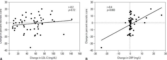
There was no significant correlation between LDL-C level reduction and decrease in CRP (r=0.2, p=0.14). Change in percent necrotic core showed a significant correlation with
change in CRP level (r=0.4, p=0.003). In contrast, changes in LDL-C level and lipid core demonstrated no significant associations (Fig. 4).
Ischemic events
None of the patients in either the TCFA or non-TCFA groups experienced ischemic events related to the target segment during the 6-month follow-up period.
DISCUSSION
Our study investigated whether early statin therapy in patients with vulnerable plaque compared with stable plaque reduces necrotic core assessed by IVUS-VH at 6 months. The results of this study clearly showed a reduction of lipid core in only patients with TCFA, which suggests that plaque stabilization
Table 4. Volume Parameters Derived from IVUS Measurement
Baseline Follow-up Percent change p value compared
with baseline
Plaque volume (mm³) 76.1±32.1 73.2±31.7 2.85±10.99 0.03
Lumen volume (mm³) 70.5±24.1 67.9±23.1 3.17±9.54 0.02
Vessel volume (mm³) 146.6±52.3 141.3±50.6 3.15±7.94 0.002
IVUS-VH, Intravascular Ultrasound; SD, standard deviation.
Values are mean±SD.
Fig. 2. Change in percentages of the four plaque components in TCFA (n=14) during 6-month follow-up. TCFA, thin-cap fibroatheroma.
Fig. 3. Representative examples of IVUS-VH images at largest necrotic core site for each plaque type. Baseline (left panel) and follow-up (right panel) images are displayed side by side. (A) Baseline image in the TCFA type. (B) Reduction of necrotic core is clearly observed af- ter 6 months. (C) Baseline image in the non-TCFA type. (D) Increase of necrotic core is clearly observed. IVUS-VH, Intravascular Ultrasound-Virtual Histology; TCFA, thin-cap fibroatheroma.
A
C
B
D
0 20 10 40 30 60 50 80 70 100 90
(%)
Fibrotic Fibrofatty Dense calcium Necrotic core p=0.14
p=0.16
p=0.88
p=0.017 Baseline Follow-up
creased in the long-term is needed.
In contrast to the observation presented here, recent data provide conflicting evidence that early statin therapy does not reduce the incidence of major cardiovascular events.15 However, there were several differences among previous studies, including population selection, baseline risk profile, and statin initiation time. In addition, some of the benefits of statin treatment in the early period after ACS may only become manifested or evident in long-term follow-up, be- cause it is likely that the beneficial effects of statins are cu- mulative. Further studies are warranted.
Demonstration of composition change
Parallel to clinical outcomes trials, imaging studies, in par- ticular IVUS-based studies, have substantially contributed to understanding the benefits of lipid lowering therapies in coronary artery disease. In fact, these studies have demon- strated slowing of atherosclerosis progression or regression as a surrogate marker of clinical outcomes after statin thera- py.16-18 However, these modalities may not provide precise histological findings. On the contrary, the use of radiofre- quency signals may be more reliable for plaque character- ization. A study using Integrated Backscatter-IVUS, which is able to depict the tissue characteristics of plaques, showed a significant reduction of lipid core volume after statin ther- apy.19 The present study showed consistent findings with those of animal studies that demonstrated a reduction of macrophage content and an increase of fibrosis after lipid lowering therapy.20,21 However, IVUS-VH further has the ability to define TCFA plaques as well as plaque composi- tion in vivo. In fact, we clearly demonstrated not only a re- duction in plaque volume, but also a remarkable change in relative lipid volume and an increase in fibrosis in TCFA.
following statin therapy might occur earlier in vulnerable plaque than in stable plaque. In addition, change in necrotic core showed a significant correlation with change in CRP lev- el. This is the first report to clarify the relationship between plaque stability and statin response, particularly during the early period of ACS, and to further examine the influence of a systemic inflammatory marker on plaque composition in vivo.
Plaque stability and early statin response
Goldstein, et al.12 suggested that plaque instability might re- flect a “pan-coronary” process. Their concept of multifocal plaque instability was supported by angiographic natural history studies in patients with ACS, in whom rapid pro- gression not only occurred in culprit lesions, but also in nonculprit lesions. In a serial angiographic evaluation of our recent data, multiple complex plaques in non-culprit le- sions were also identified in 27% of acute ST-segment ele- vation MI.13 It seems likely that overall coronary instability is responsible for the frequent recurrence rate after acute treatment of culprit lesions in ACS. In fact, it is within the early period after an ACS that patients experience the high- est rate of death and recurrent ischemic events.8 Recently, the MIRACL and PROVE-IT trials indicated that early in- tensive statin initiation during the acute phase of ACS re- duces the risk of recurrent ischemic events.9,10 However, there is little scientific basis for recommending early statin initiation to reduce recurrent ischemic events.14 Our study may provide insight into the early benefit of statin follow- ing ACS. As a matter of fact, we substantiated our specula- tion that the benefit of early statin therapy could be more pronounced in patients with vulnerable plaque than in those with stable plaque. To confirm our data, determination of whether or not percent necrotic core in non-TCFA is de-
Fig. 4. Correlation between (A) change in necrotic core percentage and change in LDL-C level, as well as between (B) change in necrotic core percentage and change in CRP level. LDL-C, low-density lipoprotein cholesterol; CRP, C-reactive protein.
A B
-40 -40
-30 -30
-20 -20
-10 -10
0 0
10 10
20 20
30 30
Change in percent necrotic core Change in percent necrotic core
0 20 40 60 80 100 120 140 160 -30 -20 -10 0 10 20 30
Change in LDL-C (mg/dL) Change in CRP (mg/L)
r=0.2
p=0.12 r=0.4
p=0.003
composition change, bifurcation lesions, lesions with se- vere angulations, heavily calcified lesions and lesions with poor quality image were excluded from this study. Thus, we precluded generalization of the findings to all patients with ACS. Second, an analysis of entire coronary segments was not performed. Measurement of the 10-mm subseg- ment with the most severe disease is substantially less rig- orous compared with that of the entire segment. In addition, image matching of the target lesion between baseline and follow-up could increase measurement variability. Third, we could not include a control group who received a place- bo; however, it was deemed ethically unacceptable in the setting of ACS. We compensated for the lack of a control group by blinding the information on the IVUS-VH. Fourth, VH-TCFA is not yet a validated surrogate for plaque prone to thrombosis. A study regarding the natural history of TCFA derived from IVUS-VH study, therefore, is needed to ad- dress this problem. It is also unclear whether TCFA detect- ed by IVUS-VH was a true vulnerable plaque. In fact, a re- cent study indicated that this modality alone is not sufficient for detecting TCFA.23 However, in an effort to reduce bi- ased selection, we demonstrated little intra-observer or in- ter-observer disagreement in the diagnosis of VH-derived TCFA. Finally, a long-term follow-up study is needed to es- tablish earlier statin responses in TCFA than in non-TCFA.
In conclusion, despite these limitations, this is the first re- port, to our knowledge, to evaluate in vivo the relationship between plaque stability and early statin response, and to examine the influence of a systemic inflammatory marker on plaque composition. In conclusion, the present study provides valuable insight into understanding plaque com- position change according to plaque stability, especially during the early period of ACS.
ACKNOWLEDGEMENTS
This work was funded by Ulsan University Hospital (Bio- medical Research Center Promotion Fund, UUH 2007-13).
The authors would like to thank Jung Yong Park for his as- sistance and important contributions.
REFERENCES
1. Virmani R, Kolodgie FD, Burke AP, Farb A, Schwartz SM. Les- sons from sudden coronary death: a comprehensive morphologi-
Mechanism
The REVERSAL and ASTEROID trials utilizing conven- tional IVUS showed that intensive lipid-lowering treatment achieved reduced progression or regression of coronary atherosclerosis in stable patients after 18 to 24 months.16,17 The recent ESTABLISH study also revealed that early lipid lowering therapy by atorvastatin significantly reduced plaque volume in ACS after only 6 months.11 Our results of volu- metric analysis are in line with these studies in terms of re- duction of plaque volume. However, there was a discordant result regarding plaque composition after statin therapy. Al- though analysis of the entire patient population in this study showed no change in percent necrotic core between base- line and follow-up, a significant reduction in necrotic core percentage was observed in TCFA, but not in non-TCFA.
The detailed mechanism of how statin treatment is more ef- fective in vulnerable plaque than stable plaque during the early period of ACS could not be identified. Interestingly, a recent investigation from the REVERSAL trial demonstrat- ed that constrictive remodeling of the arterial wall, repre- sentative of plaque stabilization, lacked an independent re- lation to LDL-C, but was positively related to CRP levels.22 These results suggest that the reduction of plaque burden associated with LDL-C lowering alone does not ensure constrictive remodeling following statin therapy. Moreover, the degree of CRP level reduction appears to be more im- portant for plaque stabilization than that of LDL-C level re- duction, especially during the early period of ACS. Further- more, in the PROVE-IT trial, CRP levels rapidly diverged between treatment groups, likely accounting for the particu- larly rapid divergence of major cardiovascular events.10 Our data also demonstrated a significant correlation between change in percent necrotic core and CRP level. Thus, our observation reemphasizes the role of inflammation in plaque stabilization by statin therapy, particularly during the high risk period after ACS. However, the exact mechanism of this early benefit according to plaque stability cannot be es- tablished solely on the basis of these aforementioned re- sults. To do so, biochemical markers or higher-resolution imaging techniques that can better define the mechanism of early stabilization after statin therapy are needed.
Study limitations
Several limitations should be taken into consideration. First, only 54 patients from a single center were enrolled; howev- er, in order to obtain the high-quality images that allowed us to accurately identify a significant difference in plaque
of multiple complex coronary plaques in patients with acute myo- cardial infarction: a study with coronary angiography. Am Heart J 2004;147:281-6.
14. Ridker PM, Cannon CP, Morrow D, Rifai N, Rose LM, McCabe CH, et al. C-reactive protein levels and outcomes after statin ther- apy. N Engl J Med 2005;352:20-8.
15. Newby LK, Kristinsson A, Bhapkar MV, Aylward PE, Dimas AP, Klein WW, et al. Early statin initiation and outcomes in patients with acute coronary syndromes. JAMA 2002;287:3087-95.
16. Nissen SE, Tuzcu EM, Schoenhagen P, Brown BG, Ganz P, Vogel RA, et al. Effect of intensive compared with moderate lipid-lower- ing therapy on progression of coronary atherosclerosis: a random- ized controlled trial. JAMA 2004;291:1071-80.
17. Nissen SE, Nicholls SJ, Sipahi I, Libby P, Raichlen JS, Ballantyne CM, et al. Effect of very high-intensity statin therapy on regres- sion of coronary atherosclerosis: the ASTEROID trial. JAMA 2006;295:1556-65.
18. Schartl M, Bocksch W, Koschyk DH, Voelker W, Karsch KR, Kreuzer J, et al. Use of intravascular ultrasound to compare effects of different strategies of lipid-lowering therapy on plaque volume and composition in patients with coronary artery disease. Circula- tion 2001;104:387-92.
19. Kawasaki M, Sano K, Okubo M, Yokoyama H, Ito Y, Murata I, et al. Volumetric quantitative analysis of tissue characteristics of cor- onary plaques after statin therapy using three-dimensional inte- grated backscatter intravascular ultrasound. J Am Coll Cardiol 2005;45:1946-53.
20. Armstrong ML, Megan MB. Arterial fibrous proteins in cynomol- gus monkeys after atherogenic and regression diets. Circ Res 1975;36:256-61.
21. Aikawa M, Rabkin E, Okada Y, Voglic SJ, Clinton SK, Brincker- hoff CE, et al. Lipid lowering by diet reduces matrix metallopro- teinase activity and increases collagen content of rabbit atheroma:
a potential mechanism of lesion stabilization. Circulation 1998;97:
2433-44.
22. Schoenhagen P, Tuzcu EM, Apperson-Hansen C, Wang C, Wolski K, Lin S, et al. Determinants of arterial wall remodeling during lipid-lowering therapy: serial intravascular ultrasound observa- tions from the Reversal of Atherosclerosis with Aggressive Lipid Lowering Therapy (REVERSAL) trial. Circulation 2006;113:
2826-34.
23. Sawada T, Shite J, Garcia-Garcia HM, Shinke T, Watanabe S, Otake H, et al. Feasibility of combined use of intravascular ultra- sound radiofrequency data analysis and optical coherence tomog- raphy for detecting thin-cap fibroatheroma. Eur Heart J 2008;29:
1136-46.
cal classification scheme for atherosclerotic lesions. Arterioscler Thromb Vasc Biol 2000;20:1262-75.
2. Davies MJ, Richardson PD, Woolf N, Katz DR, Mann J. Risk of thrombosis in human atherosclerotic plaques: role of extracellular lipid, macrophage, and smooth muscle cell content. Br Heart J 1993;69:377-81.
3. Nair A, Kuban BD, Tuzcu EM, Schoenhagen P, Nissen SE, Vince DG. Coronary plaque classification with intravascular ultrasound radiofrequency data analysis. Circulation 2002;106:2200-6.
4. Rodriguez-Granillo GA, García-García HM, Mc Fadden EP, Val- gimigli M, Aoki J, de Feyter P, et al. In vivo intravascular ultra- sound-derived thin-cap fibroatheroma detection using ultrasound radiofrequency data analysis. J Am Coll Cardiol 2005;46:2038-42.
5. Nasu K, Tsuchikane E, Katoh O, Vince DG, Virmani R, Surmely JF, et al. Accuracy of in vivo coronary plaque morphology assess- ment: a validation study of in vivo virtual histology compared with in vitro histopathology. J Am Coll Cardiol 2006;47:2405-12.
6. Randomised trial of cholesterol lowering in 4444 patients with coronary heart disease: the Scandinavian Simvastatin Survival Study (4S). Lancet 1994;344:1383-9.
7. Heart Protection Study Collaborative Group. MRC/BHF Heart Protection Study of cholesterol lowering with simvastatin in 20,536 high-risk individuals: a randomised placebo-controlled tri- al. Lancet 2002;360:7-22.
8. Invasive compared with non-invasive treatment in unstable coro- nary-artery disease: FRISC II prospective randomised multicentre study. FRagmin and Fast Revascularisation during InStability in Coronary artery disease Investigators. Lancet 1999;354:708-15.
9. Schwartz GG, Olsson AG, Ezekowitz MD, Ganz P, Oliver MF, Waters D, et al. Effects of atorvastatin on early recurrent ischemic events in acute coronary syndromes: the MIRACL study: a ran- domized controlled trial. JAMA 2001;285:1711-8.
10. Cannon CP, Braunwald E, McCabe CH, Rader DJ, Rouleau JL, Belder R, et al. Intensive versus moderate lipid lowering with statins after acute coronary syndromes. N Engl J Med 2004;350:
1495-504.
11. Okazaki S, Yokoyama T, Miyauchi K, Shimada K, Kurata T, Sato H, et al. Early statin treatment in patients with acute coronary syn- drome: demonstration of the beneficial effect on atherosclerotic lesions by serial volumetric intravascular ultrasound analysis dur- ing half a year after coronary event: the ESTABLISH Study. Cir- culation 2004;110:1061-8.
12. Goldstein JA, Demetriou D, Grines CL, Pica M, Shoukfeh M, O’Neill WW. Multiple complex coronary plaques in patients with acute myocardial infarction. N Engl J Med 2000;343:915-22.
13. Lee SG, Lee CW, Hong MK, Kim JJ, Park SW, Park SJ. Change