4)
*공동 제 저자1 .
책임저자 진동규 서울시 강남구 일원로: , 81,성균관의대 삼성서울병원 소아청소년과 Tel: 02)3410-3525, Fax: 02)3410-0043, E-mail: [email protected]
국내 6 형 뮤코다당증의 임상 양상 분자유전학적 , 특징 및 효소치료의 효과에 대한 고찰
Khunton Wichajarn 1* ㆍ 김진섭 2* ㆍ 양아람 2 ㆍ 손영배 3 이범희 4 ㆍ 유한욱 4 ㆍ 조성윤 2 ㆍ 진동규 2
Division of Medical genetics, Department of Pediatrics1, Faculty of Medicine, Khon Kaen University, Khon Kaen, Thailand
성균관대학교 의과대학 삼성서울병원 소아청소년과2
아주대학교 의과대학 아주대학교병원 의학유전학과3, 울산대학교 의과대학 서울아산병원 소아청소년과4
Clinical Features, Molecular Analysis, and Outcome of ERT in Korean Patients with Mucopolysaccharidosis Type VI
Khunton Wichajarn
1*, Jinsup Kim
2*, Aram Yang
2, Young Bae Sohn
3Beom Hee Lee
4, Han-Wook Yoo
4, Sung Yoon Cho
2, Dong-Kyu Jin
2 Division of Medical Genetics, Department of Pediatrics1, Faculty of Medicine,Khon Kaen University, Khon Kaen, Thailand Department of Pediatrics2, Samsung Medical Center, Sungkyunkwan University School of Medicine, Seoul, Korea
Department of Medical Genetics3, Ajou University School of Medicine, Suwon, Korea Department of Pediatrics4, Asan Medical Center Children's Hospital,
University of Ulsan College of Medicine, Seoul, Korea
Mucopolysaccharidosis type VI (MPS VI) is a rare disease caused by the mutation of ARSB with preva- lence range from 1/5,000 in northeast Brazil to 1/2,057,529 births in Czech Republic. In Asia, there is only one published figure in Taiwan of about 1/833,000 births. The exact prevalence in the Korean pop- ulation is unknown, but we estimated the incidence of MPS VI is about 0.03/100,000 live births. Enzyme replacement therapy (ERT) with recombinant human Arylsulfatase B (rhASB) is a modality for the treat- ment of MPS VI that reduces the excretion of urine glycosaminoglycan (GAG) and improves joint motion, pulmonary function, and endurance. We presented the clinical features, molecular analysis and outcome of ERT in three Korean MPS VI patients. All patients had the typical characteristic clinical features of MPS IV. Short stature, dysostosis multiplex, corneal opacity and valvular heart disease were found at first presentation, while restrictive lung disease and carpal tunnel syndrome developed later in all patients.
Molecular analysis demonstrated novel missense and nonsense mutation in the patients, including p.Ile 67Ser, p.Gly328Arg, p.Arg191*, p.Asp352Asn, and p.Gly17Asp. After ERT, urine GAG was decreased in all patients. Skeletal involvement, corneal opacity, heart valve abnormalities and pulmonary function were not improved with ERT, but it had a better outcome on regarding joint motion and endurance.
One patient underwent allogeneic bone marrow transplantation (BMT) prior to ERT, but their clinical re- sponse was not improved much after BMT. This study demonstrates clinical phenotypes and molecular analysis of the severe form of MPS VI in Korean patients.
Key words: Mucopolysaccharidosis, Mucopolysaccharidosis type VI, Recombinant human arylsulfatase
B, enzyme replacement therapy
Introductions
Mucopolysaccharidosis type VI (MPS VI) or Maroteaux-Lamy syndrome, an autosomal reces- sive lysosomal storage disease, is caused by the mutation of arylsulfatase B (ARSB gene), which results in the reduction or absence of N-acetyl- galactosamine-4-sulfatase (arylsulfatase B, ASB) activity. This defect causes an accumulation of dermatan sulfate (glycosaminoglycan, GAG), which affects many systems. The prevalence of this dis- ease varies by nationality. It ranges from 1 in 5,000 live births in northeast Brazil to 1 in 2,057,529 births in Czech Republic
1, 2). In Asia, one study from Taiwan reported an incidence of about 1 in 833,000 live births
3). The relative pro- portion of MPS VI to total MPS is quite high in Brazil, Portugal and Australia (18.48%, 16.12%
and 10.36%, respectively) but low in Canada, Sweden and Norway (5%, 4% and 2%, respec- tively)
4). In Asia, the relative frequency of MPS VI is about 6% in Taiwan and 10% in Israel
5, 6). In South Korea, the proportion of MPS VI to total MPS is extremely low, unpublished data from a single institute, Samsung Medical Center from 1994-2015 estimates that MPS VI account for 1.9% of total MPS (three MPS VI patients of total 156 patients with MPS).
MPS VI can be classified into three groups: the severe or infantile form, intermediate or juvenile form, and the mild or adult form
7, 8). The onset of the severe form is usually before 2 or 3 years of age and presents with short stature, coarse face, corneal opacity, dysostosis multiplex, hepatosple- nomegaly and kyphoscoliosis. All clinical symp- toms usually progress rapidly. Restrictive lung disease and valvular heart disease are serious complications and leading causes of death in the
third decade of life
4). The intermediate form usu- ally presents in late childhood with less severe skeletal involvement, valvular heart disease, cor- neal opacity and mild hepatomegaly. However, the patient can develop complications as a severe form including hip dysplasia, restrictive lung disease and carpal tunnel syndrome, but slower on pro- gression. The adult form manifests in the second decade of life with mildly clinical symptoms, slower progression and involvement in fewer sys- tems and presents with including corneal opacity, mild skeletal involvement, valvular heart disease and carpal tunnel syndrome. Height usually re- aches the normal range, and patients can survive until the fifth decade of life
4, 8). Enzyme replace- ment therapy (ERT) with recombinant human aryl- sulfatase-B (rhASB) for MPS VI has been ap- proved since 2005 in the US 2006 in the EU, and 2008 in South Korea. ERT leads to the reduction of urine GAG and improvement of joint movement, pulmonary function, and endurance
9, 10). Bone mar- row transplantation (BMT) is another treatment option for MPS VI, but it is limited by the risks of treatment-related complications, such as graft- versus-host disease
4).
In this report, we present the clinical charac- teristics, molecular analysis, and clinical course of ERT of three Korean patients with the severe form of MPS VI.
Case Reports
1. Patient 1
A 4-year-old boy was referred to Samsung
Medical Center with corneal opacity. He was the
first child from non-consanguineous Korean pa-
rents and was born at 39 weeks of gestation with
a birth weight of 3.4kg. His development was
normal for his age. The physical examination re- vealed cloudy corneas, coarse face, grade III/VI systolic murmurs, multiple joint stiffness, and ky- phoscoliosis (Table 1). The skeletal survey showed calvarial thickening (Fig. 1A), short and broad long bones (Fig. 1B), proximal pointing of the meta- carpal bones (Fig. 1C), irregular carpal and tarsal bones (Fig. 1C and 1D), paddle-shaped widened ribs (Fig. 1E), hip dysplasia (Fig. 1E), abnormal vertebral bodies and kyphoscoliosis (Fig. 1F).
The urine GAG (measured by a cetylpyridinium chloride [CPC] precipitation test) was 704.6 CPC unit/g creatinine (reference range [RR] <175 CPC unit/g creatinine). The ASB activity from cultured skin fibroblasts was 0.1 pmol/min mg protein (normal range 12-39 pmol/min mg protein). The mutational study of the ARSB gene from leuko- cytes showed novel compound heterozygous mu- tations: c.200T>G (p.Ile67Ser) and c.982G>A (p.
Gly328Arg). Echocardiography showed mild aortic
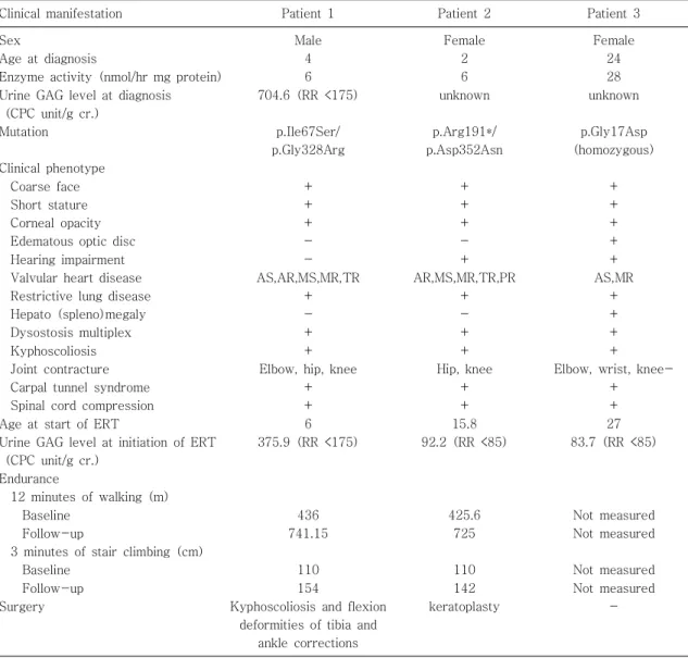
Table 1. Clinical Phenotypes and Genotypes in Three Patients
Clinical manifestation Patient 1 Patient 2 Patient 3
Sex
Age at diagnosis
Enzyme activity (nmol/hr mg protein) Urine GAG level at diagnosis
(CPC unit/g cr.) Mutation
Clinical phenotype Coarse face Short stature Corneal opacity Edematous optic disc Hearing impairment Valvular heart disease Restrictive lung disease Hepato (spleno)megaly Dysostosis multiplex Kyphoscoliosis Joint contracture Carpal tunnel syndrome Spinal cord compression Age at start of ERT
Urine GAG level at initiation of ERT (CPC unit/g cr.)
Endurance
12 minutes of walking (m) Baseline
Follow-up
3 minutes of stair climbing (cm) Baseline
Follow-up Surgery
Male 4 6 704.6 (RR <175)
p.Ile67Ser/
p.Gly328Arg
+ + + - - AS,AR,MS,MR,TR
+ - + + Elbow, hip, knee
+ + 6 375.9 (RR <175)
436 741.15
110 154
Kyphoscoliosis and flexion deformities of tibia and
ankle corrections
Female 2 6 unknown
p.Arg191*/
p.Asp352Asn
+ + + - + AR,MS,MR,TR,PR
+ - + + Hip, knee
+ + 15.8 92.2 (RR <85)
425.6 725
110 142 keratoplasty
Female 24 28 unknown
p.Gly17Asp (homozygous)
+ + + + + AS,MR
+ + + +
Elbow, wrist, knee- +
+ 27 83.7 (RR <85)
Not measured Not measured
Not measured Not measured
-
stenosis (AS) and regurgitation (AR), mild mitral stenosis (MS) and regurgitation (MR) and mild tricuspid regurgitation (TR), and the ejection frac- tion (EF) was 68.6%. The pulmonary function test (PFT) was normal with forced vital capacity (FVC) of 109% and forced expiratory volume in one second (FEV1) of 110%.
He has received ERT with a 1 mg/kg intrave- nous infusion of rhASB weekly from 6 years of age. At that time, his weight and height were 17 kg (-1.4 standard deviation score [SDS]) and 107 cm (-1.8 SDS), respectively. Urine GAG rapidly declined in the first 6 months after ERT (Fig. 2).
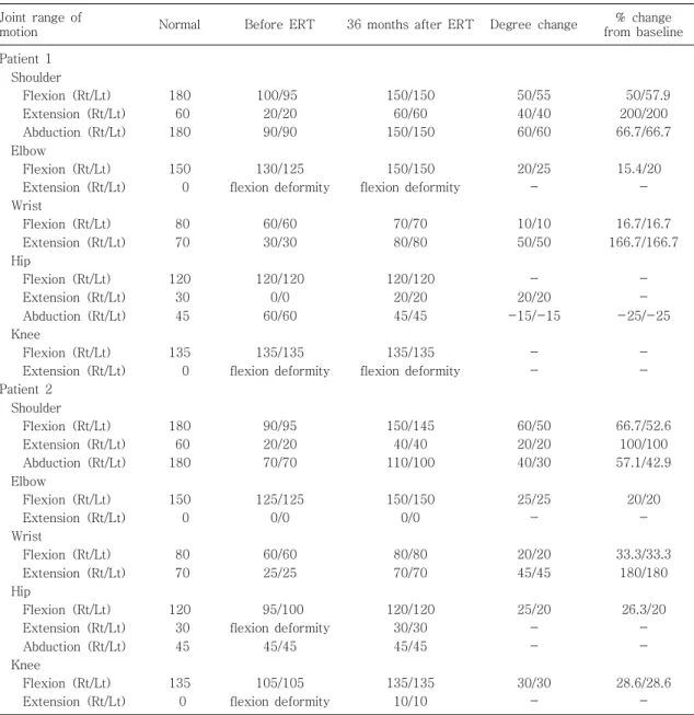
He showed no serious adverse effect except a mild transient headache that could be controlled by NSAID. He complained of decreased grip and wrist strength at 7 years. Carpal tunnel syndrome was diagnosed by electrophysiological study and he started to receive rehabilitation and occupa- tional therapy. Range-of-motion (ROM) tests were evaluated by a goniometer
11)and showed an improvement of the shoulder, elbow, and wrist of 10-60 degrees in all directions after 3 years of ERT (Table 2). AR progressed to moderate se- verity and left ventricular dilatation was found on echocardiography after 5 years of ERT, while the
Fig. 2. Urine GAG excretion before and after ERT in patients 1 and 2.
Fig. 1. Radiological findings in all patients. A-F: Skeletal survey of patient 1 when he was 4 years old (be- fore ERT). A: Calvarial thickening and J-shaped sella. B: Short and broad long bones and hypoplastic distal ulnar and radius. C: Proximal pointing of me- tacarpal bones and irregular carpal bones. D: Irre- gular tarsal bones. E: Paddle-shaped widened ribs and bilateral hip dysplasia. F: Anterior beaking of vertebral bodies and kyphoscoliosis. G-K: Skeletal survey of patient 2 when she was 15 years old (before ERT). G: Calvarial thickening and J-shaped sella. H: Short and broad long bones and hypopla- stic distal ulnar and radius. I: Short and board me- tacarpal bone and irregular carpal bone. J: Irregular tarsal bones. K: Paddle-shaped widened ribs and bilateral hip dysplasia. L: T-L spine lateral view of patient 2 demonstrated inferior beaking and mul- tiple collapsed vertebral bodies. M: MRI brain of patient 2 when she was 15 years old demonstrating cervicomedullary junction compression. N-O: Ske- letal survey of patient 3 when she was 31 years old (after 5 years of ERT) N: Proximal pointing of me- tacarpal bones and irregular carpal bones. O: Paddle- shaped widened ribs. P: MRI spine of patient 3 when she was 31 years old demonstrating cervico- medullary junction compression.
EF was normal. PFT showed decreased FVC (79
%) and FEV1 (76%) (Fig. 3), and mild restrictive lung disease developed at 10.5 years of age. His endurance was measured by a 12-minute walk test (12-MWT) and 3-minute stair climbing test (3-MSCT)
12, 13)at 6 and 18 months after ERT and showed improvement from 603.15 to 741.15 m in the 12-MWT and 118 to 154 cm in the 3-
MSCT (Table 1). Thoracolumbar spinal compres- sion was found when he was 10 years old. He had back pain, difficulty to walk and had an increased deep tendon reflex. He underwent orthopedic operation for kyphosis and scoliosis correction (T10-L3) and anterior lumbar interbody fusion (T11-12 and L1-2) without any complications.
After surgery, his back pain disappeared and his
Table 2. Range of Motion Tests of Patients 1 and 2 Compared before and after Treatment with ERT Joint range of
motion Normal Before ERT 36 months after ERT Degree change % change
from baseline Patient 1
Shoulder Flexion (Rt/Lt) Extension (Rt/Lt) Abduction (Rt/Lt) Elbow
Flexion (Rt/Lt) Extension (Rt/Lt) Wrist
Flexion (Rt/Lt) Extension (Rt/Lt) Hip
Flexion (Rt/Lt) Extension (Rt/Lt) Abduction (Rt/Lt) Knee
Flexion (Rt/Lt) Extension (Rt/Lt) Patient 2
Shoulder Flexion (Rt/Lt) Extension (Rt/Lt) Abduction (Rt/Lt) Elbow
Flexion (Rt/Lt) Extension (Rt/Lt) Wrist
Flexion (Rt/Lt) Extension (Rt/Lt) Hip
Flexion (Rt/Lt) Extension (Rt/Lt) Abduction (Rt/Lt) Knee
Flexion (Rt/Lt) Extension (Rt/Lt)
180 60 180
150 0
80 70
120 30 45
135 0
180 60 180
150 0
80 70
120 30 45
135 0
100/95 20/20 90/90
130/125 flexion deformity
60/60 30/30
120/120 0/0 60/60
135/135 flexion deformity
90/95 20/20 70/70
125/125 0/0
60/60 25/25
95/100 flexion deformity
45/45
105/105 flexion deformity
150/150 60/60 150/150
150/150 flexion deformity
70/70 80/80
120/120 20/20 45/45
135/135 flexion deformity
150/145 40/40 110/100
150/150 0/0
80/80 70/70
120/120 30/30 45/45
135/135 10/10
50/55 40/40 60/60
20/25 -
10/10 50/50
- 20/20 -15/-15
- -
60/50 20/20 40/30
25/25 -
20/20 45/45
25/20 - -
30/30 -
50/57.9 200/200 66.7/66.7
15.4/20 -
16.7/16.7 166.7/166.7
- - -25/-25
- -
66.7/52.6 100/100 57.1/42.9
20/20 -
33.3/33.3 180/180
26.3/20 - -
28.6/28.6 -
deep tendon reflex normalized. He received an- terior and medial hemiepiphysiodesis surgery for correction of the bilateral flexion and valgus de- formities of the proximal tibia and ankle when he was 11 years old. The angular deformities were improved and he could walk better after correc- tion of the angular deformities.
2. Patient 2
Patient 2 was born at full-term with a birth weight of 3 kg from non-consanguineous Korean parents. She was diagnosed with MPS VI at Soon Chun Hyang Hospital at the age of 2 years pre- senting with coarse face, joint stiffness, kyphosis and growth retardation. BMT was performed at 5 years of age with an HLA-matched sibling donor.
After the BMT, the ASB activity in the leukocytes raised to 132.5 nmol/hr mg protein (reference range 115-226 nmol/hr mg protein). At the age of 15 years, she was referred to Samsung Me- dical Center for ERT. The urine GAG was 13 g/
mol creatinine (reference range <5 g/mol crea- tinine) and the ASB activity in the leukocyte was 8.1 pmol/min protein (normal range 1.5-21). A mutational study of the ARSB gene was conducted from cultured skin fibroblasts in consideration of
post transplantation state and showed compound heterozygous mutation: c.571C>T (p.Arg191*), c.1054G>A (p.Asp352Asn). She had normal cogni- tive function, corneal opacity, coarse face, kypho- scoliosis, dysostosis multiplex and numbness both ands (Table 1). The skeletal survey showed cal- varial thickening, J-shaped sella (Fig. 1G), short and broad long bones (Fig. 1H), irregular carpal and tarsal bones (Fig. 1I and 1J), paddle-shaped ribs, hip dysplasia (Fig. 1K), kyphosis and hypo- plasia of the vertebral bodies and biconvex end- plate (Fig. 1L). Magnetic resonance imaging (MRI) of the spine demonstrated cervicomedullary junc- tion spinal compression (Fig. 1M) without any clinical symptoms. An electrophysiological study was performed due to the symptoms of numbness in both hands and the result was compatible with carpal tunnel syndrome. Echocardiography showed an EF of 68.8%, moderate AR and MR, mild MS, TR and pulmonary regurgitation (PR). PFT showed mild restrictive lung disease (FVC 65% and FEV1 55%).
ERT was started at 15.7 years of age with at a weight of 20 kg (-4.2 SDS) and height of 105 cm (-10.4 SDS). The urine GAG was 92.2 CPC unit/
g creatinine (reference range <85 CPC unit/g creatinine) at that time (Fig. 2). She started men- arche 6 months after ERT. After three years of ERT, ROM was improved by 20-60 degrees in all directions of both shoulder, elbow, wrist, hip and knee joints (Table 2). After 5 years of ERT, MR was improved on echocardiography while the other findings were persistent. PFT showed de- creased FVC and FEV1 (53% and 48%) and rest- rictive lung disease worsened to moderate seve- rity after 5 years of ERT. Her endurance showed improvement by in the 12-MWT (299.4 m) and 3-MSCT (32 cm) 18 months after ERT (Table 1). The outcome of this patient was reported
Fig. 3. Forced vital capacity (FVC) and forced expira-tory volume in one second (FEV1) before and after treatment with ERT of patients 1 and 2.
several years ago
14). Thereafter, cervicomedullary junction compression was followed-up by MRI at 18 years of age and the result had no change. She underwent keratoplasty of each eye at 18 and 19 years of age, respectively and the corneal grafts were been still clear after 5 years in thefollow- up.
3. Patient 3
Patient 3 was born at full-term with a birth weight of 3.6 kg from non-consanguineous Korean parents. She was diagnosed with MPS VI at 24 years of age at Asan Medical Center with the fol- lowing symptoms short stature, coarse face, cor- neal opacity, and multiple joint stiffness (Table 1).
The urine GAG was 83.7 CPC unit/g creatinine (reference range <85 CPC unit/g creatinine). The ASB activity in the leukocytes was 28 nmol/hr mg protein (normal range 115-226 nmol/hr mg protein). The mutational the study of ARSB gene showed a homozygous known mutation of c.512G>
A (p.Gly171Asp). Echocardiography was perfor- med at 24 years of age and the result showed moderate AS and mild MR and was managed by bisoprolol and furosemide. The PFT was conducted at 24 years of age and the result revealed re- strictive lung disease (FVC 47%, FEV1 47%).
She began ERT at Asan Medical Center at 27 years of age with a height of 127.6 cm (-6.4 SDS) and weight of 38.5 kg (-2.1 SDS). She developed weakness and radicular pain in both legs at 30 years of age that resulted in walking difficulty. However, she worked as a counselor and participated in basic activities of daily living independently. She often complained of dyspnea after running or going up stairs. She was referred to Samsung Medical Center at 31 years of age years because of foot drop and intermittent hand
weakness. The skeletal survey showed short and proximal pointing of the metacarpal bones (Fig 1N), abnormal vertebral bodies and paddle-shaped ribs (Fig. 1O). Cervical myelopathy in the cervi- comedullary junction was documented by spine MRI (Fig. 1P). Her motor power was grade IV for shoulder flexion and abduction and grade V for elbow, wrist and knee flexion, ankle dorsiflexion and plantar flexion. The electrophysiological study of both median nerves was evaluated due to in- termittent hand weakness, and the result showed carpal tunnel syndrome and was treated by reh- abilitation and occupational therapy. She had mild stiffness of shoulder flexion and abduction (160 from 180 degrees) and wrist extension (50 from 70 degree) while the hip and knee were normal after 5.5 years of ERT. Although she was shown to have severe AS, mild MS and MR on echocar- diogram, she stopped medication by herself at the age of 32 and had not been aggravated or shown chest discomfort recently. Follow-up PFT and endurance measurements were not conducted for this patient. Diffuse corneal opacity and an ede- matous optic disc were presented on eye exa- mination.
Discussion
The exact prevalence of MPS VI in South Korea is unknown, but it could be very rare because only three patients have been diagnosed with MPS VI in Korea. Unpublished data from Samsung Medical Center, one of the largest MPS clinics in South Korea, from 1994-2015 shows the relative pro- portion of MPS VI to total MPS is only about 1.4%.
In this article, we demonstrated the clinical
phenotypes of severe types of MPS VI in three
Korean patients. All patients had early-onset and
severe presentation including coarse face, short
stature, dysostosis multiplex, joint stiffness, val- vular heart disease and cloudy corneas. Skeletal involvement is usually severe and caused short stature (-1.8 SDS in patient 1, -10.4 SDS in pa- tient 2 and -6.4 SDS in patient 3). All patients have typical radiological findings of MPS (calvarial thickening, J-shaped sella, short and broad long bones, proximal pointing of the metacarpal bones, irregular carpal and tarsal bones, paddle-shaped widened ribs, hip dysplasia and abnormal vertebral bodies). In consequence of severe kyphoscoliosis, spinal compression and respiratory compromise developed in our patients. Although cervicome- dullary junction compression is a serious compli- cation and developed in all patients, but two pa- tients had clinical symptoms and only one patient with clinical symptoms that were not relieved by conservative management underwent surgery.
Joint stiffness was found in all patients, and the extremities were more affected than the trunk.
The aortic valve is the most commonly affected among valvular heart diseases. Regurgitation is more common than stenosis pathology. Moreover, we found acquired LV dilatation in patient 1 des- pite normal LV function. Other common cardiac manifestations are LV hypertrophy and pulmonary hypertension
15), but we did not find these in our patients. Restrictive lung disease and carpal tunnel syndrome were found in all patients usually pre- senting later than other clinical symptoms. Hepato- splenomegaly is also a common manifestation but it was not found in patient 2 because of previous BMT. Corneal opacity usually presents early and progresses despite ERT or BMT. GAG accumu- lates in the donor s corneal graft and causes re ’ - current corneal opacity despite BMT
16). In our study, patient 2 underwent keratoplasty, and her corneal grafts were still clear with good visual acuity over 5 years of ERT.
More than 140 mutations in the ARSB gene have been reported. There have been a few stu- dies on this service in Asia
3,17-19). Lin et al. re- ported two common mutations (p.Phe399Leu and p.Leu132Pro) in Taiwan. Kantaputra et al. reported that p.Pro445Leu and p.Trp450Cys mutations were suspected to have high prevalence in India
18). In our study, we identified novel mutations in Korean MPS VI patients. Patient 1 had compound hetero- zygous mutations with 2 novel mutations: c.200T>
G (p.Ile67Ser) and c.982G>A (p.Gly328Arg). Pa- tient 2 also had compound heterozygous mutations with one novel mutation and one known mutation:
c.571C>T (p.Arg191*), c.1054G>A (p.Asp352Asn).
Additionally, patient 3 had a homozygous known mutation: c.512G>A (p.Gly171Asp).
ERT has been used for treatment in all patients since 2008 in South Korea. Urine GAG excretion was decreased rapidly in 6 months and then gra- dually decreased to a nearly normal level. The ERT was not effective for the growth and pro- gression of kyphoscoliosis, but was effective on improvement of ROM. In this study, the ERT nei- ther resolves nor prevents cardiac involvements as found in previous studies
20, 21). Many previous studies demonstrate the improvement of PFT by ERT
10, 20-22), but restrictive lung disease in our patients worsened during the ERT period. Endu- rance by the 12-MWT and 3-MSCT showed improvement in our study but we believethis re- sulted from the improvement of ROM rather than the cardiopulmonary function.
Conclusion
Mucopolysaccharidosis type VI is very rare in
South Korea. This report presents the clinical
features including short stature, corneal opacity,
dysostosis multiplex, hepatosplenomegaly and
valvular heart disease, of three Korean patients.
Three different novel mutations were identified.
ERT in Korean patients has better outcomes in urine GAG excretion, joint movement and endu- rance. However ERT was not effective for skeletal involvement, valvular heart disease and respiratory function in this study.
요 약
뮤코다당증 형 6 (Mucopolysaccharidosis type VI) 은 ARSB 유전자의 변이로 인하여 dermatan sulfate 대사에 관여하는 arylsulfatase B 효소의 결핍으로 유 발되는 상염색체 열성 리소좀 축적 질환이다 본 연구 . 는 국내에서 뮤코다당증 형으로 진단된 명의 환자를 6 3 대상으로 하였다 명의 환자는 최초 진단 시 뮤코다당 3 증 형의 특징적인 증상인 저신장 다발성 골형성 부 6 , 전 심장 판막 이상 및 각막 혼탁을 보였으며 이후 손목 , 터널 증후군 및 제한성 폐질환의 양상을 보였다 모든 . 환자에서 소변의 GAG (glycosaminoglycan) 는 증가 되어 있었으며 피부 섬유아세포 또는 백혈구에서 측정 한 arylsulfatase B 는 모두 감소되어 있었고 중합효소 연쇄반응 염기서열분석법에 의한 - ARSB 유전자 검사 에서는 c.200T>G (p.Ile67Ser), c.982G>A (p.Gly 328Arg), c.571C>T (p.Arg191*), c.1054G>A (p.
의 이형접합체 돌연변이 및
Asp352Asn) c.512G>A
동형접합체 돌연변이를 발견할 수 있 (p.Gly171Asp)
었다. recombinant human Arylsulfatase B (rhASB) 를 통한 효소 대체 치료는 2008 년부터 국내에서도 허 가되었으며 효소대체치료 시행 후 소변 GAG (glyco- 는 감소하였으며 관절 운동 및 지구력 saminoglycan)
은 향상되었다 그러나 관절 침범 및 각막 혼탁 심장 . , 및 폐 침범 소견은 호전되지 않았다 본 연구는 뮤코다 .
당증 형의 특징적인 증상을 보였으며 생화학적 검사 6
및 분자유전학적 검사를 통해 뮤코다당증 형으로 확 6
진된 3 명의 환자를 대상으로 시행한 효소대체치료의
효과를 보고하는 바이다.
References
1) Costa-Motta FM, Acosta AX, Abe-Sandes K, Bender F, Schwartz IV, Giugliani R, et al. Genetic studies in a cluster of mucopolysaccharidosis type VI patients in Northeast Brazil. Mol Genet Metab 2011;104:603-7.
2) Poupetova H, Ledvinova J, Berna L, Dvorakova L, Kozich V, Elleder M. The birth prevalence of lyso- somal storage disorders in the Czech Republic: com- parison with data in different populations. J Inherit Metab Dis 2010;33:387-96.
3) Lin WD, Lin SP, Wang CH, Hwu WL, Chuang CK, Lin SJ, et al. Genetic analysis of mucopolysaccharidosis type VI in Taiwanese patients. Clin Chim Acta 2008;
394:89-93.
4) Valayannopoulos V, Nicely H, Harmatz P, Turbeville S. Mucopolysaccharidosis VI. Orphanet J Rare Dis 2010;5:5.
5) Lin HY, Lin SP, Chuang CK, Niu DM, Chen MR, Tsai FJ, et al. Incidence of the mucopolysaccharidoses in Taiwan, 1984-2004. Am J Med Genet A 2009;
149A:960-4.
6) Schaap T, Bach G. Incidence of mucopolysaccharidoses in Israel: is Hunter disease a "Jewish disease"? Hum Genet 1980;56:221-3.
7) Wicker G, Prill V, Brooks D, Gibson G, Hopwood J, von Figura K, et al. Mucopolysaccharidosis VI (Ma- roteaux-Lamy syndrome). An intermediate clinical phenotype caused by substitution of valine for glycine at position 137 of arylsulfatase B. J Biol Chem 1991;
266:21386-91.
8) Isbrandt D, Arlt G, Brooks DA, Hopwood JJ, von Figura K, Peters C. Mucopolysaccharidosis VI (Ma- roteaux-Lamy syndrome): six unique arylsulfatase B gene alleles causing variable disease phenotypes. Am J Hum Genet 1994;54:454-63.
9) Harmatz P, Giugliani R, Schwartz I, Guffon N, Teles EL, Miranda MC, et al. Enzyme replacement therapy for mucopolysaccharidosis VI: a phase 3, randomized, double-blind, placebo-controlled, multinational study of recombinant human N-acetylgalactosamine 4-sul- fatase (recombinant human arylsulfatase B or rhASB) and follow-on, open-label extension study. J Pediatr 2006;148:533-9.
10) Harmatz P, Yu ZF, Giugliani R, Schwartz IV, Guffon N, Teles EL, et al. Enzyme replacement therapy for mucopolysaccharidosis VI: evaluation of long-term pulmonary function in patients treated with recom- binant human N-acetylgalactosamine 4-sulfatase. J Inherit Metab Dis 2010;33:51-60.
11) Gajdosik RL, Bohannon RW. Clinical measurement of range of motion. Review of goniometry emphasizing reliability and validity. Phys Ther 1987;67:1867-72.
12) Bolton JW, Weiman DS, Haynes JL, Hornung CA, Olsen GN, Almond CH. Stair climbing as an indicator of pulmonary function. Chest 1987;92:783-8.
13) Solway S, Brooks D, Lacasse Y, Thomas S. A qualita- tive systematic overview of the measurement properties of functional walk tests used in the cardiorespiratory domain. Chest 2001;119:256-70.
14) Sohn YB, Park SW, Kim SH, Cho SY, Ji ST, Kwon EK, et al. Enzyme replacement therapy improves joint motion and outcome of the 12-min walk test in a mucopolysaccharidosis type VI patient previously treated with bone marrow transplantation. Am J Med Genet A 2012;158A:1158-63.
15) Kampmann C, Lampe C, Whybra-Trumpler C, Wie- thoff CM, Mengel E, Arash L, et al. Mucopolysac- charidosis VI: cardiac involvement and the impact of enzyme replacement therapy. J Inherit Metab Dis 2014;37:269-76.
16) Varssano D, Cohen EJ, Nelson LB, Eagle RC, Jr. Cor- neal transplantation in Maroteaux-Lamy syndrome.
Arch Ophthalmol 1997;115:428-9.
17) Furujo M, Kubo T, Kosuga M, Okuyama T. Enzyme replacement therapy attenuates disease progression in two Japanese siblings with mucopolysaccharidosis type
VI. Mol Genet Metab 2011;104:597-602.
18) Kantaputra PN, Kayserili H, Guven Y, Kantaputra W, Balci MC, Tanpaiboon P, et al. Clinical manifesta- tions of 17 patients affected with mucopolysacchari- dosis type VI and eight novel ARSB mutations. Am J Med Genet A 2014;164A:1443-53.
19) Okamura K, Munkhbat B, Batchimeg B, Tamiya G, Hozumi Y, Suzuki T. Case of a Mongolian child with extensive Mongolian spots in mucopolysaccharidosis type VI: identification of a novel mutation in the aryl- sulfatase B gene. J Dermatol 2013;40:758-9.
20) Giugliani R, Lampe C, Guffon N, Ketteridge D, Leao- Teles E, Wraith JE, et al. Natural history and galsul- fase treatment in mucopolysaccharidosis VI (MPS VI, Maroteaux-Lamy syndrome)-10-year follow-up of patients who previously participated in an MPS VI survey study. Am J Med Genet A 2014.
21) Hendriksz CJ, Giugliani R, Harmatz P, Lampe C, Martins AM, Pastores GM, et al. Design, baseline characteristics, and early findings of the MPS VI (mu- copolysaccharidosis VI) Clinical Surveillance Program (CSP). J Inherit Metab Dis 2013;36:373-84.
22) Lin HY, Chen MR, Chuang CK, Chen CP, Lin DS, Chien YH, et al. Enzyme replacement therapy for mucopolysaccharidosis VI--experience in Taiwan. J Inherit Metab Dis 2010;33 Suppl 3:S421-7.