© 2014 The Korean Ophthalmological Society
This is an Open Access article distributed under the terms of the Creative Commons Attribution Non-Commercial License (http://creativecommons.org/licenses /by-nc/3.0/) which permits unrestricted non-commercial use, distribution, and reproduction in any medium, provided the original work is properly cited.
Original Article
Effect of Macrophage Migration Inhibitory Factor on Corneal Sensitivity after Laser In Situ Keratomileusis in Rabbit
Joon Young Hyon1, Stacey Hose2, Celine Gongora3, Debasish Sinha2, Terrence O’Brien4
1Department of Ophthalmology, Seoul National University Bundang Hospital, Seoul National University College of Medicine, Seongnam, Korea
2Department of Ophthalmology, Johns Hopkins University School of Medicine, Baltimore, MD, USA
3CNRS UMR 5160, CRLC, Montpellier, France
4Bascom Palmer Eye Institute, University of Miami Miller School of Medicine, Palm Beach Gardens, FL, USA
Purpose: To investigate the effect of macrophage migration inhibitory factor (MIF) on corneal sensitivity after laser in situ keratomileusis (LASIK) surgery.
Methods: New Zealand white rabbits were used in this study. A hinged corneal flap (160-μm thick) was created with a microkeratome, and -3.0 diopter excimer laser ablation was performed. Expressions of MIF mRNA in the corneal epithelial cells and surrounding inflammatory cells were analyzed using reverse transcription poly- merase chain reaction at 48 hours after LASIK. After LASIK surgery, the rabbits were topically given either 1) a balanced salt solution (BSS), 2) MIF (100 ng/mL) alone, or 3) a combination of nerve growth factor (NGF, 100 ug/mL), neurotrophine-3 (NT-3, 100 ng/mL), interleukin-6 (IL-6, 5 ng/mL), and leukemia inhibitory factor (LIF, 5 ng/mL) four times a day for three days. Preoperative and postoperative corneal sensitivity at two weeks and at 10 weeks were assessed using the Cochet-Bonnet esthesiometer.
Results: Expression of MIF mRNA was 2.5-fold upregulated in the corneal epithelium and 1.5-fold upregulated in the surrounding inflammatory cells as compared with the control eyes. Preoperative baseline corneal sen- sitivity was 40.56 ± 2.36 mm. At two weeks after LASIK, corneal sensitivity was 9.17 ± 5.57 mm in the BSS treated group, 21.92 ± 2.44 mm in the MIF treated group, and 22.42 ± 1.59 mm in the neuronal growth fac- tors-treated group (MIF vs. BSS, p < 0.0001; neuronal growth factors vs. BSS, p < 0.0001; MIF vs. neuronal growth factors, p = 0.815). At 10 weeks after LASIK, corneal sensitivity was 15.00 ± 9.65, 35.00 ± 5.48, and 29.58 ± 4.31 mm respectively (MIF vs. BSS, p = 0.0001; neuronal growth factors vs. BSS, p = 0.002; MIF vs.
neuronal growth factors, p = 0.192). Treatment with MIF alone could achieve as much of an effect on recovery of corneal sensation as treatment with combination of NGF, NT-3, IL-6, and LIF.
Conclusions: Topically administered MIF plays a significant role in the early recovery of corneal sensitivity after LASIK in the experimental animal model.
Key Words: Corneal nerve regeneration, Corneal sensitivity, Laser in situ keratomileusis, Macrophage migra- tion-inhibitory factors
Received: July 29, 2013 Accepted: October 21, 2013
Corresponding Author: Joon Young Hyon, MD. Department of Ophthalmology, Seoul National University Bundang Hospital, Seoul National University College of Medicine, #82 Gumi-ro 173beon-gil, Bundang-gu, Seongnam 463-707, Korea. Tel: 82-31-787-7379, Fax: 82-31-787-4057, E-mail: jyhyon@snu.
ac.kr
Laser in situ keratomileusis (LASIK) is among the most frequently performed ocular surgeries and is widely applied for the correction of refractive errors. The severing of the corneal nerves during the creation of the flap in the LASIK procedure affects corneal sensitivity, which is important for tear secretion and maintenance of the normal physiology of the cornea, including natural host defense mechanisms.
Dry eye syndrome is the most commonly reported compli- cation after LASIK variably affecting up to 80% of patients and is the most common reason for dissatisfied post-opera- tive patients. Recovery of corneal sensation correlates with reinnervation of the corneal sub-basal nerve plexus. Al- though sub-basal nerve fiber bundles are known to regener- ate following LASIK, their number after one year is less than half of that before the LASIK procedure [1].
There are several cytokines that promote nerve regeneration, such as neurotrophin, nerve growth factor, and IL-6. Theses cy- tokines are expressed during the neuronal regeneration process, and some reports have demonstrated that the exogenous applica- tion of these cytokines promotes nerve regeneration in vivo [2-4].
Macrophage migration inhibitory factor (MIF) was orig- inally named as such because of its lymphokine activity in inhibiting the migration of macrophages from inflammato- ry loci [5]. However, MIF has since been shown to have various catalytic, cellular and immunological functions. It was found to be expressed in the central nervous system [6-8] and to have a protective role in neural tissues via a detoxification pathway for catecholamine products [9,10].
MIF has a potential role in peripheral nerve regeneration as well [11,12]. MIF was also found to be abundantly ex- pressed in human corneal endothelial and epithelial cells [13], and is known to play a crucial role in wound healing of the ocular surface in a mice model of chemical burn [14].
Thus, we hypothesized that MIF could play a beneficial role in the recovery of corneal sensation after LASIK. The aim of this study was to investigate the expression of MIF in the cornea and the effect of the exogenous administra- tion of MIF on corneal sensitivity after LASIK surgery.
Materials and Methods
Animals
New Zealand white adult female rabbits (3.5 to 4.5 kg of body weight) underwent LASIK surgery on the right eye.
All animals were treated according to the ARVO Regula- tions for the Use of Animals in Research and the Guide- lines for the Use of Animals in Neuroscience Research.
Laser in situ keratomileusis procedure
Intramuscular ketamine (30 mg/kg body weight; Keta- ject, Phoenix Pharmaceutical, St. Joseph, MD, USA) and intramuscular xylazine (5 mg/kg body weight; Xyla-ject, Phoenix Pharmaceutical) were used to induce anesthesia.
A Barraquer-style speculum was placed between the lids and the eye was rinsed with balanced salt solution (BSS;
Alcon Laboratories, Fort Worth, TX, USA). A pararadial linear mark with a gentian violet pencil was applied to the corneal surface. After placement of the suction ring, the intraocular pressure was verified to be greater than 65 mmHg, using a Barraquer tonometer. A nasal-based, 160-µm-thick and 8.5-mm-wide hinged corneal flap was created using an automated microkeratome (SKBM micro- keratome; Summit Technologies, Cork, Ireland). Subse- quently, the microkeratome and the suction ring were re- moved from the eye and the corneal flap was lifted and retracted against the peripheral cornea.
Excimer laser photoablation was performed on the stro- mal bed, using the Summit Apex Plus excimer laser (Sum- mit Technologies). A single zone approach (laser zone di- ameter, 6.0 mm), was used in all LASIK eyes. A myopic correction of -3.0 diopter was performed in all eyes for an approximate ablation depth of 36 µm. After the photoabla- tion, the corneal flap was carefully repositioned. A tempo- rary tarsorrhaphy was then performed by suturing of the upper and lower eyelids using a 6-0 black silk at the lateral two-thirds of the lids in order to keep the lids closed for the first 1 week. Antibiotic (Ocuflox 0.3%; Allergan, Ir- vine, CA, USA) and corticosteroid (Fluorometholone 0.1%, Allergan) eye drops were instilled four times a day for the first seven days.
Recombinant migration inhibitory factor
Recombinant MIF was produced as described earlier [9].
Reverse transcription (RT) polymerase chain reaction (PCR) of rabbit corneal RNA was used to amplify the cod- ing sequence of rabbit MIF using the primers TCC GCC CAT ATG CCT ATG TTC ATC GTG AAC ACC (5’ prim- er) and AGC GGT GGA TCC AAG TGG GGC CAG GAC
TCA AGC (3’ primer), which incorporated NdeI (5’) and BamHI (3’) restriction sites. The cDNA was first cloned into the pCRII vector (Invitrogen, Carlsbad, CA, USA) and sequenced and then subcloned into the NdeI and BamHI sites of the PET 17b vector (Novagen, Madison, WI, USA).
Following the manufacturer’s protocol, the protein was ex- pressed in BL21 (DE3) pLysS cells and induced with 0.4 mM IPTG for 3 hours at 25°C. The induced cells were washed, and the cells were stored as frozen pellets at -70°C.
The cells were lysed by thawing and sonication with a mi- crotip at a high output setting for two 10-s pulses and cen- trifuged, and the supernatant (soluble fraction) was dia- lyzed overnight. The dialyzed sample was centrifuged and concentrated. From the concentrated sample, MIF was pu- rified in the AKTA FPLC system (Amersham Pharmacia Biotech, Piscataway, NJ, USA) by ion exchange chroma- tography on a Q-Sepharose high performance column (Amersham Pharmacia Biotech), followed by gel filtration on a Superdex 75 pg column (Amersham Pharmacia Bio- tech).
Real-time reverse transcription polymerase chain reaction Five rabbits underwent LASIK as described above. For- ty-eight hours after LASIK surgery, rabbits were sacrificed by an overdose of anesthetics and the corneal epithelium above the LASIK flap and the cells under the LASIK flap (from both flap and stromal sides) were collected by gentle debridement using a crescent blade. The left eyes of the same rabbits served as the control, and the central epitheli- al cells and the cells from the superficial stromal bed were collected. The expression of MIF in corneal epithelial cells and the surrounding cells was determined using a re- al-time PCR technique [15]. Total RNA from the corneal tissues was reverse transcribed using SuperScript II Re- verse Transcriptase (Invitrogen). Random primers and dNTP mix used for the first-strand cDNA synthesis was purchased from Invitrogen. For real-time PCR analysis, a Light Cycler FastStart DNA Master SYBR Green kit (Roche Diagnostics, Penzberg, Germany) and an ABI PRISM 7700 sequence detection system (Applied Biosys- tems, Foster City, CA, USA) were used. Primer sets for MIF were: sense-CTG TCG GAG CTC ACC CAG C and antisense-CGA TGC TGT GCA GGC TGC. Hypoxanthine phosphoribosyl transferase (HPRT) [16] was used as an in- ternal control and the HPRT primers were: sense-GGG
AGG CCA TCA CAT TGT G and antisense-TCC AGC AGG TCA GCA AAG AAC. SYBR green was incorporat- ed into the reaction mixture to facilitate measurement of the product. Real-time PCR values were determined by reference to a standard curve that was generated by re- al-time PCR amplification of serially diluted cDNAs using MIF and HPRT primers. Values obtained for the levels of MIF were normalized to the levels of MIF mRNA.
Treatment
Eighteen New Zealand white adult female rabbits under- went LASIK surgery as described above and were random- ly assigned to three groups. MIF (100 ng/mL) alone (n = 6), combination of nerve growth factor (NGF; 100 ug/mL, In- vitrogen), neurotrophine-3 (NT-3; 100 ng/mL; Chemicon International, Temecula, CA, USA), interleukin-6 (IL-6; 5 ng/mL, Chemicon International), and leukemia inhibitory factor (LIF; 5 ng/mL, Chemicon International) (n = 6), or balanced salt solution (BSS, n = 6) were topically given to the rabbit cornea four times a day for three days after LASIK in each group. The concentration of each growth factor was determined based on either our previous work [17] or literature reviews regarding their activity.
Corneal sensitivity measurements
Basic ocular surface examination was performed using a portable slit-lamp to exclude other ocular pathology. Cor- neal sensitivity was measured with the Cochet-Bonnet es- thesiometer (Luneau Ophthalmologie, Chartres, France) as previously described by Joo et al. [17]. The diameter of the nylon filament was 0.12 mm, and its length could be varied from 0 to 60 mm. The pressure applied to the cornea thus ranged from 0.4 to 15.9 g/mm2. Under direct visual control, the nylon filament of the Cochet-Bonnet instrument ap- proached the center of the cornea smoothly and perpendic- ularly, until the slightest bend of the filament was ob- served. Care was taken not to touch the lid or cilia. Stimu- lus by the filament was applied a minimum of six times, and the corneal sensitivity was taken as the length of the filament in millimeters that gave a 50% positive corneal reflex (blinking reflex) response. Corneal sensitivities were checked twice at two weeks and at 10 weeks after LASIK surgery, and the mean of the two measured sensitivities were used as the corneal sensitivity.
Statistical analysis
Corneal sensitivity in each treatment group was com- pared by the Kruskal-Wallis test using PASW Statistics ver. 17 (SPSS Inc., Chicago, IL, USA). A p-value was cal- culated with the Bonferroni method.
Results
Migration inhibitory factor expression
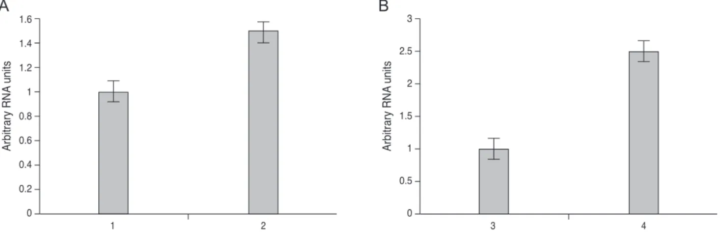
The expression of MIF mRNA was upregulated 2.5 fold in the corneal epithelium and upregulated 1.5 fold in the cells from the LASIK interface at 48 hours after LASIK surgery as compared with the control eyes (Fig. 1).
Corneal sensitivity
Preoperative baseline corneal sensitivity was 40.56 ± 2.36 mm. There was no significant difference in preopera-
tive corneal sensitivity among the three groups. In the BSS-treated group, corneal sensitivity decreased down to 9.17 ± 5.57 mm at two weeks postoperative and slightly re- covered to 15.00 ± 9.65 mm at 10 weeks postoperative.
With MIF treatment, corneal sensitivity was recovered up to 21.92 ± 2.44 mm at two weeks postoperative and 35.00 ± 5.48 mm at 10 weeks postoperative. Treatment with the combination of NGF, NT-3, IL-6, and LIF increased corne- al sensitivity to 22.42 ± 1.59 mm at two weeks postopera- tive and 29.58 ± 4.31 mm at 10 weeks postoperative (Table 1). Treatment with either MIF or neuronal growth promot- ing factors following LASIK surgery significantly en- hanced the recovery of corneal sensitivity compared to the BSS treatment at two weeks postoperative (MIF vs. BSS, p
< 0.0001; neuronal growth factors vs. BSS, p < 0.0001) and at 10 weeks postoperative (MIF vs. BSS, p = 0.0001; neu- ronal growth factors vs. BSS, p = 0.002). There was no sta- tistical significance in corneal sensitivity between the group treated with MIF and the group treated with a com- bination of the neuronal growth promoting factor either at
Table 1. Corneal sensitivity following LASIK surgery in the rabbit eye (mm, mean ± SE)
Preoperative 2 wk postoperative 10 wk postoperative p-value*
Control (BSS) (n = 6) 40.83 ± 2.58 9.17 ± 5.57 15.00 ± 9.65 0.009
MIF (n = 6) 40.00 ± 2.24 21.92 ± 2.44 35.00 ± 5.48 0.006
NGF + NT-3 + IL-6 + LIF (n = 6) 40.83 ± 2.58 22.42 ± 1.59 29.58 ± 4.31 0.003
LASIK = laser in situ keratomileusis; BSS = balanced salt solution; MIF = macrophage migration inhibitory factor; NGF = nerve growth factor; NT-3 = neurotrophine-3; IL-6 = interleukin-6; LIF = leukemia inhibitory factor.
*Friedman test.
Fig. 1. Real time reverse transcription polymerase chain reaction (PCR) of migration inhibitory factor amplification from 1) cells from the superficial stromal bed of a normal rabbit eye and 2) the cells at the flap interface from the rabbit eye following laser in situ keratomil- eusis (LASIK) (A), 3) corneal epithelium from the control eyes and 4) corneal epithelium from the rabbit eye following LASIK (B). RNA was isolated at 48 hours after LASIK. All real time PCR values were normalized to hypoxanthine phosphoribosyl transferase as the in- ternal control. Vertical lines represent standard deviations from the mean.
Arbitrary RNA units
0 0.2 0.4 0.6 0.8 1 1.2 1.4 1.6
1 2
Arbitrary RNA units
0 0.5 1 1.5 2 2.5 3
3 4
Arbitrary RNA units
0 0.2 0.4 0.6 0.8 1 1.2 1.4 1.6
1 2
Arbitrary RNA units
0 0.5 1 1.5 2 2.5 3
3 4
A B
two weeks postoperative (p = 0.815) or at 10 weeks postop- erative (p = 0.192) (Fig. 2).
Discussion
Our results showed that MIF mRNA expression in the corneal epithelium was upregulated 2.5-fold as compared with the control eyes at 48 hours after LASIK, as deter- mined by real-time PCR. Cells from the LASIK interface, which are assumed to be inflammatory cells or keratocytes, also showed upregulation in the expression of MIF mRNA by a factor of 1.5 as compared with the control eyes. These data suggest that the source of MIF in the wound healing process after LASIK could be either the corneal epithelium, keratocytes or inflammatory cells. Expression of MIF in cornea wound healing has been studied in a rat model, and Matsuda et al. [18] have shown that MIF was released with-
in three hours from the corneal epithelial cells after a pene- trating linear incision. They reported that the MIF mRNA level of the injured cornea increased from 6 to 48 hours af- ter injury and then diminished. The upregulation of MIF expression implies that MIF could have an important role in the wound healing process.
The present study demonstrates that topically-adminis- tered MIF showed a beneficial effect on the recovery of corneal sensitivity following LASIK surgery. Dry eye after LASIK results from severing of the corneal nerves and subsequent loss of corneal sensation. Decreased corneal sensitivity may trigger a cascade of events that degrade the corneal integrity by reducing the protective blinking re- flex, delaying epithelial wound healing, and decreasing aqueous tear layer production [19-21]. Recovery of corneal sensation occurs in three to seven months, but a morpho- logical study showed that recovery of corneal innervation would take more than 12 months after LASIK [1]. It has been postulated that various neuronal growth promoting factors such as a glial cell line-derived neurotrophic factor, opioid growth factor, and ciliary neurotrophic factor might enhance the recovery of corneal reinnervation after LASIK [22-24]. Our previous study has shown that nerve growth factor induced an earlier recovery in corneal sensitivity af- ter LASIK [17]. We used a combination of NGF, NT-3, IL- 6, and LIF as a positive control in this study. Our results demonstrate that the treatment with MIF alone could achieve as much of an effect on the recovery of corneal sensation as treatment with a combination of NGF, NT-3, IL-6, and LIF. The detailed mechanism of MIF on the re- generation of the corneal nerve has not been investigated in this study. It has been reported that MIF exerts an enzy- matic activity to catalyze the conversion of toxic quinone products of catecholamine neurotransmitters to indoledi- hydroxy derivatives [9] and this has raised the possibility that MIF could have a protective effect on nervous tissue by converting toxic products of catecholamine metabolism.
A preliminary study has also suggested that MIF could rescue neuronal cells from catecholamine-induced cell death (data not shown). Nishio et al. [11] also reported that MIF mRNA was up-regulated in peripheral nerves after axotomy and blocking MIF with anti-MIF antibody result- ed in delayed nerve regeneration and more apoptosis in rat peripheral nerves [12]. Thus, up-regulation of MIF in the early period of the wound healing process may protect against neuronal damage. Our study suggests that exoge- Fig. 2. Corneal sensitivity after laser in situ keratomileusis
(LASIK) surgery in the rabbit eye. Preoperative baseline corneal sensitivity was 40.83 ± 2.58 mm in the balanced salt solution (BSS)-treated group, 40.00 ± 2.24 mm in the migration inhibitory factor (MIF)-treated group, and 40.83 ± 2.58 mm in the neuronal growth factors-treated group. At two weeks after LASIK, corne- al sensitivity was 9.17 ± 5.57 mm in the BSS-treated group, 21.92
± 2.44 mm in the MIF-treated group, and 22.42 ± 1.59 mm in the neuronal growth factors-treated group. At 10 weeks after LASIK, corneal sensitivity was 15.00 ± 9.65 mm, 35.00 ± 5.48 mm, and 29.58 ± 4.31 mm, respectively. NGF = nerve growth factor; NT-3
= neurotrophine-3; IL-6 = interleukin-6; LIF = leukemia inhib- itory factor. *Kruskal-Wallis test, p-value with the Bonferroni method, MIF vs. BSS, p < 0.0001; neuronal growth factors vs.
BSS, p < 0.0001; †Kruskal-Wallis test, p-value with the Bonfer- roni method, MIF vs. BSS, p = 0.0001; neuronal growth factors vs. BSS, p = 0.002.
Corneal sensitivity (mm)
5 10 15 20 25 30 35 40 45
†
*
Baseline 2 wk 10 wk
BSS MIF NGF + NT-3 + IL-6 + LIF
nous application of MIF during the early period of wound healing may further facilitate nerve recovery.
Limitations of this study would include the small num- bers of animals, and also the short follow-up period. This study also did not include histological evidence of corneal nerve regeneration. However, rabbits assigned to a topical treatment with MIF showed a faster recovery of corneal sensation in the early postoperative period after LASIK.
This beneficial effect on earlier corneal regeneration may interrupt the cascade events that lead to deterioration of the ocular surface and may reduce dry eye symptoms in patients undergoing LASIK [25].
The results of this study suggest that the topical applica- tion of MIF plays a potential role in reinnervation of dam- aged corneal nerves after LASIK. Further studies are need- ed to elucidate the detailed mechanism of MIF on corneal nerve regeneration.
Conflict of Interest
No potential conflict of interest relevant to this article was reported.
Acknowledgements
This study was supported by grant no 04-2005-002 from the Seoul National University Bundang Hospital research fund.
References
1. Lee BH, McLaren JW, Erie JC, et al. Reinnervation in the cornea after LASIK. Invest Ophthalmol Vis Sci 2002;43:
3660-4.
2. Chaudhary S, Namavari A, Yco L, et al. Neurotrophins and nerve regeneration-associated genes are expressed in the cornea after lamellar flap surgery. Cornea 2012;31:1460-7.
3. Esquenazi S, Bazan HE, Bui V, et al. Topical combination of NGF and DHA increases rabbit corneal nerve regenera- tion after photorefractive keratectomy. Invest Ophthalmol Vis Sci 2005;46:3121-7.
4. Yang P, Wen H, Ou S, et al. IL-6 promotes regeneration and functional recovery after cortical spinal tract injury by
reactivating intrinsic growth program of neurons and en- hancing synapse formation. Exp Neurol 2012;236:19-27.
5. Nathan CF, Karnovsky ML, David JR. Alterations of mac- rophage functions by mediators from lymphocytes. J Exp Med 1971;133:1356-76.
6. Matsuda A, Tagawa Y, Matsuda H, Nishihira J. Identifica- tion and immunohistochemical localization of macrophage migration inhibitory factor in human cornea. FEBS Lett 1996;385:225-8.
7. Bacher M, Meinhardt A, Lan HY, et al. MIF expression in the rat brain: implications for neuronal function. Mol Med 1998;4:217-30.
8. Nishibori M, Nakaya N, Tahara A, et al. Presence of mac- rophage migration inhibitory factor (MIF) in ependyma, astrocytes and neurons in the bovine brain. Neurosci Lett 1996;213:193-6.
9. Matsunaga J, Sinha D, Pannell L, et al. Enzyme activity of macrophage migration inhibitory factor toward oxidized catecholamines. J Biol Chem 1999;274:3268-71.
10. Matsunaga J, Sinha D, Solano F, et al. Macrophage migra- tion inhibitory factor (MIF): its role in catecholamine me- tabolism. Cell Mol Biol (Noisy-le-grand) 1999;45:1035-40.
11. Nishio Y, Minami A, Kato H, et al. Identification of macro- phage migration inhibitory factor (MIF) in rat peripheral nerves: its possible involvement in nerve regeneration. Bio- chim Biophys Acta 1999;1453:74-82.
12. Nishio Y, Nishihira J, Ishibashi T, et al. Role of macrophage migration inhibitory factor (MIF) in peripheral nerve re- generation: anti-MIF antibody induces delay of nerve re- generation and the apoptosis of Schwann cells. Mol Med 2002;
8:509-20.
13. Matsuda A, Tagawa Y, Matsuda H, Nishihira J. Identifica- tion and immunohistochemical localization of macrophage migration inhibitory factor in human cornea. FEBS Lett 1996;
385:225-8.
14. Oh SY, Choi JS, Kim EJ, et al. The role of macrophage mi- gration inhibitory factor in ocular surface disease pathogen- esis after chemical burn in the murine eye. Mol Vis 2010;
16:2402-11.
15. Tsujimura H, Tamura T, Gongora C, et al. ICSBP/IRF-8 retrovirus transduction rescues dendritic cell development in vitro. Blood 2003;101:961-9.
16. Kuwata T, Gongora C, Kanno Y, et al. Gamma interferon triggers interaction between ICSBP (IRF-8) and TEL, re- cruiting the histone deacetylase HDAC3 to the interfer- on-responsive element. Mol Cell Biol 2002;22:7439-48.
17. Joo MJ, Yuhan KR, Hyon JY, et al. The effect of nerve growth factor on corneal sensitivity after laser in situ ker- atomileusis. Arch Ophthalmol 2004;122:1338-41.
18. Matsuda A, Tagawa Y, Matsuda H, Nishihira J. Expression of macrophage migration inhibitory factor in corneal wound healing in rats. Invest Ophthalmol Vis Sci 1997;38:
1555-62.
19. Heigle TJ, Pflugfelder SC. Aqueous tear production in pa- tients with neurotrophic keratitis. Cornea 1996;15:135-8.
20. Muller LJ, Vrensen GF, Pels L, et al. Architecture of human corneal nerves. Invest Ophthalmol Vis Sci 1997;38:985-94.
21. Stern ME, Beuerman RW, Fox RI, et al. The pathology of dry eye: the interaction between the ocular surface and lac-
rimal glands. Cornea 1998;17:584-9.
22. Koh SW. Ciliary neurotrophic factor released by corneal en- dothelium surviving oxidative stress ex vivo. Invest Oph- thalmol Vis Sci 2002;43:2887-96.
23. You L, Kruse FE, Volcker HE. Neurotrophic factors in the human cornea. Invest Ophthalmol Vis Sci 2000;41:692-702.
24. Zagon IS, Sassani JW, McLaughlin PJ. Reepithelialization of the human cornea is regulated by endogenous opioids.
Invest Ophthalmol Vis Sci 2000;41:73-81.
25. Battat L, Macri A, Dursun D, Pflugfelder SC. Effects of la- ser in situ keratomileusis on tear production, clearance, and the ocular surface. Ophthalmology 2001;108:1230-5.