http://dx.doi.org/10.5468/ogs.2016.59.2.116 pISSN 2287-8572 · eISSN 2287-8580
Introduction
Neuroendocrine cervical carcinoma (NECC) is a rare form of cervical cancer representing 0.5% to 5% of all cervical cancer cases. NECC is classified into four histologic subtypes (small cell, large, classical carcinoid and atypical carcinoid, with small cell carcinoma of cervix (SCCC) being the most frequent type [1]. Neuroendocrine carcinomas can be identified by characteristic light and electron microscopic criteria, such as small cells with hyperchromatic nuclei and scanty cytoplasm.
NECC can be present with other histologic findings, such as squamous cell and adenocarcinoma, and the presence of the NECC component defines the clinical behavior [2].
The clinical course of SCCC is aggressive, and metastasis to the bone, brain, liver, and bone marrow is common [3]. The relapse and metastasis pattern infers the nature of hematog-
enous dissemination [4]. Despite its known clinical behavior, there is no consistent method of treatment. The treatment regimen is based on small cell lung cancer, where the micro-
Prognostic factors in neuroendocrine cervical carcinoma
Da Yong Lee
1, Chul Chong
1, Maria Lee
1,2, Jae Weon Kim
1,2, Noh Hyun Park
1,2, Yong Sang Song
1,2, Sang Yoon Park
3Department of Obstetrics and Gynecology, 1Seoul National University Hospital, Seoul, 2Seoul National University College of Medicine, Seoul; 3Center for Uterine Cancer, Research Institute and Hospital, National Cancer Center, Goyang, Korea
Objective
To evaluate the clinical and pathologic factors associated with survival in patients with neuroendocrine cervical carcinoma (NECC).
Methods
The records of 61 patients with NECC diagnosed between 2000 and 2014 at Seoul National University Hospital and the National Cancer Center were retrospectively reviewed. Kaplan-Meier and Cox regression methods were used for analyses.
Results
Of the 61 patients, 67.2% were diagnosed at early stage (I to IIA) with a median age of 49 years. Of those, 78%
underwent surgery and 75.6% received postoperative adjuvant treatment. For patients diagnosed at advanced stage, 60.0% received chemotherapy only and 25.0% received concurrent chemoradiation therapy. In the univariate analysis, advanced stage (77 vs. 40 months, P=0.013), tumor size ≥2 cm (133 vs. 47 months, P=0.002) and mixed tumor (101 vs.
34 months, P=0.004) were shown to be poor prognostic factors. In the multivariate analysis, tumor stage, tumor size and tumor homology were shown to be independent prognostic factors for overall survival. Of the total, 39.3% of the patients experienced recurrence, and 54.1% of the patients had metastasis. Of the patients diagnosed at early stage, 51.2% experienced recurrence.
Conclusion
Tumor stage, tumor size and tumor homology were found to be independent prognostic factors in patients with NECC. Even in patients diagnosed at early stage, recurrence and distant metastasis were frequently observed.
Keywords: Cervical carcinoma; Neoplasm recurrence; Neuroendocrine carcinoma; Retrospective studies; Small cell carcinoma
Articles published in Obstet Gynecol Sci are open-access, distributed under the terms of the Creative Commons Attribution Non-Commercial License (http://creativecommons.
org/licenses/by-nc/3.0/) which permits unrestricted non-commercial use, distribution, and reproduction in any medium, provided the original work is properly cited.
Copyright © 2016 Korean Society of Obstetrics and Gynecology Received: 2015.5.17. Revised: 2015.9.30. Accepted: 2015.10.21.
Corresponding author: Maria Lee
Department of Obstetrics and Gynecology, Seoul National University Hospital, 101 Daehak-ro, Jongno-gu, Seoul 03080, Korea
Tel: +82-2-2072-2842 Fax: +82-2-762-3599 E-mail: [email protected]
http://orcid.org/0000-0002-8017-3176
scopic characteristics are similar. This regimen is also applied to other types of NECC. Interestingly, Hoskins et al. [5] reported a three-year failure-free survival rate of 80% in early stages (I to II) patients who received primary radiation therapy and platinum-based combination chemotherapy. A recent study showed that primary radiation therapy with at least 5 cycles of platinum-based chemotherapy resulted in a five-year over- all survival rate of 78%, better than that of 46% achieved through primary surgery alone in early stage SCCC [6]. How- ever, some studies have reported primary radical surgery fol- lowed by adjuvant chemotherapy as the preferred treatment modality with relatively favorable survival outcomes [7].
Due to the low incidence of NECC, it is very difficult to un- dertake prospective studies to elucidate the impact of treat- ment modality on survival outcome. Moreover according to our review, there are not enough studies with sufficient cases in the Korean population. Thus, to understand the prognosis in our population and improve the treatment strategy, we evaluated the clinical and pathologic factors associated with survival in patients with NECC in two of the cancer institutes in Korea.
Materials and methods
Patients diagnosed with NECC and treated between 2000 and 2014 at Seoul National University Hospital and the National Cancer Center were included in the study. One case of a pa- tient who died due to sepsis caused by aggravating myelodys- plastic syndrome was excluded. Clinicopathologic data were collected from hospital charts and scanned records. Staging was done based on International Federation of Gynecology and Obstetrics (FIGO) clinical staging criteria. Early stage was defined as stage I to IIA and advanced stage was as stages IIB and above. All patients underwent physical examination, imaging study (pelvic computed tomography, chest computed tomography and pelvic magnetic resonance imaging if indi- cated), intravenous pyelography, and cystoscopy. Patients with tumors confined to the cervix typically underwent radical hys- terectomy with pelvic lymphadenectomy. For advanced cases, radiotherapy with or without chemotherapy was performed.
Adjuvant therapy using chemotherapy or concurrent chemora- diation therapy (CCRT) was carried out except for in one case of stage 1B1. The association between risk factors and prog- nosis was evaluated using Kaplan-Meier survival analyses and log-rank tests. The independent factors found to be predictive
of survival were evaluated using Cox regression methods. P- values <0.05 were considered significant. All analyses were performed using PASW ver. 18.0 (SPSS Inc., Chicago, IL, USA) and MedCalc ver. 15.8 (MedCalc Software bvba., Acacialaan, Ostend, Belgium). The institutional review board approval number for this study is 1507-013-686.
Results
A total of 61 patients were diagnosed and treated in this study. The median age was 49 years (range, 28 to 74), and Table 1. Patients’ clinicopathologic characteristics (n=61)
Variable Value
Age (yr) 49 (28–74)
Parity 2 (0–5)
Menopause 21 (34.4)
HRT 2 (3.3)
Smoking 2 (3.3)
Stage
IB 34 (55.7)
IIA 7 (11.5)
IIB 9 (14.8)
IIIB 3 (4.9)
IVB 8 (13.1)
Histology
Small cell 41 (67.2)
Large cell 7 (11.5)
Other type 13 (21.3)
Tumor size (cm) 3.9 (0.5–9.2)
<2 10 (16.4)
≥2 51 (83.6)
Tumor homology (pathologic examination, n=36)
Pure 17 (47.2)
Mixed 19 (52.8)
LVSI (operation, n=34) 20 (58.8)
Lymph node involvement (LND, n=33) 5 (15.2) Neuroendocrine markers (n=43)
Synaptophysin 33 (76.7)
Chromogranin 30 (69.8)
CD56 30 (69.8)
Values are presented as median (range) or number (%).
HRT, hormone replacement therapy; LVSI, lymphovascular space inva- sion; LND, lymph node dissection.
41 patients had early-stage (I to IIA) disease. Patients’ demo- graphic and clinicopathologic characteristics are shown in Table 1. Small cell carcinoma was the most common histologic type, comprising 67.2% of the cases. Among early-stage pa- tients treated by radical hysterectomy, lymphovascular space invasion was found in 19 cases (59.3%). By immunohisto- chemistry, the three types of neuroendocrine marker (synap- tophysin, chromogranin, and CD 56) were stained with similar frequency.
Tables 2 and 3 show the treatment methods of the 61 patients. Of the early-stage patients, 32 (78%) underwent radical hysterectomy as a primary treatment. Among them, 22 (68.8%) patients underwent pelvic lymph node dissection and of those, eight patients underwent additional para-aortic lymph node dissection. Adjuvant therapy using chemotherapy or CCRT was conducted in most (31/32) cases. There was no specific difference in median survival between chemotherapy and CCRT. For advanced-stage patients, the main treatment modalities were chemotherapy (60%) and CCRT (25%). There was no significant difference in prognosis between the two groups. Two patients underwent primary surgery. One stage IIB patient underwent pelvic exenteration and another stage
IIB patient underwent pelvic and para-aortic lymph node dis- section before CCRT.
Post-treatment surveillance was conducted every three months. For patients experiencing recurrence, radiation and/
or chemotherapy were employed. As a first-line chemothera- peutic agent, etoposide was used most frequently (Table 3).
The efficacy of the different regimens was not clearly proved in this study.
We evaluated the significance of various clinical factors that may influence the prognosis and overall survival (Table 4, Fig. 1). Early-stage patients had a median survival time of 77 months compared with 40 months in the advanced- stage group (P=0.013). The estimated five-year survival rate was 42.1% for early-stage patients and 23.7% for advanced- stage patients. In all cases, patients with tumor size ≥2 cm had a median survival time of 47 months compared with 133 months in those with tumor size <2 cm. To remove the stage factor, a subgroup analysis on the early-stage group was done. Early-stage patients with tumor size ≥2 cm had a me- dian survival time of 44 months compared with 130 months in the group with tumor size <2 cm. Other pathologic factors that may influence prognosis (e.g., residual tumor, tumor ho- mology, lymph node involvement, lymphovascular space inva- sion, neuroendocrine markers) were also evaluated. In all of the radical hysterectomy cases, the resection margin was free from carcinoma. Of the histologic factors, only mixed histol- ogy proved to be a poor prognostic factor (P=0.004). Age had no significant effect on survival. All the preceding factors were re-evaluated only in small cell neuroendocrine carcinoma sub- type cases and the statistical results were similar with identical prognosis factors. In the multivariate analysis, tumor stage, Table 2. Treatment methods of neuroendocrine cervical carcinoma by tumor stage
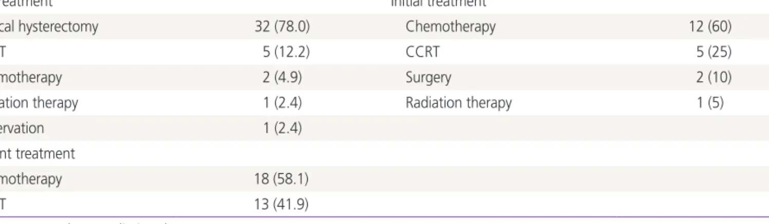
Early stage (n=41) n (%) Advanced stage (n=20) n (%)
Initial treatment Initial treatment
Radical hysterectomy 32 (78.0) Chemotherapy 12 (60)
CCRT 5 (12.2) CCRT 5 (25)
Chemotherapy 2 (4.9) Surgery 2 (10)
Radiation therapy 1 (2.4) Radiation therapy 1 (5)
Observation 1 (2.4)
Adjuvant treatment
Chemotherapy 18 (58.1)
CCRT 13 (41.9)
CCRT, concurrent chemoradiation therapy.
Table 3. Initial chemotherapy regimen of neuroendocrine cervical carcinoma
Initial chemotherapy regimen No.
Etoposide-cisplatin 31
Taxol-carboplatin 7
Etoposide-carboplatin 3
5Fluorouracil-cisplatin 3
Cyclophosphamide-adriamycin-cisplatin 1
tumor size and tumor homology proved to be significant in- dependent prognostic factors. Immunohistochemical analysis was performed in 43 of 61 cases. In the remaining 18 cases, only microscopic diagnosis was performed. Thus, subgroup analysis was performed particularly in immunohistochemically confirmed cases and the statistical results were similar.
Sites of metastasis at diagnosis and frequent recurrence sites after initial treatment are shown in Table 5. Major prima- ry metastatic sites were the liver and bone. Tumor recurrence
was found in 39.3% of all patients. Despite the large early- stage group, the recurrence rate was 51.2%. Main sites of distant recurrence were the lung and liver.
Discussion
NECC is a rare form of cervical carcinoma and consequently, prospective randomized trials to evaluate the impact of differ- Table 4. Univariate and multivariate analysis for overall survival
Variables
Univariate Multivariate
Median survival
(mo) P-value Hazard ratio Hazard ratio P-value Stage
I–IIA 77 - - - -
IIB–IV 40 0.013 2.24 9.369 0.015
Tumor size (all stage, cm)
<2 133 - - - -
≥2 47 0.002 6.61 5.556 0.022
Tumor size (early stage, cm)
<2 130 - - - -
≥2 44 0.006 5.77 - -
Tumor homology
Pure 101 - - - -
Mixed 34 0.004 3.24 2.860 0.020
Lymph node involvement (early stage)
No 80 - - - -
Yes 59 0.334 - - -
Lymphovascular space invasion
No 90 - - - -
Yes 57 0.298 - - -
Chromogranin
Negative 27 - - - -
Positive 76 0.114 - - -
Age (yr)
<49 77 - - - -
≥49 51 0.380 - - -
Menopause
No 70 - - - -
Yes 61 0.778 - - -
Etoposide containing regimen vs. others
Etoposide containing 51 - - - -
Others (primary regimen) 66 0.518 - - -
ent treatments on patient outcome have not been possible. In recent years, novel NECC treatment methods have attempted to replicate successful treatments for small cell lung carci- noma. Currently, surgical resection only plays a limited role in patients with early-stage small cell lung cancer [8]. Moreover, a report has suggested that radical hysterectomy does not prolong the survival of SCCC patients [5]. However, our results showed that radical surgery is an important component in the multimodal treatment of NECC. The survival rate in this study was better than that in previous studies. For example, Cohen et al. [9] previously analyzed the survival outcomes of 188 patients from 1979 to 2005 and found the estimated five- year survival rate to be 36.8% for early-stage patients and Fig. 1. Overall survival estimates stratified by (A) tumor stage, (B) tumor size in all stage, (C) tumor size in early stage, and (D) tumor ho- mology.
0 50 100 150 200
0 50 100 150 200
0 50 100 150 200
0 50 100 150 200 I-IIA
<2 cm
<2 cm
Pure
≥2 cm
≥2 cm
Mixed 1.0
0.8
0.6
0.4
0.2
0.0
1.0
0.8
0.6
0.4
0.2
0.0
1.0
0.8
0.6
0.4
0.2
0.0
1.0
0.8
0.6
0.4
0.2
0.0 IIB-IV
Survival time (mo)
Survival time (mo)
Survival time (mo)
Survival time (mo)
Survival probabilitySurvival probability Survival probabilitySurvival probability
A B
C D
Table 5. Site of metastasis at diagnosis and recurrence site after treatment
M etastasis at
diagnosis No. Recurrence site No.
Liver 5 Lung 17
Bone 5 Liver 8
Lung 3 Vagina (stump) 6
SCLN 1 Brain 3
Bone 3
Pelvic wall 2
Othersa) 4
SCLN, supraclavicular lymph node.
a)Omentum 1 case, kidney 1 case, left ovary 1 case, and vulva 1 case.
only 8.9% for advanced-stage patients. In our study, the rates were 42.1% and 23.7% respectively. Some factors may have contributed to the improvement of survival. First, in all surgi- cal cases the resection margin was free from cancer. Improved surgical skills and the use of frozen section examination dur- ing the surgery may have contributed to this progression. Sec- ond, Deacon et al. [10] and Chan et al. [11] have suggested smoking as a poor prognostic factor in cervical cancer and SCCC respectively. In our case, there were only two patients with history of smoking and that may be attributed to the low female smoking rate in the Korean population. Third, due to the limited randomized controlled clinical trial for NECC, some therapeutic guidelines (e.g., multimodality therapy on early- stage patients, using etoposide-containing chemotherapeutic regimens) were suggested [9,12]. These may have led to the improved outcome.
Several studies have reported disease stage as the strongest predictor of outcome, and that other factors (age, tumor size, depth of stromal invasion, and vascular space invasion) as prognostic factors in SCCC [7]. However, in our study, FIGO stage, tumor size, and tumor homology were independent prognostic factors of survival, and our univariate and multi- variate analyses found no association between age, depth of stromal invasion, or vascular space invasion and NECC patient outcome. In a previous study, patients with a mixed histologic pattern had a better prognosis than those with a homogenous pettern [11]. However, others found no relationship between tissue homology and prognosis [12,13]. In our study, patients with a homogenous histologic pattern had a better prognosis, but racial and etiologic differences (e.g., low smoking rates, human papillomavirus infection) may be confounding factors, and further studies with more cases are required to clarify this correlation.
A previous study reported the effectiveness of etoposide with platinum-based chemotherapy for NECC but not for well-differentiated carcinoid tumors [2]. Chang et al. [12]
found that chemotherapies containing cisplatin and etoposide could be effective in patients with early-stage SCCC following radical hysterectomy. A recent study showed that platinum- based combination chemotherapy was an independent prog- nostic factor for improved survival in patients with SCCC [14].
In our current study, there were no prognostic differences between etoposide and other regimens. More well-designed case-control studies are needed to verify effect of the regi- men. The overall tumor recurrence rate was 39.3%, and the
early-stage recurrence rate was as high as 51.2%. Because of the limited number of patients in our study, we could not detect a significant survival benefit in patients who received adjuvant chemotherapy. However, considering the features of early recurrence and distant metastasis with NECC, it is likely that adjuvant chemotherapy would enhance survival relative to radiation alone.
In conclusion, tumor stage and tumor size may act as surro- gates for factors prognostic of survival in patients with NECC.
Moreover, our results indicate that primary radical surgery fol- lowed by adjuvant chemotherapy may be the preferred treat- ment modality for patients with early-stage NECC.
Conflict of interest
No potential conflict of interest relevant to this article was reported.
References
1. Albores-Saavedra J, Gersell D, Gilks CB, Henson DE, Lindberg G, Santiago H, et al. Terminology of endo- crine tumors of the uterine cervix: results of a workshop sponsored by the College of American Pathologists and the National Cancer Institute. Arch Pathol Lab Med 1997;121:34-9.
2. Gardner GJ, Reidy-Lagunes D, Gehrig PA. Neuroendocrine tumors of the gynecologic tract: a Society of Gyneco- logic Oncology (SGO) clinical document. Gynecol Oncol 2011;122:190-8.
3. Delaloge S, Pautier P, Kerbrat P, Castaigne D, Haie-Meder C, Duvillard P, et al. Neuroendocrine small cell carcinoma of the uterine cervix: what disease? What treatment? Re- port of ten cases and a review of the literature. Clin Oncol (R Coll Radiol) 2000;12:357-62.
4. Viswanathan AN, Deavers MT, Jhingran A, Ramirez PT, Levenback C, Eifel PJ. Small cell neuroendocrine carci- noma of the cervix: outcome and patterns of recurrence.
Gynecol Oncol 2004;93:27-33.
5. Hoskins PJ, Swenerton KD, Pike JA, Lim P, Aquino-Parsons C, Wong F, et al. Small-cell carcinoma of the cervix: four- teen years of experience at a single institution using a combined-modality regimen of involved-field irradiation
and platinum-based combination chemotherapy. J Clin Oncol 2003;21:3495-501.
6. Chen TC, Huang HJ, Wang TY, Yang LY, Chen CH, Cheng YM, et al. Primary surgery versus primary radiation thera- py for FIGO stages I-II small cell carcinoma of the uterine cervix: a retrospective Taiwanese Gynecologic Oncology Group study. Gynecol Oncol 2015;137:468-73.
7. Lee JM, Lee KB, Nam JH, Ryu SY, Bae DS, Park JT, et al.
Prognostic factors in FIGO stage IB-IIA small cell neuroen- docrine carcinoma of the uterine cervix treated surgically:
results of a multi-center retrospective Korean study. Ann Oncol 2008;19:321-6.
8. Tsuchiya R, Suzuki K, Ichinose Y, Watanabe Y, Yasumitsu T, Ishizuka N, et al. Phase II trial of postoperative adjuvant cisplatin and etoposide in patients with completely re- sected stage I-IIIa small cell lung cancer: the Japan Clinical Oncology Lung Cancer Study Group Trial (JCOG9101). J Thorac Cardiovasc Surg 2005;129:977-83.
9. Cohen JG, Kapp DS, Shin JY, Urban R, Sherman AE, Chen LM, et al. Small cell carcinoma of the cervix: treatment and survival outcomes of 188 patients. Am J Obstet Gy-
necol 2010;203:347.e1-6.
10. Deacon JM, Evans CD, Yule R, Desai M, Binns W, Taylor C, et al. Sexual behaviour and smoking as determinants of cervical HPV infection and of CIN3 among those infected:
a case-control study nested within the Manchester cohort.
Br J Cancer 2000;83:1565-72.
11. Chan JK, Loizzi V, Burger RA, Rutgers J, Monk BJ. Prog- nostic factors in neuroendocrine small cell cervical carci- noma: a multivariate analysis. Cancer 2003;97:568-74.
12. Chang TC, Lai CH, Tseng CJ, Hsueh S, Huang KG, Chou HH. Prognostic factors in surgically treated small cell cervi- cal carcinoma followed by adjuvant chemotherapy. Can- cer 1998;83:712-8.
13. Bermudez A, Vighi S, Garcia A, Sardi J. Neuroendocrine cervical carcinoma: a diagnostic and therapeutic chal- lenge. Gynecol Oncol 2001;82:32-9.
14. Huang L, Liao LM, Liu AW, Wu JB, Cheng XL, Lin JX, et al. Analysis of the impact of platinum-based combination chemotherapy in small cell cervical carcinoma: a multi- center retrospective study in Chinese patients. BMC Can- cer 2014;14:140.