39
Production of recombinant nucleocapsid protein of Newcastle disease virus in Escherichia coli for a diagnostic ELISA
Hyun-il Kim
1,†, Kyoung-Phil Park
1,‡, Chan-Hee Park
1, Hyun-Ah Cho
1, Ho-Suk Yang
1, Tae-Wook Hahn
2,*
1Lab of Clinical Pathology, Institute of Cheilbio, Ansan 457-7, Korea
2School of Veterinary Medicine, Kangwon National University, Chuncheon 200-701, Korea (Accepted: March 26, 2009)
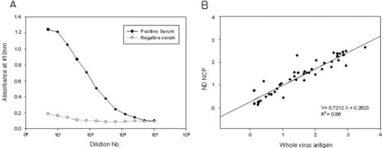
Abstract : Transmission of avian viruses both bird-to-bird and from birds to non-avian species is a major health concern. Newcastle disease virus (NDV) is an economically important avian virus that poses substantial risks to the poultry industry. Rapid and sensitive diagnostic methods, such as the enzyme- linked immunosorbent assay (ELISA), are required to track such infections. To develop an ELISA for detecting anti-NDV antibody in avian sera, the nucleocapsid protein (NCP) gene of the NDV La Sota strain was cloned and expressed in Escherichia coli and the 513-amino acid recombinant NCP was purified by Ni-NTA affinity chromatography. To evaluate its ability to replace NDV whole virus antigen as a coating antigen, NCP-coated and whole NDV-coated ELISAs were tested and compared using a panel of NDV positive antisera from chickens. Results using purified NCP were highly correlated with those obtained using whole NDV (r= 0.927), demonstrating that recombinant NCP expressed in Escherichia coli is a suitable substitute antigen for whole NDV in a diagnostic ELISA.
Keywords : E. coli expression, ELISA, Newcastle disease virus, nucleocapsid protein
Introduction
Newcastle disease virus (NDV) is an economically important avian virus that poses substantial risks to the poultry industry [1]. The most widely used methods for detection of anti-NDV antibody in birds have been serum neutralization and hemagglutination inhibition (HI) tests, but the enzyme-linked immunosorbent assay (ELISA) has emerged as a convenient method for monitoring both immune status and viral infection [9, 14]. Several commercial ELISA kits are available for detection of NDV-specific antibodies. Most of these kits use whole, inactivated NDV as a coating antigen.
However, propagation and purification of NDV is time- consuming and expensive. Unlike full virions, recom- binant proteins obtained via gene engineering are safe, and both easy and inexpensive to produce.
NDV is an enveloped virus with a single-stranded, negative-sense RNA genome. The total genome is about 15 kb in length and encodes 6 structural proteins:
hemagglutinin-neuraminidase (HN), fusion (F) protein,
matrix protein, nucleocapsid protein (NCP), phospho- protein, and large protein [1]. Of these, NCP has been the antigen of choice for anti-NDV titer diagnostic systems [3]. Nucleocapsid proteins in general are highly immunogenic in nature and have been used as antigens for detection of other viruses, including measles virus [13] and avian influenza virus [16]. The NCP subunit is a single polypeptide of 489 amino acids with a molecular weight of about 53 kDa. The viral RNA is located at the core of the virus particle surrounded by 2,200 to 2,600 NCP subunits [2].
HI test is still the most widely used serological method for measuring antibodies to NDV and is considered standard method for ND. However, ELISA seems to be more convenient than HI when very large number of sera is examined. It was also reported that sera from other species tend to show high incidence of false-positive results [14].
NDV ELISA using NCP antigen expressed in baculovirus systems were found to perform just as well as the HI test or a commercial ELISA [3, 6]. Unfor-
*Corresponding author: Tae-Wook Hahn
School of Veterinary Medicine, Kangwon National University, Chuncheon 200-701, Korea [Tel: +82-33-250-8671, Fax: +82-33-244-2367, E-mail: [email protected]]
Present address:
†Optifarm solution center, Seoul 135-937, Korea.
‡Asan Pharmaceutics, Seoul 130-070, Korea
tunately, NCP production in baculovirus suffers from low yield, and is expensive and time-consuming.
Recently, NCP antigen of NDV tagged with histidine residue was produced in
Escherichia(
E.)
coliand purified by histidine residue affinity chromatography [10, 11]. However, ELISA using the purified NCP has not been compared with whole NDV coated ELISA.
The purpose of the present study was to express the NCP gene of the NDV La Sota strain in
E. coliand to evaluate an ELISA based on the recombinant NCP and compare NCP based ELISA with whole NDV based ELISA.
Materials and Methods
Virus and RNA extraction
NDV strain La Sota was propagated in 9- to 10-day- old specific pathogen free chicken embryonated eggs.
Total RNA was extracted from 100
µl of allantoic fluid using a MagExtractor nucleic acid purification kit (Toyobo, Japan) and was dissolved in 30
µl of diethyl pyrocarbonate (DEPC)-treated distilled water (DDW).
For preparation of whole NDV antigens, the collected allantoic fluid was centrifuged at 38,000
× gfor 2 h at 4
oC. The precipitated virus was purified through a discontinuous sucrose gradient (50%, 30% and 20%) and the purified virus was inactivated with 0.3%
formalin for 30 min. The protein concentration of the purified virus was determined by Lowry’s method [5].
Reverse transcriptase - polymerase chain reaction (RT-PCR)
Complementary DNA was synthesized from genomic NDV RNA using random hexamer included in the cDNA synthesis kit (Promega, USA).
Total NDV RNA was incubated with 2
µg of random hexamer (Promega, USA) at 72
oC for 10 min and the mixture was immediately chilled on the ice. The synthesis of the first strand cDNA was carried out in a 20
µl of reaction mixture containing 0.3 mM of each deoxynucleoside triphosphate (dNTP; Promega, USA), 10 U AMV reverse transcriptase (Promega, USA), 20 U of Rnasin ribonuclease inhibitor (Promega, USA) and 1X reaction buffer [50 mM Tris-HCl, 50 mM KCl, 10 mM MgCl
2, 0.5 mM spermidine, 10 mM Dithiothreitol (DTT)], The mixture was incubated at 42
oC for 90 min and then heated at 94
oC for 5 min. Two primers, NCP sense (5'-AAG CCT TCT GCC AAC ATG TC-3') and
NCP antisense (5'-TTT GTC CA T CA A TAC CCC CA -3') were designed and used to amplify the NCP gene.
The antisense primer contains a stop codon (TCA, in bold) that was incorporated into the PCR product. A PCR was carried out in a 50
µl mixture containing 3 U
TaqDNA polymerase (Promega, USA), 3 mM MgCl
2and 1X reaction buffer [5 mM Tris-HCl (pH 8.0), 10 mM NaCl, 0.01 mM EDTA, 0.1 mM DTT, 5% glycerol, 0.1% Triton X-100], 0.3 mM of each dNTP (Promega, USA) and 5
µl of cDNA template synthesized above.
The PCR mixture was subjected to a 35-cycle PCR profile of 94
oC/30 sec, 53
oC/90 sec, 72
oC/1 min, followed by a final extension step of 72
oC/5 min.
Cloning and sequencing
The purified PCR product was ligated into a pGEM- T easy vector (Promega, USA). The ligation mixture was then transformed into
E. coliJM109 (Promega, USA). A positive clone was isolated and plasmid extracted with a DNA purification kit (Bioneer, Korea). The 1.48 kb
EcoRI fragment containing the NCP gene was ligated into the
EcoRI site of a pET28b vector (Invitrogen, USA). The ligation mixture was transformed into
E. coliBL21 (Promega, USA). Three positive clones were selected and the complete sequence of the NCP gene was obtained by reading both strands of the insert DNA.
Expression, purification and analysis of recombi- nant NCP
The verified clone was cultured in 1 l of LB medium containing 25
µg/ml kanamycin at 37
oC with vigorous shaking. When the absorbance of the cultures at 600 nm reached 0.6, NCP was induced by adding 1 mM isopropylthio-
β-galactoside (IPTG) and incubating for another 3 h. Cells were harvested and broken by sonication. The cell lysate was centrifuged at 10,000
× g