─ 125 ─
ⓒ The Korean Society of Limnology. All rights reserved.
This is an open-access article distributed under the terms of the Creative Commons Attribution Non-Commercial License (http://creativecommons.org/licenses/by-nc/3.0/), which permits unrestricted non-commercial use, distribution, and reproduction in any medium, provide the original work is properly cited.
INTRODUCTION
Chironomids have an important role in the food network of aquatic communities, representing a major link between pro- ducers and secondary consumers (Tokeshi, 1995). Chirono- midae is a large group of invertebrates found in various water worldwide, with a reported diversity of 8,000~20,000 species (Armitage, 2012). Although adult chironomids inhabit areas near the riparian zone of rivers or lakes, the larvae are aquatic organisms distributed in diverse aquatic habitat patches (Pin- der 1986, 1995). Chironomid larvae was potentially represent more than 70% abundance of the total arthropod numbers in hypersaline waters and are opportunistic omnivores, ingesting
a wide variety of food sources (Cummins and Klug, 1979). In general, these larvae ingest five kinds of food such as algae, detritus and associated microorganisms, macrophytes, wood debris, and other invertebrates (Berg, 1995). Moreover, recent eDNA metabarcoding-based gut content analysis of chirono- mid larvae indicated the presence of various small planktons, such as Desmodesmus armatus, Eolimna minima, and Tet- radesmus dimorphus, in the weirs (Jo et al., 2020).
Kwak (2015) introduced 24 genera and 43 species of chi- ronomid larvae based on visibly identified taxonomical adult stages in South Korea. The morphological studies of larvae are scarce: Yoon and Chun (1992) described the abdomen characteristics through simple drawings of eight Chironomus larval species. Ree (1981, 1998) described the larva of Crico- topus oryzaphagos. In this study, we provide the detailed pic- torial features and morphological keys to 16 genera present in Korea.
Fundamental Morphological Study of 16 Genera of Chironomid Larvae in Korea
Dong Ju Lee (0000-0001-9133-5639) , Jae-won Park 1 (0000-0002-4067-7089) and Ihn-Sil Kwak 1, * (0000-0002-1010-3965) Department of Life Science, Silla University, Busan 46958, Republic of Korea
1
Department of Ocean Intergrated Science, Chonnam National University, Yeosu 59626, Republic of Korea
Abstract Chironomids are a large group of invertebrates that live in various aquatic habitats. The distribution range of these invertebrates has become varied due to anthropogenic impacts; as such, their distribution can be used as an indicator of environmental health. Adult chironomids are well known in South Korea; however, the larvae have rarely been studied due to difficulties associated with morphological classification. To address this lack of information, we collected larvae from four important rivers in South Korea and summarized their taxonomic morphological characteristics. The antennae, mandible, and mentum were used for larval taxonomic characterization. In this study, we describe the basic morphological features and key pictorial features of 20 species of chironomids, representing 16 genera.
Key words: chironomid larvae, morphological character, antenna, mentum, mandible, Chironomidae
Manuscript received 25 May 2021, revised 16 June 2021, revision accepted 21 June 2021
* Corresponding author: Tel: +82-61-659-7148, Fax: +82-61-659-7149 E-mail: [email protected]
ISSN: 2288-1115 (Print), 2288-1123 (Online)
Original article
16.9 ℃ (range: 8.76~23.35 ℃) the field water temperature was measured using portable equipment (Model: YSI Profes- sional Plus, Ohio, USA).
Specimens were dissected and the dissected parts were mounted on slides using a glycerol mounting medium. The dissection was performed on a glass slide under a microscope (Olympus SZX12). The body was dissected with a needle and appendages were used to prepare the slides. The sand- wich method was used for setting the position and rotating the specimen. The permanent mount slide was sealed with transparent nail varnish. All drawings were made using a drawing tube on an Olympus BX51 differential interference contrast microscope. The descriptive terminology follows Epler (2001).
RESULTS AND DISCUSSION
A total of 16 genera and 20 species were identified from four major rivers in Korea (Table 1). The classification crite- ria were determined according to the morphological cha ract- eristics. Chironomid larvae have a cylindrical body shape and are well-segmented. Some appendages located on head cap- sule and posterior part is very important for identification.
teristic is used for identification by segment number and ratio. This is the length of the basal segment divided by the remaining segments. The placement and shape of the Lauter- born organ sensory structures often located on the second seg-
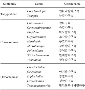
Table 1. List of the 16 genus of Chironomids in South Korea.
Subfamily Genus Korean name
Tanypodinae Conchapelopia 민다리깔따구속
Tanypus 늪깔따구속
Chironominae
Chironomus 깔따구속
Cryptochironomus 음깔따구속
Einfeldia 다모깔따구속
Glyptotendipes 조각깔따구속
Harnischia 무깔따구속
Microtendipes 꼬마깔따구속
Polypedilum 무늬깔따구속
Stictochironomus 반지깔따구속
Tanytarsus 장부깔따구속
Orthocladinae
Chaetocladius -
Cricotopus 아기딸따구속
Diplocladius 쌍깔따구속
Orthocladius 깃깔따구속
Tokunagayusurika 빨간도꾸나가깔따구
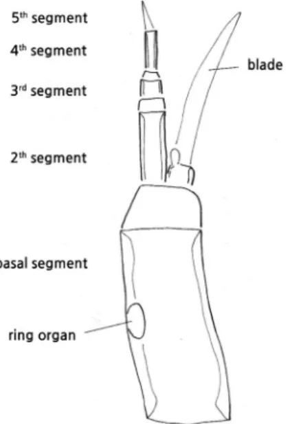
Fig. 1. The general morphological structure of chironomid larvae (A) and the main locations of the ventral head capsule (B).
(A) (B)
ment or at its apex are important, as is the location of the ring organ.
2) Mandible (Fig. 3)
The mandible is located on the right and left side of the body at the front of the head capsule (Fig. 1). Strong teeth are located foremost, which used for holding on tightly to food.
Several complex characteristics was used for identification, including the number and shape of the inner apical teeth, the
present or absence of a seta interna, the morpho-type of seta subdentalis, and the pectin mandibularis.
3) Mentum (Fig. 4)
The strong teeth with ventromental plates are located at the center of the head capsule. The shape and number of teeth are the most important characteristics for identification. The shape of the ventromental plate and the presence or absence of a beard are noticeable features.
2. Morphological key for the 16 genera of the chironomid larvae in South Korea
1. Posterior parapods long, relatively; the ratio of length/
width of the head capsule is 1.5~2.0 times ...
... (Tanypodinae) ������������������� 2 - Posterior parapods short; the ratio of length/width of the
head capsule is 1.0 to 1.3 times ...
... (Chironominae / Orthocladinae) ������������������� 3 2. The shape of the ligular teeth is V-shaped type (Fig. 5A)
�������������������������������������������������������� genus Conchapelopia - The shape of the ligular teeth is aligned (Fig. 5B) ������������
������������������������������������������������������������������ genus Tanypus 3. Antenna extremely elongated (Fig. 6A); Mandible with 1
to 2 apical teeth �������������������������������������������������������������� 4 - Antenna not elongated (Fig. 6B) ������������������������������������ 5 4. The first segment of the antenna with a seta, located in
the middle part of the segment ������������� genus Tanytartus - First segment of the antenna without seta �������������������������
������������������������������������������������������������ Tokunagayu surika 5. Labrum with naked or simple seta ��������������������������������� 6 - Labrum with plumose seta ��������������������������������������������� 8 6. Ring organ in antenna located above 1/2 (Fig. 6C) �������� 7 - Ring organ in antenna located under 1/2 (Fig. 6D) �����������
������������������������������������������������������������������������ genus Cha- etocladius, genus Cricotopus*, genus Diplocladius, Einfeld- ia dissideus, genus Glyptotendipes, Orthocladius suspensus
* - The ratio of the first segment of the antenna is not long Fig. 2. Basic structure of the antenna in the head capsule (Einfeldia
dissidnus).
Fig. 3. Basic structure of the mandible in the head capsule (Chirono- mus sp.).
Fig. 4. Basic structure of the mentum in the head capsule (Dipocla-
dius sp.).
(about 3.5 times) ������������������������������������������ C. sylvestris - The ratio of the first segment of the antenna is long (about
10 times) ������������������������������������������������� C. oryzaphages 7. The mentum is a plate-type (Fig. 7A) � genus Harnischia - The mentum is a V-shaped type (Fig. 7B) �������������������������
������������������������������������������������� genus Cryptochironomus 8. Antenna with 5-segmented ������������������������������������������� 9 - Antenna with 6-segmented ����������������������������������������� 10 9. Tubules presented; Mentum teeth is 6 : 3 : 6 (Fig. 8A) �������
������������������������������������������������������� genus Chironomus**
- Tubules absent; Mentum teeth is 6 : 2 : 6 (Fig. 8B) ����������
���������������������������������������������������� Polypodium scalaenum
** includes Chironomus kiiensis, C. flaviplumus, C. sp.
10. Median part of mentum is pale ������� genus Microtendipes - Median part of mentum is dark ��� genus Stictochironomus
3. Taxonomic account 1) Subgenus Tanypodinae
Diagnosis. Posterior parapods long. The ratio of length/
width of the head capsule is 1.5 to 2.0 times. Antenna 4-seg- mented, retractile into the head capsule, Anal tubules usually well developed in Korean freshwater species. Genus Concha- pelopia and Tanypus can easily be distinguished by the shape of the ligular teeth.
Ecology. This subfamily consists of free-swimming or crawling predators; some burrow in the bottom mud. Concha- pelopia and Tanypus are identified and recognized from differ- ent water temperature. Conchapelopia is found at a high tem- perature (19.80 ℃), Tanypus punctipennis is a low (9.30 ℃).
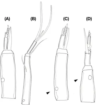
Fig. 6. Representative structure of the antenna in the head capsule.
(A) Chironomus sp., (B) Tanytartus sp., (C) Chaetocladius sp., (D) Cryptochironomus sp.
(A) (B) (C) (D)
Fig. 5. Representative structure of the ligular in the head capsule.
(A) Conchapelopia sp., (B) Tanypus sp.
Fig. 8. Representative structure of the mentum in the head capsule.
(A) Polypodium scalaenum, (B) Chironomus sp.
(A)
(B)
Fig. 7. Representative structure of the mentum in the head capsule.
(A) Harnischia sp., (B) Cryptochironomus sp.
2) Subgenus Chironominae
Diagnosis. Posterior parapods short. The head capsule is mostly of the same aspect ratio (1.0 to 1.3 times). Antenna usually 4 to 6-segmented. Mentum teeth is 6 : 3 : 6, well scle- rotic teeth, sometimes reduced. Anal tubules usually present.
Ecology. The most abundant subfamily in South Korea.
9 genera and 11 species are recognized. According to Epler (2001), this group also inhabits brackish and marine water.
Most larvae build tubes using sediment. The genus Chirono- mus has hemoglobin, exhibits a red color, and could survive in low oxygen conditions. The Chironomus species occur under ordinary temperature conditions (15~22 ℃) in April and May.
Three species were identified C. kiiensis, C. flaviplumus, C. sp. Species of genera Microtendipes and Tanytarsus could occur under low temperature conditions in early spring.
3) Subgenus Orthocladinae
Diagnosis. Posterior parapods short, sometimes reduced.
Antenna with 3~7 segments; sometimes strongly reduced or could be longer than the head capsule. Mentum well sclero- tized. There are two recognized Cricotopus species that could be easily identified by the ratio of the first antennal segment.
Ecology. Orthoclad larvae are found in a variety of freshwa- ter habitats. Most larvae are scrapers, collectors. Five genera and 7 species have been recognized in this study. These spe- cies are found in the spring season (March to June) and can survive at 13 ℃.
저자정보
이동주 (신라대학교 생명과학과 겸임교수), 박재
원 (전남대학교 해양융합과학과 석사과정), 곽인실 (전남대
학교 해양융합과학과 교수)
저자기여도
개념설정: 곽인실, 방법론: 곽인실, 이동주, 박재 원, 분석: 곽인실, 이동주, 자료제공: 곽인실, 자료관리: 곽인실, 원고 초안작성: 곽인실, 이동주, 박재원, 원고 교정: 곽인실, 이 동주, 박재원, 원고 편집 및 검토: 곽인실, 이동주, 박재원, 과제 관리: 곽인실, 연구비 수주: 곽인실
이해관계
이 논문에는 이해관계 충돌의 여지가 없음.
연구비