Incisional hernia after minimally invasive gastrectomy in Incisional hernia after minimally invasive gastrectomy in gastric cancer patients
gastric cancer patients
Sung Chun Cho
1, Bang Wool Eom
1,2, Hong Man Yoon
1,2, Young-Woo Kim
1,2,3, Keun Won Ryu
1,21
Department of Surgery, National Cancer Center, Goyang, Korea
2
Center for Gastric Cancer, National Cancer Center, Goyang, Korea
3
Department of Cancer Control and Population Health, National Cancer Center Graduate School of Cancer Science and Policy, Goyang, Korea
Purpose: Although there are several studies on the incidence and risk factors for incisional hernia (IH) after open surgery, data about IH after minimally invasive surgery (MIS) for gastric cancer is rare. This study aimed to identify the incidence and risk factors for IH after MIS in gastric cancer patients.
Methods: We analyzed the clinicopathologic data of patients who had laparoscopic or robotic gastric cancer surgeries between January 2006 and July 2019 at National Cancer Center, South Korea. Risk factors for development of IH were investigated with univariate and multivariate analyses.
Results: A total of 2,769 patients underwent laparoscopic-assisted or robot-assisted gastrectomy with extracorporeal gastric resection and reconstruction, while 1,469 underwent totally laparoscopic or totally robotic gastrectomy (TLRG) with intracorporeal gastric resection and reconstruction. IH repair was performed in 23 patients (0.5%) after gastric cancer surgery. In the multivariate analysis, female sex (odds ratio [OR], 5.23; 95% confidence interval [CI], 2.03–13.43; p = 0.001), high body mass index (BMI) of ≥25 kg/m2 (OR, 4.23; 95% CI, 1.73–10.35; p = 0.002), larger tumor size (OR, 21.67; 95% CI, 5.37–87.34; p < 0.001), and intracorporeal procedure (OR, 5.63; 95% CI, 2.15–14.61; p < 0.001) were independent significant risk factors for IH.
Conclusion: IH after MIS for gastric cancer is not common. Female sex, high BMI, large tumor size, and intracorporeal procedure were significant risk factors for it in this study. Therefore, in patients with risk factors, surgeons should cautiously close the abdominal wall access wound after MIS for gastric cancer, to prevent IH.
Keywords: Stomach neoplasms, Minimally invasive surgical procedures, Incisional hernia
Received September 10, 2020 Revised 1st October 28, 2020
2nd November 12, 2020 3rd November 13, 2020 Accepted November 13, 2020 Corresponding author Keun Won Ryu
Center for Gastric Cancer, National Cancer Center, 323 Ilsan-ro, Ilsandong-gu, Goyang 10408, Korea Tel: +82-31-920-1628
Fax: +82-31-920-0069 E-mail: [email protected] ORCID:
https://orcid.org/0000-0002-5935-9777
This is an Open Access article distributed under the terms of the Creative Commons Attribution Non-Commercial License (http://
creativecommons.org/licenses/by-nc/4.0/) which permits unrestricted non-commercial use, distribution, and reproduction in any medium, provided the original work is properly cited.
Copyright © The Korean Society of Endo- scopic and Laparoscopic Surgeons.
Journal of Minimally Invasive Surgery Journal of Minimally Invasive Surgery
J Minim Invasive Surg 2021;24(2):84-90
INTRODUCTION
Incisional hernia (IH) is a common long-term complication after abdominal surgery. Its incidence rate after midline laparotomy ranges widely from 9% to 20% [1–3]. Since it uses smaller incisions than open surgery, laparoscopic surgery has many advantages such as reduced blood loss, less pain, and faster recovery, and it is being gradually used in a wide range of applications [4–6]. Fur-
thermore, a systematic review and meta-analysis showed that the rate of IH after laparoscopic surgery is significantly lower than after open surgery [7].
Recently, laparoscopic gastrectomy has become more widely
used for gastric cancer surgery. Several large-scale randomized
clinical trials that compared laparoscopic gastrectomy and open
gastrectomy in gastric cancer patients showed the non-inferiority
of laparoscopic surgery with regard to short-term and long-term
surgical outcomes [8–10]. Initially, laparoscopic-assisted gastrecto- my (LAG), with extracorporeal gastric resection and reconstruc- tion through additional minilaparotomy incision, was mainly performed. However, nowadays, advancement in laparoscopic surgical equipment and surgical technique has led to increased use of totally laparoscopic gastrectomy (TLG), with intracorpo- real gastric resection and reconstruction, and specimen removal through extension of umbilical port. TLG has several advantages over LAG, including smaller wounds and less invasiveness [11,12].
Furthermore, robotic surgery is also performed as a modality of minimally invasive surgery (MIS), and the number of cases man- aged this way has recently increased [13,14].
Several variables, such as specimen extraction site, high body mass index (BMI), and comorbidity, have been identified as independent risk factors associated with IH after laparoscopic colorectal surgery [15–17]. However, studies about the incidence and risk factors for IH after MIS for gastric cancer are few.
The purpose of this study was to identify the incidence rate and risk factors for IH after MIS in gastric cancer patients.
MATERIALS AND METHODS
Study design
This study was a retrospective case-control study. We analyzed the clinicopathologic data of gastric cancer patients who had laparoscopic or robotic gastric cancer surgeries between January 2006 and July 2019 in National Cancer Center, South Korea. Pa- tients were divided into two groups depending on occurrence of IH after the gastric cancer surgery. Risk factors for development of IH in these patients were investigated by univariate and multi- variate analyses.
Surgical procedures
In this study, two types of surgery were defined as extracorpo- real or intracorporeal procedure, depending on gastric resec- tion and reconstruction method. Laparoscopic or robot-assisted gastrectomy (LRAG) consisted of gastric resection and specimen removal and reconstruction through additional minilaparotomy, and it included distal gastrectomy, total gastrectomy, proximal gastrectomy, and pylorus-preserving gastrectomy [18]. Totally laparoscopic or robotic gastrectomy (TLRG) consisted of intra- corporeal gastric resection and reconstruction, and the specimen was removed by extension of the umbilical port; it included the same types of gastrectomy as are done in extracorporeal anas- tomosis. In extracorporeal procedure, we made one camera port at the umbilicus and four trocar ports at the left upper quad- rant, left lower quadrant, right upper quadrant, and right lower quadrant. All surgical procedures for dissection, except gastric
resection and reconstruction and specimen extraction, were per- formed using laparoscopy or robotics. Surgeons performed ad- ditional vertical or transverse minilaparotomy in the epigastric area for gastric resection, specimen extraction, and extracorpo- real anastomosis. In intracorporeal procedure, port sites were the same as for extracorporeal anastomosis, but surgeons performed a minilaparotomy by extending the umbilical port site instead of making another epigastric incision. All surgical procedures, including gastric resection and reconstruction, were performed intracorporeally, and the specimen was extracted through the minilaparotomy.
The minilaparotomy incisions of LRAG and umbilical exten- sion wounds of TLRG were closed either layer by layer or as one layer, using absorbable suture materials such as Vicryl (Ethicon, Somerville, NJ, USA) or Maxon (Medtronic, Minneapolis, MN, USA), and with continuous or simple interrupted suture tech- nique according to each surgeon’s preference.
Statistical analysis
For the BMI, we divided the group by <25 kg/m
2and ≥25 kg/m
2. And for the tumor size, we divided the group by <10 cm and ≥10 cm. In general, statistical analysis was performed based on the median or mean tumor size, but we considered the clinically meaningful size as 10 cm and analyzed it based on this.
Continuous variables were evaluated with Student t test. Cat- egorical data were compared with chi-square test or Fisher exact test. Multivariate analysis of risk factors for IH was performed with logistic regression.
All analyses were performed with SAS version 9.1.3 for Win- dows (SAS Institute, Cary, NC, USA). The p values of <0.05 were considered statistically significant.
RESULTS
Between January 1, 2006 and July 31, 2020, 4,238 patients un- derwent laparoscopic or robotic gastric cancer surgery in the study center. Among them, 2,769 underwent LRAG, while 1,469 underwent TLRG. IH repair was performed in 23 patients (0.5%) after gastric cancer surgery, with median follow-up period of 43 months.
The baseline clinicopathologic characteristics of enrolled patients are shown in Table 1. Male patients were dominant in the non-IH group (n = 2,608, 61.9%), while female patients were dominant in the IH group (n = 17, 73.9%). Median BMI was 23.9 kg/m
2in non-IH group and 26.6 kg/m
2in IH group. Median age was 59.1 years in non-IH group and 61.3 years in IH group.
The incidence rates of IH in extracorporeal and intracorporeal
procedures were 0.2% and 1%, respectively (n = 8 and n = 15, re-
spectively; p = 0.002). The median interval from gastric cancer
Table 1.
Table 1. Demographics of enrolled patients Variable
Variable Non-IH group Non-IH group IH group IH group pp value value
No. of patients 4,215 23
Age (yr) 59.1 ± 12.0 61.3 ± 13.4 0.367
Sex <0.001
Male 2,608 (61.9) 6 (26.1)
Female 1,607 (38.1) 17 (73.9)
Body mass index (kg/m2) 23.9 ± 3.3 26.6 ± 4.4 <0.001
ASA PS classification 0.849
I 1,433 (34.0) 7 (30.4)
II 2,543 (60.3) 15 (65.2)
≥III 239 (5.7) 1 (4.3)
Tumor size (cm) 3.29 ± 2.0 4.5 ± 3.4 <0.001
Histology 0.222
WD 774 (18.4) 2 (8.7)
MD 751 (17.8) 5 (21.7)
PD 1,126 (26.7) 4 (17.4)
SRC 1,506 (35.7) 12 (52.2)
Other 58 (1.4) 0 (0)
Pathologic stage 0.914
I 3,680 (87.3) 20 (87.0)
II 391 (9.3) 2 (8.7)
III 134 (3.2) 1 (4.3)
IV 10 (0.2) 0 (0)
Surgical approach 0.511
Laparoscopic 3,874 (91.9) 22 (95.7)
Robotic 341 (8.1) 1 (4.3)
Gastric resection 0.178
PPG 272 (6.5) 0 (0)
DG 3,416 (81.0) 19 (82.6)
PG 118 (2.8) 0 (0)
TG 409 (9.7) 4 (17.4)
Anastomosis method 0.002
Extracorporeal 2,761 (65.5) 8 (34.8)
Intracorporeal 1,454 (34.5) 15 (65.2)
LN dissection 0.016
<D2 1,803 (42.8) 9 (39.1)
≥D2 2,412 (57.2) 14 (60.9)
LN harvest 36.0 ± 13.5 39.3 ± 13.1 0.717
Adjuvant chemotherapy 445 (10.6) 3 (13.0) 0.149
Days on admission 8.3 ± 5.9 8.4 ± 3.9 0.934
Postoperative complicationa) 159 (3.8) 2 (8.7) 0.217
Values are presented as number only, mean ± standard deviation, or number (%).
IH, incisional hernia; ASA PS, American Society of Anesthesiologists physical status; WD, well-differentiated; MD, moderately-differentiated; PD, poorly- differentiated; SRC, signet ring cell; PPG, pylorus preserving gastrectomy; DG, distal gastrectomy; PG, proximal gastrectomy; TG, total gastrectomy; LN, lymph node.
a)Clavien-Dindo classification ≥ 3.
surgery to IH repair surgery was 14 months (14 ± 12 months).
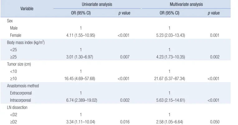
Table 2 shows univariate and multivariate analyses of risk fac- tors for IH after gastric cancer surgery. Incidence rate of IH was significantly higher among female patients and those who had intracorporeal procedure (p = 0.001 and p < 0.001, respectively).
BMI was significantly higher and tumor size was significantly larger in the IH group (p = 0.002 and p < 0.001, respectively). In the multivariate analysis, female sex, higher BMI (≥25 kg/m
2), larger tumor size (≥10 cm), and intracorporeal procedure were independent significant risk factors for IH. D2 lymph node dis- section was a significant risk factor for IH in univariate analysis, but it was not significantly different between the IH and non-IH groups in multivariate analysis.
Of the patients who had extracorporeal procedure, two had IH in the umbilical area, two in the epigastric area, and five at other port sites. All of the patients who had intracorporeal procedure had IH in the umbilical area (Table 3). Application of mesh dur- ing IH repair was based on the surgeon’s preference; thus, 14 pa- tients received mesh reinforcement, while nine patients did not.
There were two cases of reoperation for IH repair and both oc- curred in patients who did not have mesh reinforcement during the first hernia repair.
Table 2.
Table 2. Risk factors for incisional hernia after laparoscopic gastric cancer surgery Variable
Variable Univariate analysis Univariate analysis Multivariate analysis Multivariate analysis OR (95% CI)
OR (95% CI) pp value value OR (95% CI) OR (95% CI) pp value value
SexMale 1 1
Female 4.11 (1.55–10.95) <0.001 5.23 (2.03–13.43) 0.001
Body mass index (kg/m2)
<25 1 1
≥25 3.01 (1.30–6.97) 0.007 4.23 (1.73–10.35) 0.002
Tumor size (cm)
<10 1 1
≥10 16.45 (4.69–57.68) <0.001 21.67 (5.37–87.34) <0.001
Anastomosis method
Extracorporeal 1 1
Intracorporeal 6.74 (2.389–19.02) 0.002 5.63 (2.15–14.61) <0.001
LN dissection
<D2 1 1
≥D2 3.34 (1.11–10.04) 0.016 2.58 (1.05–6.64) 0.050
OR, odds ratio; CI, confidence interval; LN, lymph node.
Table 3.
Table 3. Characteristics of incisional hernia in the present study Variable
Variable Patient Patient (n = 23) (n = 23)
Hernia siteLaparoscopic or robot-assisted gastrectomy 9
Umbilical area 5 (55.6)
Epigastric area 2 (22.2)
Other port sites 2 (22.2)
Totally laparoscopic or robotic gastrectomy 14
Umbilical area 14 (100)
Other port sites 0 (0)
Mesh application during incisional hernia repair 23
Mesh reinforcement 14 (60.9)
No mesh reinforcement 9 (39.1)
Reoperation of recurrent incisional hernia 2 Mesh reinforcement during the first hernia repair 0 (0) No mesh reinforcement during the first hernia repair 2 (100) Values are presented as number (%).
DISCUSSION
MIS is becoming popular for stomach surgeries. Incidence and risk factors for IH after open surgery are well known, but study of IH after MIS for gastric cancer is limited [19–21]. Recently, although not with respect to gastric cancer surgery, there have been reports that IH occurs more frequently in single-incision laparoscopic surgery (SILS) than in laparoscopic surgery us- ing multiple ports [22–26]. This is presumed to be due to the large umbilical port made in SILS. In recent years, TLRG, with intracorporeal gastric resection and reconstruction and speci- men removal through extension of the umbilical port, tends to be preferred because it eliminates the need for additional mini- laparotomy. Moreover, SILS is also increasingly being attempted by many surgeons. Overall, umbilical port extension is becoming common; and therefore, the risk for IH, especially in the umbili- cal area, is increasing. However, only few studies have shown tendency of increased IH rate after intracorporeal procedure using extended umbilical incision, especially in gastric cancer surgery [27]. In this present study, it was clearly observed that IH occurred more frequently after intracorporeal procedure than after extracorporeal procedure. Furthermore, incidence rate of IH was significantly higher with female sex, high BMI, and large tumor.
For gastric cancer surgery, high BMI have been shown as risk factors for IH in previous studies [27]. High BMI has also been consistently identified as a risk factor for IH irrespective of the type of surgery, site of operation, and use of open surgery or laparoscopic surgery. The results of our study also support exist- ing studies [27].
Most existing studies evaluated risk factors for IH differently by sex, but a previous study on gastric cancer surgery reported female sex as a significant risk factor [27]. IH through umbilical incision was frequently encountered in this study, but the reason for its high incidence among females is unclear. It is supposed that the fat distribution of females is more accumulated in the subcutaneous layer than in intra-abdominal viscera as found in males. Thick subcutaneous fat of obese female patients therefore makes the closure of fascia difficult and results in IH [28].
Large tumor size was also a significant risk factor for IH in this study. Surgeons attempted to make the smallest possible in- cisions for extraction of the specimens. The larger the tumor, the larger the specimen, and the longer the incision needed for speci- men removal. Hence, extended umbilical incision for removing large specimen during TLRG would have increased the risk of IH [29].
As the result of this study, umbilical wound is more vulnerable to IH than epigastric wound, especially in patients who have risk factors like high BMI, female sex, and large tumor size. There- fore, surgeons should take care to prevent IH in such patients and should be cautious during closure of surgical wound.
Mesh reinforcement could be considered to prevent the recur- rence of IH after repair. There were two patients in this study who underwent reoperation due to recurrence of IH after repair.
The first patient was 74-year old female patient and she under- went LADG. IH was found at umbilical wound and her BMI was 23.7 kg/m
2. The second operation for recurred hernia was done 6 months after the first hernia operation. The second patient was 52-year old female patient and she underwent TLDG. IH was found at umbilical wound and her BMI was 30.7 kg/m
2. The sec- ond operation for recurred hernia was done 1 year after the first hernia operation. In a previous study, mesh reinforcement re- duced the recurrence rate of IH [30]. Therefore, surgeons should be mindful of the possibility of IH recurrence and consider mesh reinforcement during hernia repair in order to prevent recur- rence.
This study has several limitations. First, it has the inherent limitations of a retrospective study from one center even though the data volume is large; accordingly, some potential risk factors for IH, such as specific suture technique and wound infection, could not be analyzed. Second, diagnosis of and decision to re- pair IH were up to surgeons’ experiences and preferences. Some surgeons opted for surgical hernia repair, while other surgeons decided on observation for similar cases. Therefore, selection bias could have existed, and the number of IH cases could have been underestimated. Third, these results may not accurately represent the real incidence of IH because not all patients were followed up postoperatively in our institution. Fourth, wound infection and suture material or method could affect IH but those were excluded to analyze because of lack of data. It should be included to analyze in further study.
In conclusion, IH after MIS for gastric cancer patients is not very common. Female sex, high BMI, large tumor size, and in- tracorporeal procedure were significant risk factors for IH after MIS for gastric cancer in this study. Therefore, in patients with risk factors, surgeons should cautiously close the surgical wound after MIS for gastric cancer, in order to prevent IH.
NOTES
Ethical statements
The Institutional Review Board of the National Cancer Center approved this study (No. NCC2020-0182). The study was con- ducted according to the principles of the Declaration of Helsinki.
Informed consent was waived because of the retrospective nature of the study.
Authors ’ contributions
Conceptualization: SCC, KWR
Formal analysis: All authors Methodology: SCC, KWR Writing–original draft: SCC
Writing–review & editing: All authors
All authors read and approved the final manuscript.
Conflict of interest
All authors have no conflicts of interest to declare.
ORCID
Sung Chun Cho, https://orcid.org/0000-0003-2667-4225 Bang Wool Eom, https://orcid.org/0000-0002-0332-2051 Hong Man Yoon, https://orcid.org/0000-0002-6218-7080 Young-Woo Kim, https://orcid.org/0000-0002-1559-9672 Keun Won Ryu, https://orcid.org/0000-0002-5935-9777
REFERENCES
1. Diener MK, Voss S, Jensen K, Büchler MW, Seiler CM. Elective mid- line laparotomy closure: the INLINE systematic review and meta- analysis. Ann Surg 2010;251:843-856.
2. Fink C, Baumann P, Wente MN, et al. Incisional hernia rate 3 years after midline laparotomy. Br J Surg 2014;101:51-54.
3. van’t Riet M, Steyerberg EW, Nellensteyn J, Bonjer HJ, Jeekel J. Meta- analysis of techniques for closure of midline abdominal incisions. Br J Surg 2002;89:1350-1356.
4. Athanasiou CD, Robinson J, Yiasemidou M, Lockwood S, Markides GA. Laparoscopic vs open approach for transverse colon cancer. A systematic review and meta-analysis of short and long term out- comes. Int J Surg 2017;41:78-85.
5. Cheng Y, Xiong XZ, Wu SJ, Lin YX, Cheng NS. Laparoscopic vs.
open cholecystectomy for cirrhotic patients: a systematic review and meta-analysis. Hepatogastroenterology 2012;59:1727-1734.
6. Xiong JJ, Nunes QM, Huang W, et al. Laparoscopic vs open total gas- trectomy for gastric cancer: a meta-analysis. World J Gastroenterol 2013;19:8114-8132.
7. Kössler-Ebs JB, Grummich K, Jensen K, et al. Incisional hernia rates after laparoscopic or open abdominal surgery: a systematic review and meta-analysis. World J Surg 2016;40:2319-2330.
8. Kim HH, Hyung WJ, Cho GS, et al. Morbidity and mortality of laparoscopic gastrectomy versus open gastrectomy for gastric cancer:
an interim report. A phase III multicenter, prospective, randomized Trial (KLASS Trial). Ann Surg 2010;251:417-420.
9. Kim YW, Baik YH, Yun YH, et al. Improved quality of life outcomes after laparoscopy-assisted distal gastrectomy for early gastric cancer:
results of a prospective randomized clinical trial. Ann Surg 2008;248:
721-727.
10. Lee HJ, Hyung WJ, Yang HK, et al. Short-term outcomes of a mul-
ticenter randomized controlled trial comparing laparoscopic distal gastrectomy with D2 lymphadenectomy to open distal gastrectomy for locally advanced gastric cancer (KLASS-02-RCT). Ann Surg 2019;
270:983-991.
11. Han WH, Yehuda AB, Kim DH, et al. A comparative study of totally laparoscopic distal gastrectomy versus laparoscopic-assisted distal gastrectomy in gastric cancer patients: short-term operative outcomes at a high-volume center. Chin J Cancer Res 2018;30:537-545.
12. Han WH, Oh YJ, Eom BW, Yoon HM, Kim YW, Ryu KW. A com- parative study of the short-term operative outcome between intra- corporeal and extracorporeal anastomoses during laparoscopic total gastrectomy. Surg Endosc 2020;35:1602-1609.
13. Obama K, Kim YM, Kang DR, et al. Long-term oncologic outcomes of robotic gastrectomy for gastric cancer compared with laparoscopic gastrectomy. Gastric Cancer 2018;21:285-295.
14. Parisi A, Reim D, Borghi F, et al. Minimally invasive surgery for gastric cancer: a comparison between robotic, laparoscopic and open surgery. World J Gastroenterol 2017;23:2376-2384.
15. Ooms LS, Verhelst J, Jeekel J, Ijzermans JN, Lange JF, Terkivatan T.
Incidence, risk factors, and treatment of incisional hernia after kid- ney transplantation: an analysis of 1,564 consecutive patients. Surgery 2016;159:1407-1411.
16. Seo GH, Choe EK, Park KJ, Chai YJ. Incidence of clinically relevant incisional hernia after colon cancer surgery and its risk factors: a na- tionwide claims study. World J Surg 2018;42:1192-1199.
17. Walming S, Angenete E, Block M, Bock D, Gessler B, Haglind E. Ret- rospective review of risk factors for surgical wound dehiscence and incisional hernia. BMC Surg 2017;17:19.
18. Guideline Committee of the Korean Gastric Cancer Association (KGCA); Development Working Group & Review Panel. Korean Practice Guideline for Gastric Cancer 2018: an evidence-based, multi- disciplinary approach. J Gastric Cancer 2019;19:1-48.
19. Jensen KK, Krarup PM, Scheike T, Jorgensen LN, Mynster T. In- cisional hernias after open versus laparoscopic surgery for colonic cancer: a nationwide cohort study. Surg Endosc 2016;30:4469-4479.
20. Lee L, Abou-Khalil M, Liberman S, Boutros M, Fried GM, Feldman LS. Incidence of incisional hernia in the specimen extraction site for laparoscopic colorectal surgery: systematic review and meta-analysis.
Surg Endosc 2017;31:5083-5093.
21. Lee L, Mata J, Droeser RA, et al. Incisional hernia after midline versus transverse specimen extraction incision: a randomized trial in pa- tients undergoing laparoscopic colectomy. Ann Surg 2018;268:41-47.
22. Julliard O, Hauters P, Possoz J, Malvaux P, Landenne J, Gherardi D.
Incisional hernia after single-incision laparoscopic cholecystectomy:
incidence and predictive factors. Surg Endosc 2016;30:4539-4543.
23. Agaba EA, Rainville H, Ikedilo O, Vemulapali P. Incidence of port- site incisional hernia after single-incision laparoscopic surgery. JSLS 2014;18:204-210.
24. Alhambra-Rodríguez de Guzmán C, Morandeira-Rivas AJ, Herrero- Bogajo ML, Moreno-Sanz C. Incidence and risk factors of incisional
hernia after single-incision endoscopic surgery. J Laparoendosc Adv Surg Tech A 2020;30:251-255.
25. Dhaou MB, Zouari M, Chtourou R, Zitouni H, Jallouli M, Mhiri R.
Incidence of incisional hernia after single-incision laparoscopic sur- gery in children. J Minim Access Surg 2017;13:240-241.
26. Hoyuela C, Juvany M, Guillaumes S, et al. Long-term incisional hernia rate after single-incision laparoscopic cholecystectomy is sig- nificantly higher than that after standard three-port laparoscopy: a cohort study. Hernia 2019;23:1205-1213.
27. Jang EJ, Kim MC, Nam SH. Risk factors for the development of in- cisional hernia in mini-laparotomy wounds following laparoscopic distal gastrectomy in patients with gastric cancer. J Gastric Cancer
2018;18:392-399.
28. Vermillion ST, Lamoutte C, Soper DE, Verdeja A. Wound infection after cesarean: effect of subcutaneous tissue thickness. Obstet Gyne- col 2000;95(6 Pt 1):923-926.
29. Comajuncosas J, Hermoso J, Gris P, et al. Risk factors for umbilical trocar site incisional hernia in laparoscopic cholecystectomy: a pro- spective 3-year follow-up study. Am J Surg 2014;207:1-6.
30. Jairam AP, Timmermans L, Eker HH, et al. Prevention of incisional hernia with prophylactic onlay and sublay mesh reinforcement ver- sus primary suture only in midline laparotomies (PRIMA): 2-year follow-up of a multicentre, double-blind, randomised controlled trial.
Lancet 2017;390:567-576.